
Abstract
Abstract
Purpose:
To achieve a safer alternative to intravitreal injection of corticosteroids, we developed and characterized triamcinolone acetonide-loaded liposomes formulations (TA-LFs) to be used topically for vitreoretinal drug delivery.
Methods:
Four different 0.2% TA-LFs (TA-LF1 to TA-LF4) were generated and submitted to physicochemical characterization. Posteriorly, an ex vivo diffusion assay was performed using rabbit corneas as membranes. Finally, concentrations of triamcinolone acetonide (TA) were determined by high-performance liquid chromatography in ocular tissues from New Zealand white rabbits after multiple topical doses of TA-LF2 (6 times per day, 14 days). In addition, toxicity and tolerability of TA-LF2 was evaluated by cell viability assay and eye examination of study animals, respectively.
Results:
TA-LF2 was the most stable formulation maintaining a stable hidrogenion potential (pH) at 30 and 40°C and even improving encapsulation with higher temperature. TA-LF2 and TA-LF3 presented the best diffusion performance in vitro reaching the highest TA concentrations after 8 h of follow-up. In vivo diffusion and pharmacokinetics analysis showed that concentrations of TA in retina and vitreous reached the highest peak at 12 h after topical administration of TA-LF2 (252.10 ± 90.00 ng/g and 32.6 ± 10.27 ng/g, respectively) and subsequently decline to 24.0 ± 11.72 ng/g and 19.5 ± 13.14 ng/g, respectively, at 14 days of follow-up. Finally, cell viability was unaffected by TA-LF2, and no increase in intraocular pressure nor ocular alterations were observed after topical administration of this formulation in rabbits.
Conclusion:
TA-loaded liposomes, administered topically, can deliver TA in the vitreous cavity and reach the retina efficiently.
Introduction
C
Although, intravitreal injection of TA is a well-described and capable route to release this corticosteroid into the posterior pole of the eye, this procedure is not exempt of potential severe complications such as endophthalmitis, lens injury, and retinal detachment.6–8 In addition, clinical studies have related the use of intravitreal TA with intraocular pressure (IOP) increase, cataract formation or progression, and noninfectious endophthalmitis.9–11 As expected, the risk for these complications is higher in patients with ophthalmic diseases that require repeated intravitreal injections of TA over a period of time, such as, macular edema or uveitis.
To diminish ocular hazards related to intravitreal injections of TA, it is necessary to develop alternative strategies for drug delivery. Among drug delivery methods, liposomes (LPs) represent a promising strategy. LPs are particles composed of an aqueous core and delimited by a membrane-like lipid bilayer that works as carriers for water-soluble, lipid-soluble, and amphiphilic drugs.12–15 Their size, lipid composition, permeability, and electric charge can be easily manipulated.13,16,17 Besides, LPs are nontoxic, low antigenic, metabolized, and biodegradable. 18 LPs in ocular diseases as vehicles for topical administration of anti-inflammatory drugs was first reported in 1982. 19 Since then, LPs have been used to improve the drug transport and bioavailability in ocular tissues.20,21
Different reports have demonstrated the efficacy of liposomal formulations for drug transport into ocular posterior segment; releasing drugs in a controlled manner, increasing their half-life (t1/2) and avoiding toxicity by requiring lower doses. However, in these studies LPs are delivered by intravitreal injection. The drugs used in these studies have included ganciclovir, 22 amikacin, 23 amphotericin B, 24 antisense oligonucleotides, 25 bevacizumab, 26 and cyclosporine among others. 27
On the contrary, information about pharmacokinetics of topical TA in posterior ocular tissues is limited, whereas pharmacokinetics of intravitreal TA has been constantly revised. For instance; in a group of white New Zealand rabbits with a single intravitreal injection of 6 mg of TA and 8 months of follow-up, 28 the highest concentration of TA in vitreous was recorded at day 1 (14,434.0 ± 10,768 μg/L), while the highest concentrations in the anterior chamber was reported at day 3 (28.9 ± 24.5 μg/L). A 2-compartment model of elimination, in both parts of the eye, was observed: first a burst period with exponential reduction within the first 4 weeks, then a stable drop over the following months. For the 8th month, concentrations were roughly 200 times lower compared with the burst period: in the anterior chamber were 3.3 ± 1.6 μg/L and 70.7 ± 37.0 μg/L in the vitreous. In an additional comparable work, 29 that studied the TA pharmacokinetics of the intravitreal injection of 4 and 8 mg of the drug in rabbits, TA concentration showed an initial peak (burst period) and diminished progressively over time (duration period). TA half-life was 24 days for the 4 mg injection and 34 days for the 8 mg injection. Importantly, no substantial change in IOP was identified nor signs of impairment in the retina on any study.
Thus, to achieve a safer alternative to intravitreal injection of corticosteroids for disorders that affect retina and vitreous, we developed triamcinolone acetonide-loaded liposomes formulations (TA-LFs) to be used topically for vitreoretinal drug delivery. In the methods section, we show the physicochemical characterization and pharmacokinetics of these TA-LFs. In brief, we found that TA-LFs are suitable for ophthalmic use and can release TA efficiently in the vitreous and retina.
Methods
Preparation of liposomal formulation
OPKO Health, Inc. (Guadalajara, Mexico) provided 4 TA-LFs (TA-LF1 to TA-LF4). Self-forming, thermodynamically stable loaded LPs (QuSomes®) were generated spontaneously upon adding polyethylene glycol (PEG)-12 glyceryl dimyristate to an aqueous solution containing TA. Composition of TA-LFs are described in Table 1. LFs were extruded through a size-controlled 0.22-μm pore size polycarbonate membrane for 10 cycles under nitrogen pressure (≤200 psi) to obtain lipid vesicles with a unimodal size distribution. Final TA concentration in the resultant dispersion was 2 mg/mL (0.2%). Control liposome formulations (cLFs) were made with the same composition of experimental LFs, except for the TA content (cLF1 to cLF4).
PEG, polyethylene glycol; TA-LF, triamcinolone acetonide-loaded liposomes formulation.
Characterization of liposomal formulation
To evaluate the efficiency of encapsulation, we measured the concentration of TA in the filtered solution obtained after TA-LFs, and their cLFs were extruded through a size-controlled 0.22-μm pore size polycarbonate membrane under nitrogen pressure (≤200 psi). The concentration of TA on TA-LFs samples was subtracted to the concentration of TA in cLFs, and the result was expressed as percentage of encapsulation. The analysis of the TA concentration was performed by high performance liquid chromatography (HPLC) using a Varian 920 LC (Agilent Technologies, Santa Clara, CA) with a Zorbax Eclipse Plus C18, 4.6 × 100 mm and 3.5-μm column (Agilent Technologies) at 30°C. The samples (20 μL) were eluted from the column in a mobile phase comprised water:methanol (30:70) at a flow rate of 1 mL/min. Detection was performed at 254 nm. Retention time and detection limit were 6.8 min and 0.004 mg/mL, respectively. The TA standard curve was linear from 0.004 to 0.100 mg/mL (correlative >0.99). In vitreous, concentrations of TA were determined for the recovery and intra- and interday reproducibility. 30
Hidrogenion potential (pH) of TA-LFs was analyzed by a pH meter in triplicate at room temperature. Osmolarity was measured by a vapor pressure osmometer and performed in triplicate at 33°C (the ocular surface temperature). 31 Viscosity was measured also in triplicate at 33°C. Viscosity was measured using a thermostatically controlled rheometer when the steady state was reached with shear rates increasing from 0 to 1,000 s−1. Particle size of the TA-LFs was analyzed by means of Dynamic Light Scattering and zeta potential (ζ) was calculated by measuring the velocity of the particles using Laser Doppler Velocimetry at 25°C (Zetasizer Nano ZS; Malvern Instruments, Malvern, UK). The Z-average (mean particle diameter) and polydispersity index (PDI) were calculated from the particle size distribution.
To evaluate the stability of the TA-LFs, a thermic stress assay was performed. LFs samples were kept at 30°C, 40°C, and 60°C for 3 weeks. pH and encapsulation efficiency were evaluated on days 0 (baseline) 7, 14, and 21 of thermal incubation as previously described.
In vitro diffusion analysis of TA-loaded LPs
Diffusion chambers and rabbit corneas were used to conduct diffusion experiments (Chemotaxis Chambers BW200S; Neuro Probe, Gaithersburg, MD). Rabbit corneas from New Zealand white rabbits used for experiments were obtained from Centro de Investigación Biomedica de Occidente (CIBO) at Guadalajara, Mexico. We excised a circle of 6 mm in diameter from the central cornea using a trephine. This corneal tissue was located between the top and bottom compartments of the diffusion chambers to act as a TA diffusion barrier. The top compartment was filled with 180 μL of balanced salt solution (BSS), while the bottom compartment was filled with 200 μL of TA-LFs (TA-LF1 to TA-LF4). To avoid evaporation, the diffusion chambers were located into a 37°C humidity camera. The TA concentration analysis of solutions, obtained from the top compartment at 2, 4, 6, and 8 h after starting the diffusion assay, was performed by HPLC as previously described.
Cell viability assay for TA-LF2
Cell viability assay was performed to evaluate toxicity for TA-LF2. This formulation resulted as the most suitable for topical use, due to its physicochemical characteristics, stability, and diffusion profile in vitro. For this experiment, primary human corneal fibroblasts (HCFs) were isolated and cultured as previously described. 32 HCFs were derived from the stroma of human corneal rims obtained after removal of central tissue for transplantation. Tissues were provided by Corneal Transplantation Unit of Instituto Mexicano del Seguro Social (Guadalajara, Jalisco, Mexico). All procedures used in the cell viability assay adhered to the tenets of the Declaration of Helsinki.
Once HCFs were isolated, they were expanded in Dulbecco's modified Eagle's medium (DMEM; Sigma-Aldrich, St. Louis, MO) supplemented with 10% fetal bovine serum (FBS; Corning), 1% glutamine (Sigma-Aldrich), and 1% penicillin/streptomycin (Sigma-Aldrich). Cells were used at passage 3. After harvesting, using trypsin-EDTA, the cells were diluted in DMEM supplemented with FBS at 3.0 × 10⁵/mL, 100 μL of the suspension were seeded onto 96-well plate. After 24 h of cell inoculation, a visual validation was performed to check the monolayer formation by using an inverted microscopy (Nikon). Later, HCFs cultures were exposed to 2 different concentrations of TA-LF2, 5% and 20%. The lowest concentration corresponds to that found in the final tear volume after instillation of a drop of TA-LF2 (5 μL/10 μg), and the subsequent concentration was increased 4 times (20 μL/40 μg). One hundred microliters of the 5% suspension or 20% suspension were added to different plates of the cell culture to be exposed for 1, 5, 15, and 20 min, respectively. Exposure time was based on the normal tear flow rate (0.5–2.2 μL/min). Each culture condition was performed by triplicate.
Finally, after each time of exposure was reached, MTT assay was applied (Sigma-Aldrich) based on the protocol described by Mosmann. 33 A positive control was added. Absorbance was recorded at 650 nm using the microplate spectrophotometer system (Bio-Rad, Hercules, CA). The absorbance obtained from the positive controls was subtracted to exposed wells for analysis. Results of the cell viability are presented as percentage of the control values and with its respective standard deviation.
In vivo diffusion analysis of TA-LF2 and tolerability assessment
Animals
The study received approval by the Bioethics Committee of the Centro Universitario de Ciencias Exactas e Ingenierias, Universidad de Guadalajara, Guadalajara, Jalisco, Mexico; it was also conducted according to the Association for Research in Vision and Ophthalmology Statement for the Use of Animal in Ophthalmic and Vision Research. Twenty New Zealand white rabbits (2–2.5 kg each) acquired from CIBO were acclimatized and housed in clean cages for at least 1 week before they were enrolled in the study.
Treatment protocol
Rabbits were randomly distributed into 4 groups. One-drop TA-LF2 solution (50 μL) was applied to 1 eye every 2 h 6 times during 14 days. Five rabbits were sacrificed after starting the instillation of TA-LF2 at 12 h, 1, 7, and 14 days. Before tissue collecting, an eye examination was performed under anesthesia (intramuscular injection of ketamine hydrochloride 30 mg/kg and chlorpromazine hydrochloride 15 mg/kg). This evaluation included slit-lamp biomicroscopy, fluorescein staining, funduscopy with direct ophthalmoscope, and IOP measurement (iCare Tonometer i350, Vantaa, Finland). In addition, ocular irritability test was evaluated according to pharmacopeia of Estados Unidos Mexicanos. A positive irritant reaction is considered when more than 1 rabbit presented the following: cornel ulceration revealed by fluorescein staining, corneal opacity, iris or conjunctival inflammation, and dilatation of conjunctival vessels, especially around the cornea. After enucleation, conjunctiva, cornea, retina, 150 μL of aqueous humor, and 200 μL of vitreous were collected. The solid tissues were washed in phosphate-buffered saline. Then, tissues were homogenized with 0.3 mL of acetonitrile (Sigma-Aldrich, Toluca, Mexico). Posteriorly, each sample was centrifuged at 15,294g for 5 min. The supernatants were evaporated to add 100 μL of methanol. Another centrifugation was performed and 20 μL of the resultant supernatants were used for analysis of TA concentration by HPLC, performed as previously described.
Pharmacokinetics analysis
A compartmental and a noncompartmental models were used to determine pharmacokinetics of TA-loaded LPs in ocular tissues. Linear-trapezoidal method was used to evaluate the area under the curve. The half-life was calculated by linear regression of the concentration at different times.
Statistical analysis
Values are presented as means ± standard deviations of the mean. To compare 2 groups, Student's test (paired groups) was used in case of data normally distributed or the Wilcoxon signed-rank test for non-normally distributed. For more than 2 groups, we performed analysis of variance for correlated samples. Then, Bonferroni posttests were used for determination of statistical significance. Significance was defined as a P value <0.05.
Results
TA-LFs shown diverse physicochemical characteristics
The physicochemical characteristics, of the 4 TA-LFs are summarized in Table 2. The pH of the TA-LFs was acid in all cases, and nearby 6 (from 5.8 to 6.24). Physiologic pH range is between 6.5 and 7.6, 34 hence TA-LF1 is near normal pH of tears and is more recommendable for use in human eyes. Then again, viscosity values between 15 and 25 centipoise (cP) are considered optimal for ophthalmic applications, hence, TA-LF3 is adequate for eye drops.35,36 TA-LF1 and TA-LF2 were found in isotonic margins according to the criteria of the Pharmacopoeia of the United Mexican States where a substance is considered as nonirritating to the eye between 205 and 684 mOsm/L, although it allows hypertonic ophthalmic solutions to promote absorption and provide a suitable concentration of active ingredients for an effective therapeutic action. TA-LF3 showed the smallest particle diameter, whereas zeta potential (ζ) of all formulation was near to 0, therefore, there is a risk of crystallization of the TA encapsulated in the micelles.
Values represent the average of 3 measures.
ζ, zeta potential; cP, centipoise; PDI, polydispersity index; pH, hidrogenion potential.
TA-LF2 was the most stable
After 21 days of thermic stress, TA-LF2 exhibited the best performance, keeping pH stable at 30 and 40°C and even improving encapsulation with higher temperature (60°C). TA-LF1 was the most unstable with very low encapsulation efficiency at any temperature. TA-LF3 and TA-LF4 presented elevated percentage of encapsulation, however, presented macroscopic decomposition after 21 days of thermal stress (data not shown). pH and encapsulation efficiency values of LFs in the stability assay are presented in Table 3.
Values represent the average of 3 measures.
TA, triamcinolone acetonide.
TA-LF2 and TA-LF3 presented the best diffusion performance in vitro
To evaluate the diffusion of TA-LFs in vitro, diffusion chambers were used. These devices analyze the diffusion of pharmacological agents through animal or human tissue. In this case, we analyze diffusion of TA-LFs through rabbit corneas. We observed that TA-LF2 and TA-LF3 presented the best diffusion performance, reaching the highest TA concentrations after 8 h of follow-up. Table 4 summarizes the TA values observed during the in vitro (ex vivo) diffusion analysis.
Values represent the average of 3 measures.
Formulation 3 has a more uniform particle size distribution than the others, which explains its greater permeability through the cornea than the others. Although formulation 4 has a particle size slightly greater than that of formulation 3, its diffusion through the sclera is possibly superior due to its chemical structure and its compatibility with the chemical structure of the sclera.
Topical TA-LF2 reached vitreous and retina
To evaluate the diffusion in vivo and characterize the pharmacokinetic behavior of the formulation, concentrations of TA were determined by HPLC in ocular tissues from New Zealand white rabbits after multiple doses of TA-LF2, the only formulation suitable for topical. In addition, tolerability of TA-LF2 was evaluated by eye examination of study animals.
The concentrations of TA in retina and vitreous reached the highest peak at 12 h (252.1 ± 90.00 ng/g and 32.6 ± 10.27 ng/g, respectively) to subsequently decline to 24.0 ± 11.72 ng/g and 19.5 ± 13.14 ng/g, respectively, at 14 days of follow-up. TA concentration versus time in different ocular tissues is presented in Fig. 1 and Table 5.

Concentration of TA in ocular tissues after topical administration of TA-LF2 in rabbit eyes across the time. TA, triamcinolone acetonide; TA-LF, triamcinolone acetonide-loaded liposomes formulation.
Values represent the average ± standard deviation of the mean of 4 samples.
Pharmacokinetic parameters are shown in Table 6. Cmax was 2,156.07 ± 1,055.41 ng/g in cornea, 1,886.33 ± 398.95 ng/g in conjunctiva, 9.9 ± 1.95 ng/g in aqueous humor, 83.3 ± 30.49 ng/g in lens, 32.6 ± 10.27 ng/g in vitreous, and 252.10 ± 90.00 ng/g in retina.
AUC0−∞, area under the curve from 0 to infinity; AUC0−t, area under the curve until the last measurable; Cmax, observed maximum concentration; Cmin, observed minimum concentration; k12, rate of transfer from central to peripheral compartment; k21, rate of transfer from peripheral to central compartment; ke, elimination rate constant; t1/2, elimination half-life.
TA-LF2 did not affect human cell fibroblast viability in vitro, and it was well tolerated in vivo
Cell viability assay was performed to evaluate cytotoxicity of TA-LF2. Any significant change in cell viability was recorded after exposition of HFCs to 2 different concentrations of TA-LF2 (5% and 20%) for 1–20 min (Fig. 2). Finally, no increase in IOP was observed in any of the study subjects (normal IOP in this species is 12–28 mmHg). Staining with fluorescein sodium and bengal rose showed superficial punctate keratitis in the first 6 h after instillation of the formulation. This condition was resolved in all cases in the examination at 12 h after the administration of the formulation. Therefore, according to pharmacopeia of Estados Unidos Mexicanos, ocular irritability test was satisfactory, and TA-LF2 is considered nonirritant.
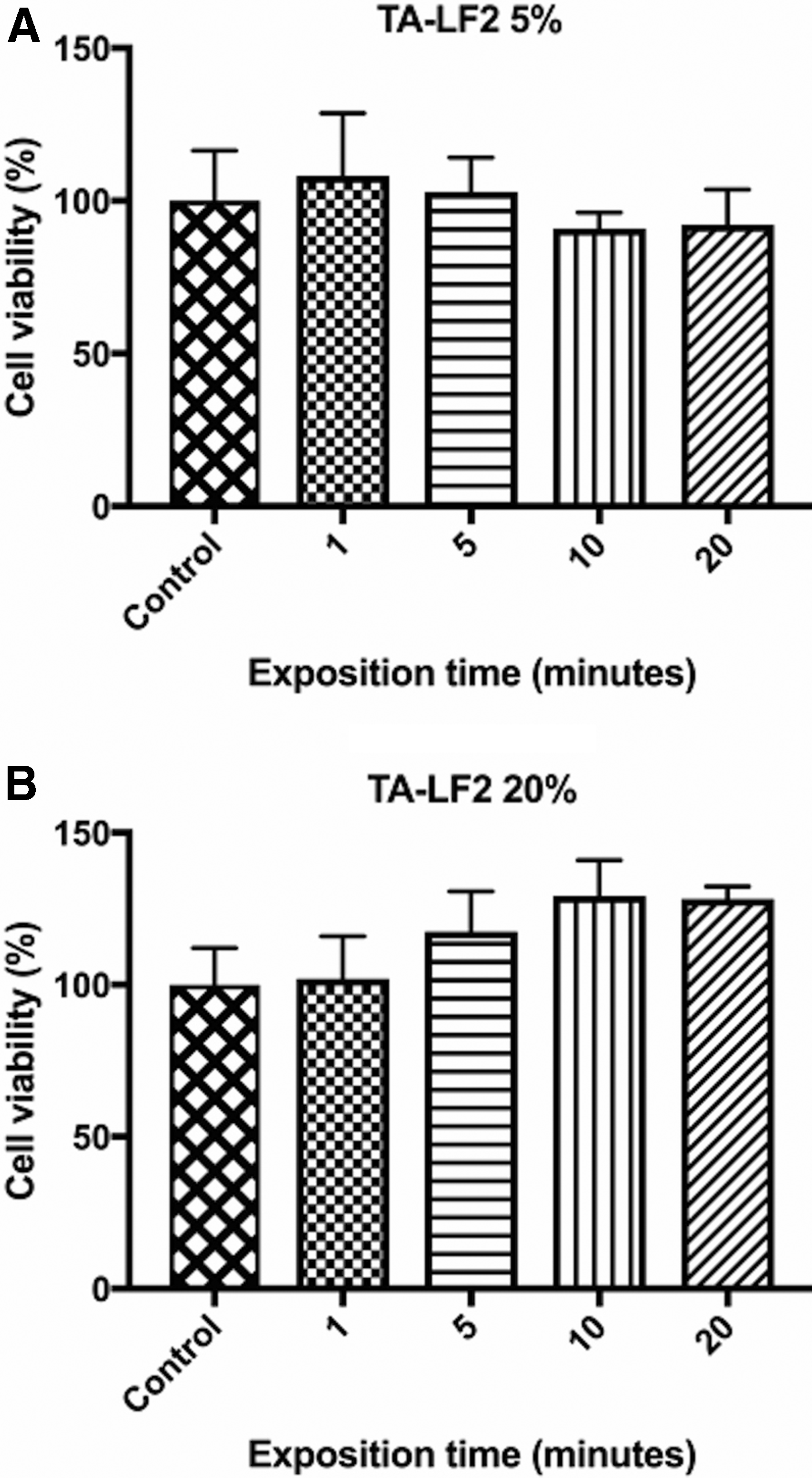
Cell viability assay. After exposition of HCFs for 1–20 min to
Discussion
Corticosteroids play a major role in the management of different vitreoretinal diseases due to its ability to regulate angiogenic and inflammatory genes; for example, downregulating expression of vascular endothelial growth factor (VEGF) and interleukin 6.37,38 Nevertheless, because of both outer and inner blood–retinal barriers, oral or parenteral corticosteroids scarcely get into the vitreous cavity. Thus, to increase corticosteroids penetration, intravitreal injections have been widely used. 1 TA is an injectable synthetic corticosteroid formulated as suspension with a powerful anti-inflammatory activity (7.5 times more potent than cortisone). 39 Because TA cannot be dissolved in water, it is capable of remaining in the vitreous longer than other corticosteroids, which are eliminated within a few days. Due to its properties, TA is commonly used for the management of posterior ocular illnesses unresponsive to topical corticosteroids.3–5
TA is usually used by ophthalmologist in intravitreal injections at concentration of 4 mg/0.1 mL.40,41 However, despite its low cost, the risk of severe potential complications, inherent to intravitreal injection, such as infectious and noninfectious endophthalmitis, retinal detachment, cataract formation, and/or vitreous hemorrhage,9–11,42 as well as the risk of increased IOP, 43 have limited its use for long periods. In fact, severe 44 and intractable45,46 IOP elevation represents the major obstacle to steroid use. Besides, the discomfort caused to the patient by the intraocular injection itself, could cause pour adherence to the treatment. Consequently, it is imperative to offer alternatives to deliver TA into the vitreous cavity.
With this work, we probed that TA-loaded LPs, administered topically, can release TA into the vitreous cavity and reach the retina efficiently. Also, we proved that TA-LF2 is associated with higher TA concentration in ex vivo diffusion assay and shows adequate tolerability in the in vivo analysis. However, it is important to consider that components of TA-LF2 formulation other than LPs such as Kolliphor® HS 15, could increase the efficiency of the vitreous release of TA. The PEG (15)-hydroxystearate or Kolliphor HS 15 is a potent nonionic solubilizer and emulsifying agent, with low toxicity proposed to act as a permeability enhancer, promoting drug transport across cell membranes (increasing the endocytosis rate) and promoting drug translocation through the paracellular route (affects actin organization on the cell cytoskeleton with the subsequent tight junction opening). 47
It is worth mentioning that, although the concentration of TA achieved in vitreous by topical administration of TA-loaded LPs was lower in contrast to TA concentration reached by intravitreal injection in rabbits (252.10 ± 90.00 ng/g and vs. 14,434.0 ± 10,768 μg/L), 28 steroids efficiency is not only determined by the potency and dosage but also on the bioavailability in the ocular tissue. An increment in dose not necessarily boosts the effectiveness.48,49 As it was presented in the Results section, concentration of TA in vitreous was constant, while in retina remained above vitreous concentration at all time-points. Therefore, LFs deliver TA in vitreous and retina efficiently, maintaining drug availability, however, at low concentration. This could be of benefit, because low concentrations diminish the risk of toxicity 50 and remain therapeutic. For instance, in the fluocinolone acetonide (FA) intravitreal implant of 0.2 μg/day study, the Cmax was barely 1.26 ng/g in vitreous and 12.2 ng/g in retina of rabbits, 51 however, the implant proved to be efficient in patients with diabetic macular edema through 36 months of follow-up (improvement of ≥15 letters).48,52
Concerning the pharmacokinetics of TA-loaded LPs, after analysis of the results, we propose a model that explains the distribution and elimination of TA. According to this model, TA absorption is carried out mainly through cornea and conjunctiva with posterior disposition of the drug to aqueous humor; however, the aqueous humor replacement capability allows its removal quickly from the anterior ocular chamber. Successively aqueous transfers this concentration to lens and from there to the vitreous. Vitreous humor apparently behaves like a reservoir, where TA concentrations are constant during treatment. Retina, which is the compartment of interest, reaches higher concentrations than those observed in the vitreous humor. In this tissue, a replacement phenomenon occurs, and it is in this compartment, that the disappearance of the drug takes place with a first order elimination kinetics. Although this model represents the expected pharmacokinetics, different considerations must be made. For instance, in vitrectomized rabbit eyes, concentration of TA declined 1.5 times faster versus nonvitrectomized rabbit eyes, similarly half-life is shorter in vitrectomized eyes.53,54 Therefore, effects of vitrectomy on TA pharmacokinetics should be further investigated and incorporated to the proposed model. In addition, when using animal models for pharmacokinetic analyzing, anatomy and physiology differences between species should be taken into account.
Regarding pharmacodynamics, presumably the metabolism of TA in retina is carried out by the CYP3A family of enzymes, the most important CYP enzymes associated in glucocorticoids (GCs) metabolism. This family includes the proteins CYP3A4, −5, −7, and −43. The expression of this enzymes is distinguished by tissue and age.55,56 GCs are able to induce CYP3A expression, and messenger RNAs (mRNAs) of all isoforms.57–59 In the eye, the expression of CYP3A genes has been identified. CYP3A4 has been recognized in human cornea and retina/choroid tissues, 60 whereas CYP3A8 (an homologous to human CYP3A4) has been identified in retina of monkeys, which is upregulated by dexamethasone. 61 In the liver, CYP3A enzymes' regulation, when in treatment with GCs, had been broadly described, but not for the eye. In liver cells, CYP3A stimulation is facilitated by pregnane X receptor (PXR)62,63 or via glucocorticoid receptor (GR) and constitutive androstane receptors (CAR).64,65 Consistently, when in the cytosol, GCs bind to GR and form a homodimer that translocates into the nucleus, which in turn increases transcription of CAR. Then, CAR binds to retinoid X receptor alpha (RXRα), forming a heterodimer that interacts with RXR-response element, inducing the expression of CYP3A to metabolize GCs, including TA. 64
On the contrary, although TA-LF2 did not affect cell viability in the in vitro experiment and it was well tolerated in the in vivo assay, other issues should be considered when planning its use in humans. For instance, blurred vision and ocular comfort could be an important issue to be addressed; due to LF2's elevated viscosity and slightly acid pH.
Finally, different studies have established the effectiveness of liposomal formulations delivery by intravitreal injection.22–24,26 However, to the best of our knowledge, this is the first report that analyze topical LPs use for delivering drugs into the posterior ocular segment. As we have shown, liberation of drugs into vitreous cavity and retina using topical loaded LPs is feasible. Hence, the use of topical formulations of LPs, instead of intravitreal injections for drug delivery in ocular diseases of the posterior segment, could result in an accessible and safer therapy, since neither special infrastructure nor trained professionals are required for its administration. Moreover, complication related to intravitreal injection would be avoided.
In conclusion, TA-LFs are suitable for ophthalmic use and are capable to release TA efficiently into the vitreous cavity and retina. TA-LFs have the potential to treat many vitreoretinal diseases due to its capacity to regulate angiogenic and inflammatory genes,37,38 however, biological and therapeutic activity should be confirmed in further studies. In addition, we probed that LPs could be an alternative to intravitreal injection as a drug delivery system, resulting in a safer and more accessible option of treatment.
Footnotes
Author Disclosure Statement
This research is sponsored by OPKO Health, Inc. and may lead to the development of products.