
Abstract
Abstract
Purpose:
To investigate the ocular penetration of natamycin (NAT) and voriconazole (VRC) after topical instillation in New Zealand white rabbits using simplified liquid chromatography–tandem mass spectrometry (LC-MS/MS) and high-performance liquid chromatography.
Methods:
Seventy-eight healthy rabbits were randomly divided into 3 groups. In the first 2 groups, 72 rabbits were used for single-dose testing (36 for NAT, 36 for VRC), in which 50 μL of 5.0% NAT or 1.0% VRC was instilled into the rabbits' left eyes. In the 3rd group, 6 rabbits were used for repeated-dose testing in which 50 μL of 5.0% NAT was instilled into their left eyes 12 times (once per hour) during the daytime. These animals were sacrificed immediately to collect their aqueous humors and corneas.
Results:
After a single topical instillation, the highest concentrations in the cornea and aqueous humor for VRC were 34.1 μg/g and 14.7 μg/mL, respectively. The permeability ratios of aqueous/cornea were from 0.1 to 1.26. The highest concentrations in cornea and aqueous humor for NAT were 299.3 ng/g and 27.1 ng/mL, respectively. The permeability ratios of aqueous/cornea were from 0.02 to 0.23. In the repeated-dose group, the NAT concentrations in the cornea and aqueous humor were 10,569 ng/g and 54.4 ng/mL, respectively. The permeability ratio was as low as 0.0051.
Conclusion:
The better corneal penetration of VRC suggests that it is more suitable for deep corneal fungal infections than NAT via topical ocular administration.
Introduction
F
Natacyn® is presently the only commercially available topical antifungal drug approved by the Food and Drug Administration (FDA). Although effective against the filamentous fungi, particularly Fusarium and Aspergillus species, it is less effective against Candida. 6 A new generation of broad-spectrum antifungal voriconazole (VRC) is widely used in systemic treatment of filamentous fungal infections.7–10 Recent studies have confirmed that the drug is safe for intraocular use by intravitreal injection. 11 Our study investigated the penetration and pharmacokinetics of VRC eye drops compared with Natacyn by topical administration in rabbits. Moreover, we developed sensitive and reliable liquid chromatography–tandem mass spectrometry (LC-MS/MS) and high-performance liquid chromatography (HPLC) methods to accurately determine NAT and VRC concentrations in rabbit ocular tissue. According to what we have learnt, only 2 articles reported using the LC-MS/MS bioanalytical method for analysis of NAT. One was for ocular pharmacokinetics, the other for an estimation of NAT in rabbit and human plasma.12,13 They both applied solid-phase extraction for sample preparation, which is a high-cost and rate-limiting step to achieve high-throughput analysis. Our method not only has a simpler sample pretreatment, higher test sensitivity (2 ng/mL), and wider linear dynamic range (500-fold) but also requests a smaller sample volume (50 μL).
Methods
Chemicals and reagents
Pimaricin and its internal standard (IS, amphotericin) were purchased from Dr. Ehrenstorfer (Germany). VRC and its IS (ethylparaben) were obtained from the National Institute for the Control of Pharmaceutical and Biological Products (Beijing, P.R. China) and Natacyn from Alcon Laboratories, Inc. (Cambridge, MA). VRC eye drops (1.0%, for research only) were prepared by Shenyang Xingqi Pharmaceutical Co., Ltd. HPLC-grade methanol and acetonitrile were purchased from Sigma-Aldrich Co. (Saint Louis, MO). Acetic acid (analytical grade) was supplied by Dikma Technology Co., Ltd (Beijing, P.R. China).
Instrumentation and analytical conditions
Sample analyses were performed on a Shimadzu 2010AHT liquid chromatography (Kyoto, Japan) equipped with an AB SCIEX QTRAP 5500 mass spectrometer, and Analyst®1.6.1 software was used for data acquisition and processing (Redwood, CA). HPLC separation for NAT was carried on a Dikma Diamond C18 (150 × 4.6 mm, 5 μm) column with gradient elution and the temperature was kept at 35°C. The mobile phase consisted of acetonitrile/water with 4 mM ammonium acetate delivered at 0.8 mL/min. The eluate from the column was introduced into the inlet of the MS spectrometer after 3.0 min. Electrospray ionization (ESI) was operated in the positive mode. The instrument uses nitrogen as nebulization and desolvation gas. The parameter settings were as follows: Curtain gas, 20 psi; Collision Gas, Medium; Ionspray Voltage, 5500 V; Temperature, 400°C; Ion Source Gas1 and Gas 2, 65 psi. Quantitation was performed using multiple reaction monitoring (MRM) of precursor/product ion transitions of 666.2 → 503.2 for NAT and m/z 924.5 → 906.5 for IS, with 15, 18 eV and 90, 120 V as the collision energies and declustering potential, respectively. For VRC, the mobile phase of the HPLC system was 0.05 M sodium dihydrogen phosphate in acetonitrile/water using isocratic elution and pumped at a flow rate of 1.0 mL/min. The UV detector was set to 254 nm.
Preparation of calibration standards and quality control samples
Stock solutions of NAT and its IS were separately prepared at 1.0 mg/mL in methanol. A series of NAT standard working solutions were made in concentrations of 2–1,000 ng/mL and the IS working solution was prepared at a concentration of 200 ng/mL amphotericin in methanol. Quality control (QC) working solutions at 5, 100, and 800 ng/mL were made from a separately prepared 1.0 mg/mL stock solution of NAT.
VRC and its IS stock solutions were separately prepared in acetonitrile at concentrations of 1.0 mg/mL. A series of VRC working solutions were prepared at 0.1, 0.2, 0.5, 1, 2, 5, and 10 μg/mL and the IS working solution was 0.5 μg/mL ethylparaben. QC working solutions of VRC at 0.1, 1, and 8 μg/mL were prepared.
All these solutions were stored at 4°C and brought to room temperature before use.
Animals, drug administration, and sample collection
Seventy-eight New Zealand white rabbits, in which half were males and half were females, weighing between 2.0 and 2.5 kg were raised under standard controlled conditions and were allowed free access to water and food. All care and handling of animals adhered to the Association for Research in Vision and Ophthalmology (ARVO) Statement for the Use of Animals in Ophthalmic and Vision Research. The animals were divided into 3 groups randomly (with an equal number of males and females). Group 1 included 36 rabbits for NAT single-dose testing, group 2 included 36 for VRC single-dose testing, and group 3 included 6 rabbits for NAT repeated-dose testing. A single dose of 50 μL of 5.0% Natacyn and 1.0% VRC was carefully instilled to group 1 and 2, respectively. These 2 groups were then divided evenly into 6 subgroups. In each subgroup, the animals were sacrificed at the following time points after the instillation of drugs: 5, 10, 15, 30, 60, and 90 min. About 100 μL of aqueous humor was collected from the anterior chamber using a 1.0 mL insulin syringe, then the eyes were enucleated, and the corneas were separated. For group 3, 50 μL of Natacyn was instilled into the left eyes 12 times (once per hour) during the daytime. The animals were sacrificed at 10 min after the last instillation and ocular samples were collected the same way as the single-dose group.
The corneal tissues were rinsed immediately with distilled water 3 times, blotted on filter paper, and weighted. All collected samples were stored at −80°C until analysis.
Sample preparation
The collected samples of cornea were individually grinded with a Bertin Precellys Evolution homogenizer (Montigny, France) in double-distilled water (1:10, w/v).
For group 1 and 3, 50 μL of corneal homogenate or aqueous humor was aliquoted into a 1.5-mL tube, followed by adding 150 μL of amphotericin IS working solution and 50 μL of methanol. After vortex mixing for 1 min and centrifugating for 10 min (10,000 rpm), an aliquot of 10 μL of the supernatant was injected into the LC-MS/MS system. For group 2, the process was the same as above except that the precipitator was acetonitrile, the IS was ethylparaben, and the injection volume was set at 20 μL.
The NAT and VRC amount in corneal tissues was calculated as follows:
c: concentration (ng/mL); v: volume (mL); m: corneal weight (g)
Method validation
The method was validated for selectivity, linearity, reliable lower limit of quantification, precision, accuracy, matrix effect, extraction recovery, and stability according to the US FDA Bioanalytical Method Validation guidance. 14
Results
Method validation
Selectivity was investigated by analyzing blank rabbit aqueous humor and corneal samples from 6 different sources to evaluate the presence of interference at the retention time of the analyte and IS. No interference from endogenous peaks was found. Corneal samples were taken as examples to show selectivity (Figs. 1 and 2).
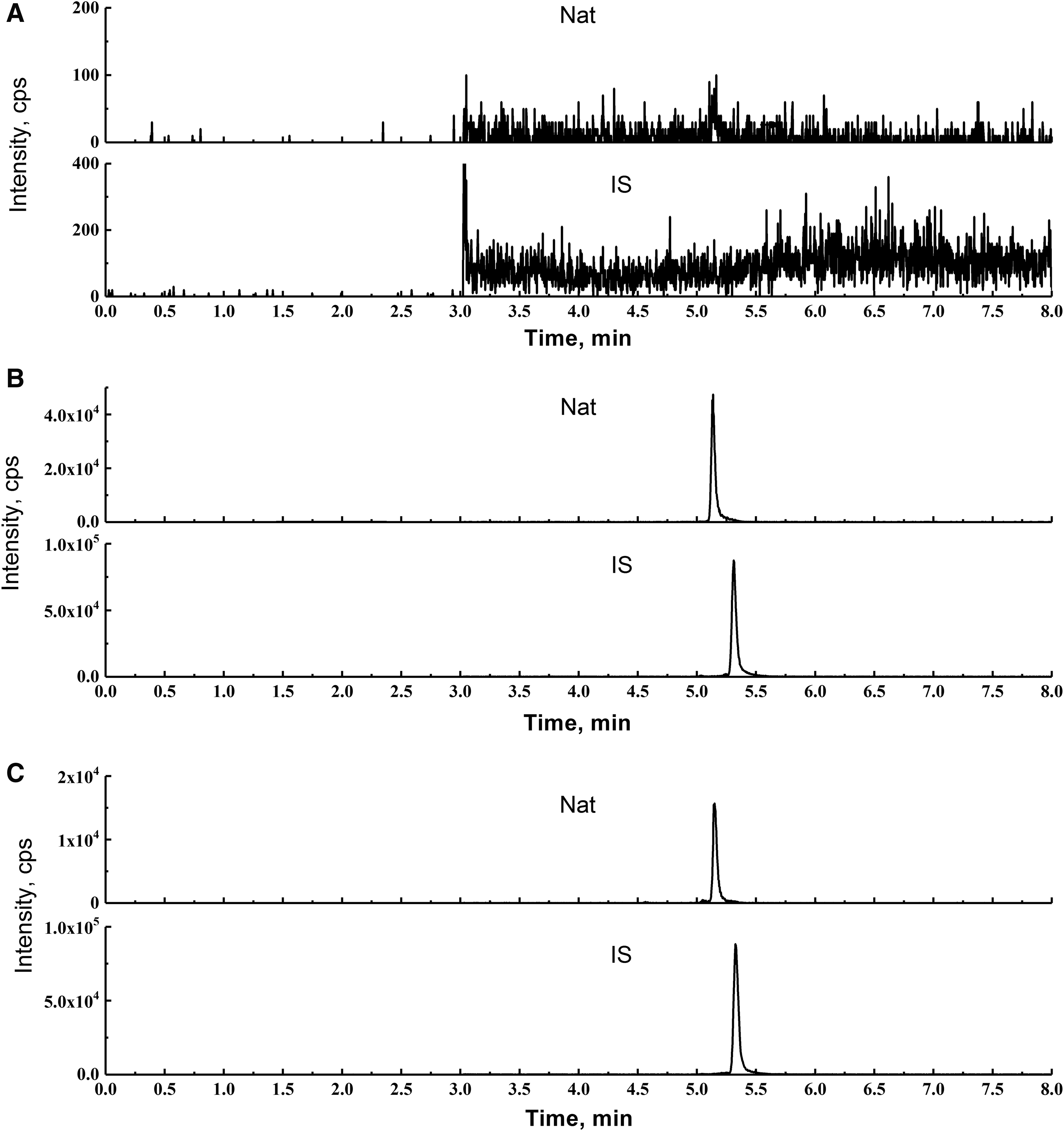
Representative MRM chromatograms of a blank corneal homogenate

Typical chromatograms of a blank corneal homogenate
Linearity was assessed by analyzing calibration standards in duplicate on 3 different days. Calibration curves were created by analyzing the peak/area ratio of the analyte to IS (analyte/IS) versus the concentrations of the analyte. All calibration curves in Table 1 showed good linearity and their correlation coefficients (r) were 0.99 or better.
NAT, natamycin; VRC, voriconazole.
The precision and accuracy of NAT and VRC were assessed by analyzing LLOQ and 3 levels of QC samples in 6 replicates on 3 validation days. The relative standard deviations (RSDs) of intraday and interday assays were 4.74%–8.16% for NAT, and 4.88%–8.17% for VRC. The accuracy was expressed using relative error (RE), which for NAT and VRC ranged from −1.15% to 3.00% and from −0.96% to −3.33%, respectively (Tables 2 and 3).
RSD, relative standard deviation.
The recovery of NAT or VRC was evaluated by comparing the peak area obtained from extracted QC samples with those extracted from blank biosamples reconstituted at the same QC levels. The ranges of NAT and VRC from homogenate were 88.7%–98.9% and 73.0%–105.4%, respectively (Tables 2 and 3). Matrix effect was determined by comparing the peak ratio of NAT obtained from extracted blank biosamples spiked with the analytes with those dissolved in mobile phase. The peak ratios ranged from 98.7% to 104.5% with RSDs of less than 6.77% (Table 2), which indicates no matrix effect for NAT and IS.
NAT and VRC were found to be stable in rabbit aqueous humor and corneal homogenate at room temperature for 4 h (RE<−6.93%), after 2 freeze–thaw cycles (RE<−6.90%) and at −80°C for 30 days (RE<−7.00%). The results are shown in Table 4.
Pharmacokinetic and ocular tissue distribution study
The mean concentrations of NAT and VRC in aqueous humor and cornea are presented in Fig. 3 and the main pharmacokinetic parameters are listed in Table 5.
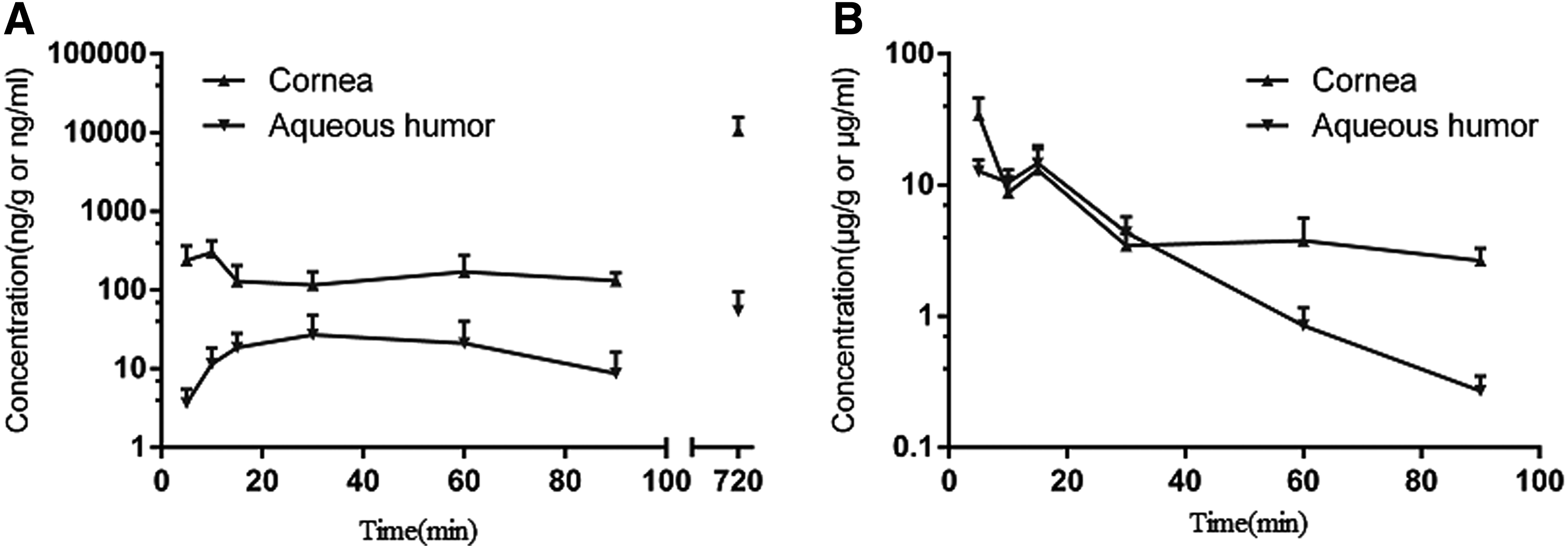
The plots of mean concentration versus time for NAT
In group 1, NAT reached the highest concentration in cornea at 10 min (Cmax:299.3 ng/g) and the concentration almost reduced by half at 90 min (131.7 ng/g). NAT was detected at all the time points in aqueous humor, and the highest concentration was 27.1 ng/mL at 30 min due to poor permeability through the corneal barrier. The permeability ratios of aqueous/cornea ranged from 0.02 to 0.23 (Fig. 4). In group 2 after a single administration of VRC, the highest concentration was found in the cornea at 5 min (Cmax: 34.1 μg/g), while the lowest was 2.7 μg/g at 90 min, which decreased more than 10 times from the highest concentration. Distribution of VRC was lower in the aqueous humor. The highest concentration was 14.7 μg/mL at 15 min and the lowest was 0.3 μg/mL at 90 min, which decreased more quickly than in the cornea. The permeability ratios of aqueous/cornea ranged from 0.1 to 1.26. In group 3, the highest concentration of NAT in cornea increased to 10,569 ng/g after a repeated-dose administration of NAT, while in aqueous humor, NAT was found as low as 54.4 ng/mL. The permeability through the cornea was not improved by repeated administration, and the permeability ratio of aqueous/cornea was 0.0051.

The permeability ratios of aqueous/cornea versus time for NAT and VRC after a single topical administration.
Discussion
The concentration of VRC in cornea and aqueous humor might be too high so that the mass could not achieve the linear range when the concentration is over 5 μg/mL due to mass spectrometry response saturation. In our experiment, the more available HPLC was chosen to determine VRC in the ocular tissues of rabbits, which allowed VRC quantification as low as 100 ng/mL (R > 0.99) with a sample run time of 18 min, for up to 90 min after a single administration of 1.0% VRC to the eye. Due to the low concentrations of NAT in aqueous humor, we had to choose the more sensitive LC-MS/MS for quantification. It was found that using positive ion detection under ESI sources could provide a stronger signal than using negative ion. Figure 5 shows the product ion mass spectra of NAT and its IS.

The product ion mass spectra of the [M+H]+ of NAT
Protein precipitation, the simplest and fastest method for biological sample preparation, was used in our study. Methanol was chosen as the precipitant of NAT to obtain a good peak shape without solvent effect, and acetonitrile was more suitable for VRC for better extraction recovery and less endogenous interferences.
Fungal keratitis is a prevalent and leading cause of ocular disease. Fungal infections are often more difficult to treat than bacteria since fungi can perforate cornea and are more virulent.15,16 NAT is almost insoluble in water and has a relatively high molecular weight (665 Da), which could limit its corneal penetration. 17 We attempted to increase the dose frequency to hourly, but unfortunately NAT only accumulated in the cornea and little could penetrate into the aqueous humor. Contrarily, VRC could reach a higher level in both cornea and aqueous humor in 5 min due to its smaller molecular weight (349 Da). An in vitro experiment showed that the mean minimum inhibitory concentration (MIC) of NAT and VRC was found, respectively, to be 3.4 and 1.91 μg/mL for Aspergillus, 3.0 and 2.54 for Fusarium, and 0.68 and 0.29 μg/mL for Candida. 18 In our study, the concentration of NAT in aqueous humor was even lower than the MIC in vitro for various filamentous fungi with hourly topical administration. Whereas for VRC, aqueous humor Cmax/MIC and AUC0-t/MIC could be higher than 10 and 125, respectively, which could reach effective antimicrobial activity.19–21 The concentration of VRC decreased quickly in aqueous humor (t1/2: 15.7 min), which suggests that optimization of the formulation is needed to increase the viscosity and extend the release time.
In conclusion, the relatively poor corneal penetration of NAT suggests that it is more suitable for superficial corneal fungal infections since even with repeated administration, high-level distribution of NAT was only observed in the cornea. High distribution of VRC in cornea and aqueous humor suggests an increased efficacy for deep corneal fungal infections via topical ocular administration.
Footnotes
Acknowledgments
The authors thank Miss Xiqin Yang and Miss Kun Gao for their valuable technical assistance.
Author Disclosure Statement
All authors have no conflict of interest to declare.