




Abstract
Abstract
Purpose:
Given that porcine and human retinas have similar structures and characteristics, ex vivo culture of porcine neuroretina provides an attractive model for studying mechanisms of human retinal injury and degenerative disease. Here, we describe the method that was used to establish and characterize an adult porcine retina culture system as a rapid screening tool for retinal survival in real time.
Methods:
Neuroretina explants 8 mm in diameter were harvested from adult swine and cultured on porous cell culture inserts with adjustable heights. Retina explant viability was evaluated at 1, 4, 7, 11, and 14 days of culture using a resazurin-based metabolic assay. The explants were analyzed morphologically through immunohistochemistry for glial activation and apoptosis. Morphometric analysis was also performed on hematoxylin and eosin-stained retina sections from each time point.
Results:
The viability of retina explants gradually decreased over time in culture. The laminar structure of the neuroretina was well preserved during the first 7 days. However, by day 14, most explants showed significant loss of cells in each laminar layer and obvious thinning. Overall, the progressive loss of retinal lamination and thickness, and increase in apoptotic nuclei with activated hypertrophic Müller cells were well correlated with the metabolic activity of the ex vivo neuroretina explants.
Conclusions:
This study was the first report to describe the use of a high-throughput and quantitative method for monitoring retina explant viability in real time. Ex vivo neuroretina cultures closely mimic the functional dynamics of the organ, and can be used efficiently to screen novel therapeutics for retinal neurodegenerative disease.
Introduction
R
The most common method used to initiate retinal organotypic cultures involves dissecting the retinal sheet free from the underlying retinal pigment epithelium (RPE)/choroid, and placing it with the photoreceptor layer facing downward onto porous cell culture inserts.15–18 Despite reports of promising data obtained from various retina explant culture systems, it is often difficult to interpret findings from these studies. Protocols used to harvest and culture the explants differ widely between different groups. Aside from the experimental species used, the composition of culture medium, either with high concentrations of serum or serum free, the length of culture, and laminar orientation of the tissue in regard to the substrate could all cause subtle variations in the tissue behavior. More importantly, most assessments performed by various investigators to evaluate the behavior of retinal tissue have largely been qualitative or at best semiquantitative. A standard approach typically involves fixing and processing retina explants for histology and immunohistochemistry for an array of neuronal or retinal markers. Semiquantitative analysis of the percentage of immunoreactive cells per field of view is then performed. In terms of tissue viability, caspase-3 or terminal deoxynucleotidyl transferase-dUTP nick-end labeling (TUNEL) immunostaining on fixed transverse retina explant sections has been regularly performed. To our best knowledge, there are no reports of the use of a quantitative method to evaluate the viability of retina explants over time.
Tetrazolium or resazurin-based metabolic assays have been frequently used to assess the viability and proliferation of a wide variety of adherent and nonadherent cells.19–21 MTT (3-[4-5-dimethylthiazol-2-yl]-2-5-diphenyl bromide tetrazolium bromide) and other tetrazolium assays are based on the cleavage and reduction of the tetrazolium ring to blue formazan crystals by the mitochondrial dehydrogenases.19–21 These crystals can be solubilized and quantified in the cell lysate as a measure for metabolic cell activity. Despite the recurrent use of these assays, the fact that this methodology can only be used as an endpoint analysis is a clear disadvantage. The resazurin-based AlamarBlue and PrestoBlue (PB) assays, in contrast, also based on the redox activity of the cells, use the mitochondrial activity to reduce the nonfluorescent blue resazurin to the fluorescent pink resorufin.19–21 This nontoxic water-soluble dye enables continuous live-cell monitoring, and has shown great potential for assessing the viability of human glioma and porcine dermal explants in ex vivo culture.22,23 We have modified the PB assay and our ex vivo organotypic culture setup to quantitatively evaluate the viability of retina explants at different time points.
Here, we describe the use of a high-throughput and quantitative method to assess retina explant viability in real time for up to 14 days in culture. Ex vivo organotypic cultures of porcine retina were initiated, and the behavior of the explants was further characterized through standard techniques such as immunohistochemistry for glial cell markers and TUNEL for apoptotic cells. Retinal morphometric analysis was also performed on hematoxylin and eosin (H&E)-stained transverse explant section images to determine the total retinal thickness and thickness of each laminar retinal layer. We hypothesized that the degenerative patterns established through standard morphological assessments would correlate with the metabolic activity of the ex vivo neuroretina explants measured using the PB assay.
Methods
Retina explant harvest and culture
Postmortem eyes without known ocular defects and injuries were obtained from female adult Yorkshire swine 6 to 9 months old. After euthanasia, porcine eyes were enucleated, immersed in betadine solution during transport to the laboratory, and processed within 2 h postmortem in room temperature. Under aseptic conditions, the eyes were rinsed in phosphate-buffered saline (PBS), and excess fat and connective tissue were trimmed off. A circumferential incision was made around the limbus of each eye, followed by removal of the anterior segment, lens, and vitreous body. An 8-mm dermal punch was used to mark 4 equal-sized explants per eye from the central region of the retina distal to the optic disk. The retinal sheet was carefully separated from the RPE using a surgical spatula.
Retina explants were transferred to permeable Nunc™ cell culture inserts (12-mm diameter, 0.4-μm pore size, polycarbonate; Thermo Fisher Scientific, Waltham, MA) and maintained using Nunc's Carrier Plate System (Thermo Fisher Scientific). This unique carrier plate system was designed to hold cell culture inserts at 3 positions above the growth surface of the multiwell dish containing the inserts (high position at 6.3 mm above the well, medium position at 3.3 mm, or low position at 0.9 mm). The explants were cultured with the ganglion cell layer (GCL) facing up in serum-free media composed of Dulbecco's Modified Eagle Medium (DMEM)/F12 and DMEM (1:1), nonessential amino acids (NEAA), 2% B-27 (proprietary serum-free supplement), and 1% antibiotic–antimycotic mixture (Thermo Fisher Scientific) at 37°C with 5% CO2 and 95% humidity. The culture medium level was maintained in contact with the support membrane beneath the explant. Additional medium was added to the top of the insert to keep the explant moist during culture. Cell culture media were carefully replaced every other day without disturbing the explants.
Assessing retina explant viability through the PB cell viability assay
The viability of porcine retina explants was assessed using the PB cell viability reagent (Thermo Fisher Scientific). The PB reagent contains a cell-permeable blue compound resazurin that is virtually nonfluorescent, but is converted to a pink/red-colored and highly red fluorescent resorufin by viable cells. The conversion is proportional to the number of metabolically active cells, and, therefore, can be measured quantitatively. This change can be detected through absorbance or fluorescence measurements, whereas the latter is more sensitive and is the preferred detection method.
After 1, 4, 7, 11, or 14 days in culture, retina explants were incubated in 1 × PB working solution prepared in explant maintenance media for 8 h at 37°C with 5% CO2 (or 16 h for overnight incubation). After incubation with the PB reagent, 100 μL of the resultant buffer was collected from each sample and transferred to a 96-well cell culture plate in triplicates. Fluorescence readings were collected using a spectrophotometer set to the excitation and emission peaks for resorufin (excitation 560 nm and emission 590 nm with 10-nm bandwidth). No cell controls (wells containing cell culture media only) were included whose background fluorescence values were averaged and subtracted from the fluorescence value of each experimental well. Normalized fluorescence versus experimental condition (i.e., days in culture in this case) was analyzed and plotted. Retina explants were washed with PBS, replenished with fresh media, and returned to culture until the next time point.
Histology and H&E staining
Retina explant samples were fixed in 10% neutral buffered formalin and processed for routine histological evaluation using an automatic tissue processor (Leica ASP300S; Leica Biosystems, Buffalo Grove, IL). Processed tissue specimens were embedded in paraffin blocks and stored at room temperature until use. Sections 4–5 μm thick were cut from each sample and placed on treated glass slides (Superfrost Plus; Thermo Fisher Scientific), dried for 1 h in an oven set to 60°C, and then stored in room temperature until use. Sections were deparaffinized and rehydrated in graded alcohols, stained with H&E using an autostainer (Leica Autostainer XL; Leica Biosystems), dehydrated, cleared, and mounted. Slides stained with H&E were imaged using a slide scanner (Zeiss Axio Scan.Z1; Carl Zeiss, Oberkochen, Germany) and used for routine histological analysis.
Retinal morphometric analysis
H&E was used to investigate structural changes as well as changes in the thickness of the retinal layers. Three H&E-stained retina sections from each time point were analyzed using the measurement tool through the image analysis software provided by ZEN (Carl Zeiss Microscopy; Carl Zeiss). Measurement of the thickness of the whole retina (excluding the outer segments), GCL, inner plexiform layer (IPL), inner nuclear layer (INL), outer plexiform layer (OPL), and outer nuclear layer (ONL), was performed. For each layer, 3 measurements per image were averaged.
Immunohistochemistry
Immunohistochemistry was used to visualize glial fibrillary acidic protein (GFAP). In brief, tissue sections were deparaffinized in xylene, rehydrated through graded alcohol washes, and incubated in 1 × antigen retrieval buffer/citrate buffer pH 6.0 from Abcam (Abcam, Cambridge, MA) for 15 min in a vegetable steamer for heat-induced epitope retrieval. After several washes in 1 × tris-buffered saline with Tween 20 (TBST), tissue sections were blocked with 10% goat serum for 1 h in room temperature to minimize nonspecific binding, and subsequently incubated with the primary antibody (Anti-GFAP, 1:100, CAT# 14-9892; eBioscience, San Diego, CA) overnight at 4°C. Alexa fluor-conjugated secondary antibody (Alexa Fluor 488, 1:500, CAT# A-11001; Thermo Fisher Scientific) was used for immunofluorescence detection and counterstained with 4′,6-diamidino-2-phenylindole (DAPI). Negative controls were obtained by omitting incubation with the primary antibody. Fluorescent images of immunostained tissue sections were acquired using an Olympus FLUOVIEW FV1000 confocal microscope (Olympus America, Inc., Center Valley, PA).
TUNEL assay
The TUNEL assay was performed to visualize apoptotic cells using an in situ cell death detection kit from Roche, following the manufacturer's recommendations (fluorescein No. 11684795910; Roche Diagnostics, Basel, Switzerland). The assay preferentially marks DNA strand breaks during apoptosis by labeling free 3′-OH termini with modified nucleotides in an enzymatic reaction. For controls, terminal deoxynucleotidyl transferase enzyme was omitted from the labeling solution (negative control). Moreover, cell nuclei were counterstained with DAPI (1 μg/mL; Invitrogen, Carlsbad, CA). The specimens were imaged with a laser-scanning confocal microscope (Olympus America, Inc.).
Statistical analysis
All data are represented as mean value ± standard deviation. Data sets were examined for statistical significance using one-way ANOVA, with Tukey's multiple comparison test for PB fluorescence readings, TUNEL staining quantification, and retinal thickness measurements (GraphPad Prism Version 6.05; GraphPad Software, Inc., La Jolla, CA). A P value ≤0.05 was considered significant.
Results
Retina explant harvest and culture
We have successfully identified optimal conditions to maintain porcine retina explants in ex vivo culture for up to 14 days. Four 8-mm explants were generated from each eye and cultured using the Nunc Cell Culture Inserts/Carrier Plate Systems, which allowed the inserts to be held at 3 different positions above the growth surface, resulting in different media height and volume requirements at each position. The explants were cultured at the highest position, allowing a greater volume of media to be used, but were moved to the lowest position to reduce the amount of buffer required during incubation with the PB reagent. A serum-free media formulation containing DMEM/F12, DMEM, NEAA, and B-27 plus antibiotics was used to maintain the explants at the air–liquid interface. There were no apparent differences in the gross morphology of retina explants immediately after explantation and over time ex vivo. The general appearance of the tissue did not seem to change over time in culture. Figure 1 depicts the general procedure of porcine neuroretina harvest and preparation, along with a schematic of the Nunc Cell Culture Inserts/Carrier Plate Systems used to cultivate the retina explants.
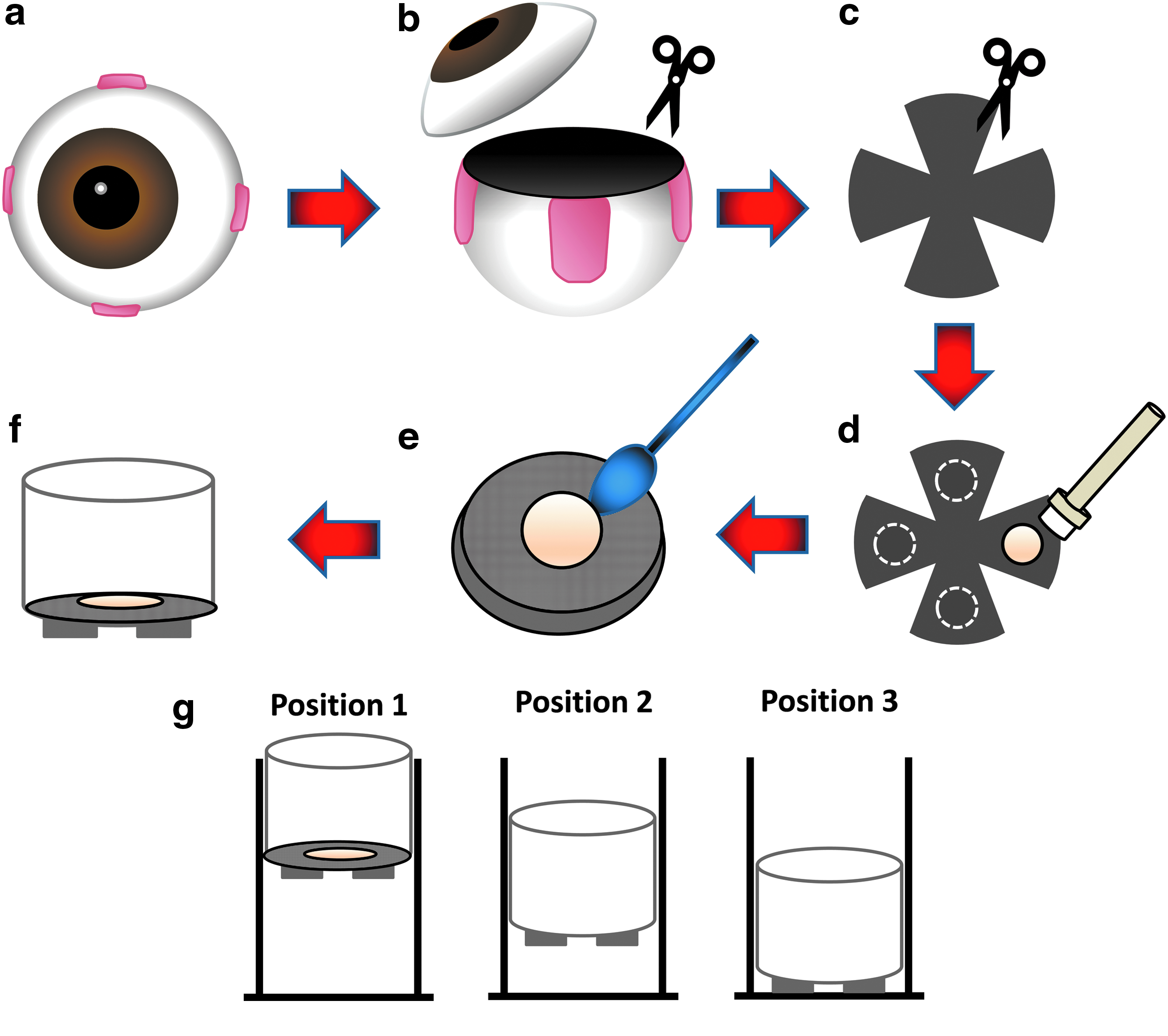
Schematic of retina explant harvest and culture. After euthanasia, porcine eyes were enucleated and excess fat and connective tissue were trimmed off
Quantitative assessment of retina explant viability through the PB cell viability assay
The viability of porcine retina explants was assessed quantitatively using the PB cell viability reagent. The assay has been predominantly used to assess the viability of a variety of adherent or nonadherent cells; however, recent studies have described the use of PB to examine the viability of porcine dermal explants 22 and human glioma 23 in ex vivo culture. In these studies, the tissue explants were transferred to a smaller multiwell dish during incubation with PB, which was not possible with thin fragile samples such as the neuroretina. In our study, the retina explants remained on the cell culture insert that was moved to the lowest position during incubation with the PB buffer at each time point. The explants were rinsed with PBS once before returning to culture with fresh cell culture media.
As indicated in Fig. 2a, retina explants remained viable throughout the entire duration of the experiment. However, as expected, the viability of the explants gradually decreased over time in culture as shown in Fig. 2a. Figure 2b shows the corresponding color change at different time points.
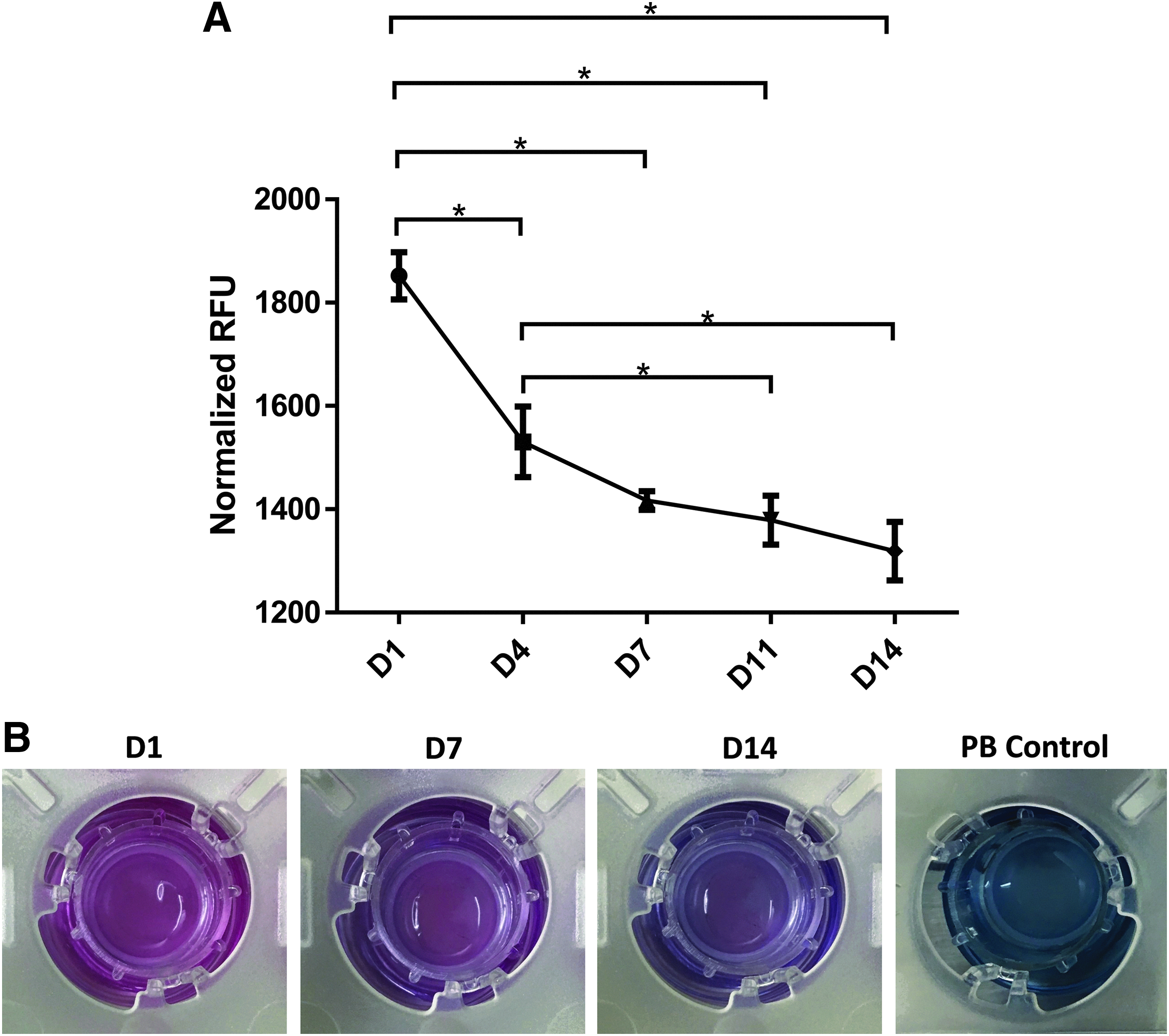
Assessment of retina explant viability and apoptotic processes
The TUNEL assay was performed on paraffin-embedded sections of the neuroretina to determine the population of apoptotic cells in retina explants over time in culture, where a temporal appearance of TUNEL+ cells was observed. Sections of retina explants showed a few TUNEL+ nuclei mostly in the ONL after 4 days in culture. Retina explants harvested after 7 days in culture showed a considerable number of TUNEL+ nuclei in both the ONL and the INL. By day 14, the total number of TUNEL+ apoptotic cells appeared to have decreased, perhaps due to significant cell loss from each of the nuclear layers in the tissue (Fig. 3a, b).

TUNEL assay results.
Although most reports in the literature suggest that solutions containing resazurin, the active ingredient in PB reagent, are not toxic to cells, other reports show that cell viability is affected depending on the length of exposure and concentration of resazurin to which they are subjected.24–27 According to the manufacturer's recommendations, it seems unlikely that cells or tissues can be adversely affected by the exposure to PB if assay conditions remain within the suggested incubation times (i.e., up to 24 h for low number of cells). To verify that the PB reagent was not cytotoxic to the retina explants after long incubation, we treated early time point (day 4) neuroretina samples with PB for 24 h, fixed and processed for TUNEL staining. We observed negligible differences in the number of TUNEL+ apoptotic cells in the PB-treated retina specimens versus untreated controls as shown in Supplementary Fig. S1a–c (Supplementary Data available online at www.libertpub.com/jop). Multiple PB assessments over time did not appear to have an adverse effect on the retinal tissue as illustrated in Supplementary Fig. 2a, b. There were negligible differences in the number of TUNEL+ cells in retina explants that have undergone 5 PB assessments on days 1, 4, 7, 11, and 14 (PB 5 × ) versus control retina explants that have never been treated with PB (No PB) on day 14.
Preservation of retinal microarchitecture and changes in retinal thickness
We used H&E-stained sections to examine the overall appearance of the plexiform and nuclear layers in retina explant cultures from different time points (Fig. 4a). Overall, the laminar structure of the neuroretina was well preserved in retina explants cultured in serum-free media during the first 4–7 days, but was gradually lost in the following week. Not all retinal layers were clearly detectable between days 11 and 14. By day 14, most explants in culture showed significant loss of cells in each of the nuclear layers. Furthermore, there was obvious thinning of the retina explant especially in the second week of culture, likely due to photoreceptor death and condensation in the plexiform layers, which was most evident in the IPL. On average, cells at both nuclear layers appeared densely packed during the first week of culture, with only a moderate reduction in the number of nuclei within each of the nuclear layers. By the end of the second week of culture, the total retinal thickness was significantly reduced, that is, around 103.183 ± 8.087 μm compared with about 184.978 ± 6.611 μm before culture (Fig. 4g). Figure 4b–f shows thickness of the INL, ONL, GCL, OPL, and IPL from day 0 through day 14.
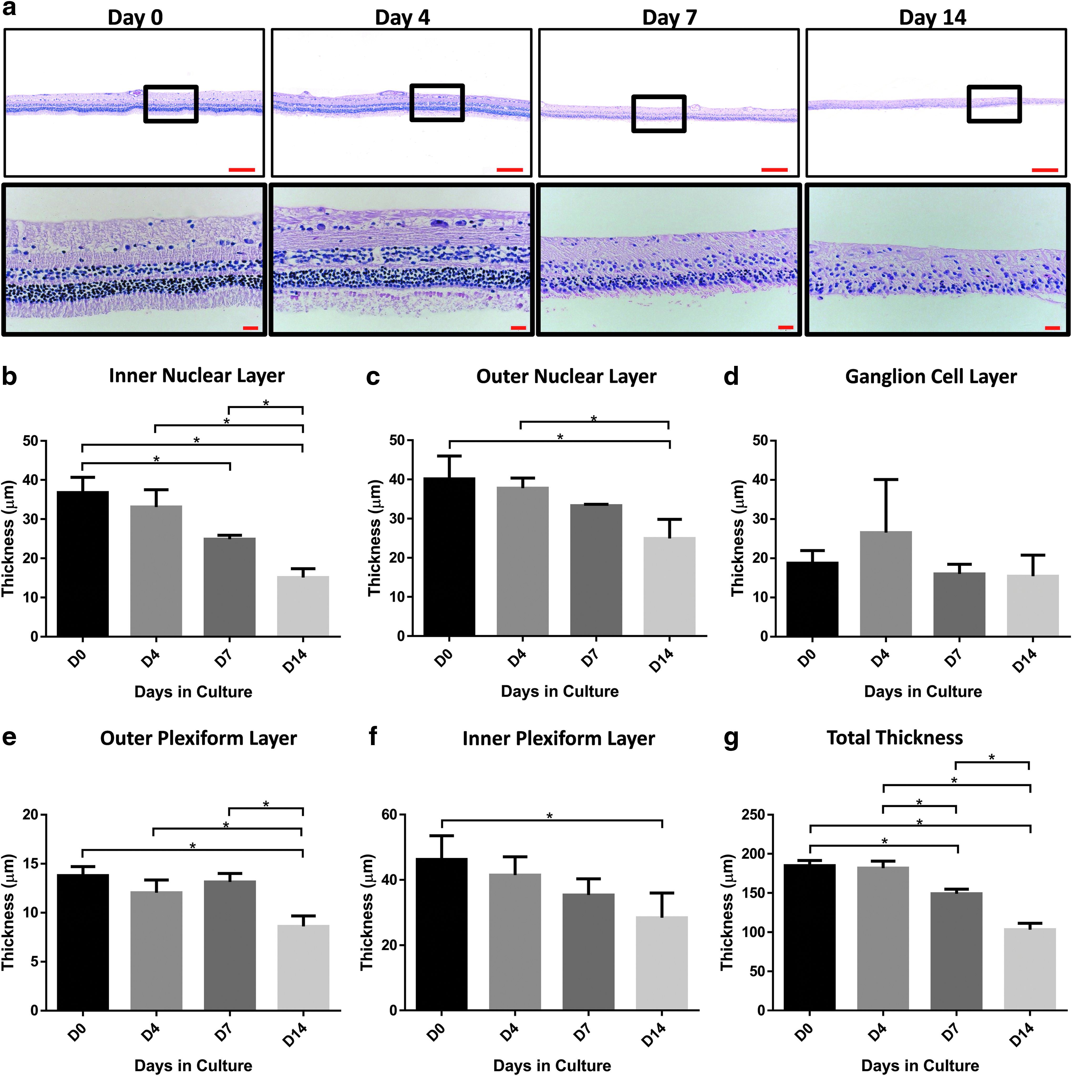
Immunohistochemical characterization of glial activation
To address the degree of glial cell activation, retina samples were immunostained with antibodies against GFAP, an intermediate filament protein present in glial cells. Figure 5 shows a compilation of representative GFAP immunohistochemistry images of retina explants harvested from different time points. In baseline control samples (freshly isolated, not cultured), GFAP-labeled cells were sparse and localized to the GCL and nerve fiber layer, with a few processes extended into the OPL. After 4 days in culture, retina explant specimens showed an upregulation of GFAP that has now been extended to the ONL. After 7 days in culture, increased and widespread GFAP immunoreactivity or reactive gliosis was present in all of the retinal cell layers. By day 14, the retina explants also showed a high general upregulation of GFAP throughout the specimen, with the Muller cell fibers in a disorganized array. In addition, signs of retinal degeneration were evident, including loss of retinal lamination and distortion.
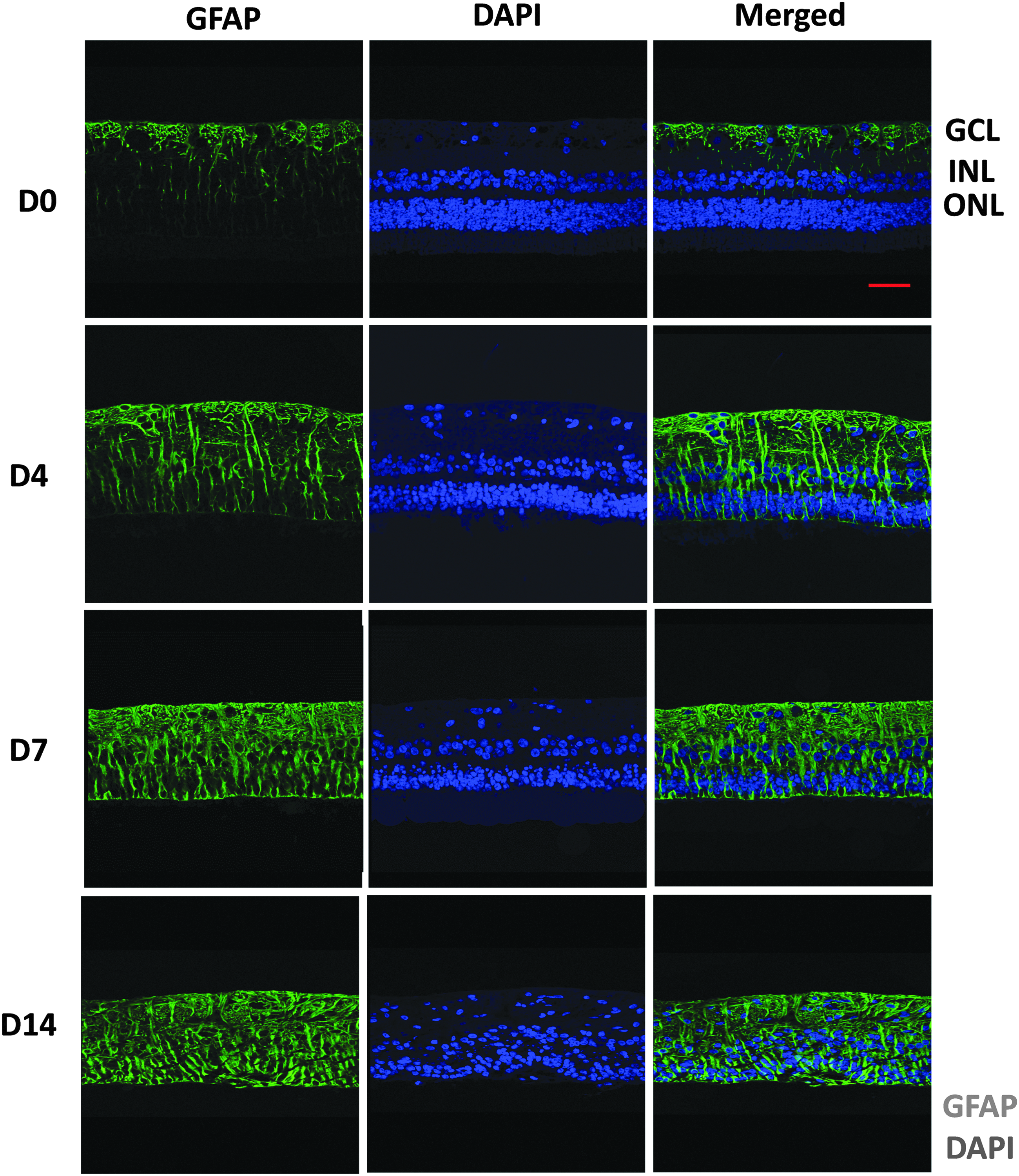
Representative immunohistochemistry images for GFAP in retina explants at baseline (noncultured sample, harvested and processed right away at D0), D4, D7, and D14. Transverse retina explant sections were immunohistochemically labeled with GFAP (shown in green), whereas the nuclei were counterstained with DAPI (in blue). Scale bar = 50 μm. GFAP, glial fibrillary acidic protein.
Discussion
Retina explant cultures have been widely investigated to understand the mechanisms responsible for retinal development, neurodegeneration, and neuroprotection. At first glance, the explant cultures may seem relatively easy to establish, since after dissection, the piece of tissue is merely transferred to a culture well or insert with minimal processing. Nevertheless, the harvest of retina explants is not a simple procedure. As the retina is thin and extremely fragile, careful handling of the specimen is critical. Erroneous handling can lead to variability in tissue viability, even before an experiment is conducted.
The most common method used to culture retina explants has been a variation of the technique first described by Caffe et al. in 1989, in which the mouse neuroretina was placed with the photoreceptor layer facing downward on rafts made of nitrocellulose filters and polyamide gauze grids. 15 The polarity or orientation of the retinal tissue was most likely chosen due to the high metabolic rate of the photoreceptors, with the rationale that survival of these cells would be enhanced by being in proximity to the culture medium. Recently, Taylor et al. cultured adult porcine retina explants with the inner retinal surface facing down against the membrane (opposite orientation to conventional methods), and reported better preservation of the retinal laminar architecture, along with significant attenuation of photoreceptor and ganglion cell death. 28 The investigators attributed these observations to the physical support and stability provided by the nonelastic cell culture membrane to the adult inner retina. Another study performed by Wang et al. cultured porcine neuroretina explants on sterile Whatman™ filter paper (with GCL adhered to the paper) placed on a cell culture insert that was elevated from the bottom of the multiwell dish through a custom-made stand, and reported preservation of all retinal layers for up to 7 days. 29 In our earlier attempts to set up an ex vivo neuroretina organotypic culture system, we also explanted the retinal tissue onto a filter paper and cultured it in a similar setup. However, the filter paper method did not appear to be suitable for long-term culture as it was common for the explants to detach from the filter substrate over time. In this study, we adhered to the conventional method of culturing retina explants with the inner retinal surface facing up on a commercially available porous cell culture insert. We believe this is a better representation of the in vivo conditions, in which the neuroretina is supported and fed by nutrients from the underlying RPE/choroid.
It has been shown in the literature that an adult mammalian retina is difficult to maintain in vitro, mainly due to the high metabolic demands of the photoreceptors. Koizumi et al. described a rabbit retina organotypic culture system in which the oxygen supply for photoreceptors was ameliorated by raising the height of the culture insert using a custom-made stand (2-cm diameter, 1-cm high) made from a 1-mL Monojet Tuberculin syringe and agitating the culture medium. 30 The Nunc Cell Culture Insert/Carrier Plate System used in our study allowed us to raise the height of the culture insert to ensure adequate nutrient exchange during culture, while giving us the flexibility to lower the insert to reduce the amount of buffer required for other assays.
Different media formulations have been described in the literature that were used to culture retina explants; some were serum free or contained high concentrations of serum. Johnson and Martin found that serum-free media supplemented with B-27 and N2 were superior in maintaining the laminar microarchitecture of the retina, forestalling necrosis and apoptosis, and preserving physiological expression of a wide array of retinal protein markers. 4 This finding is important, as serum-free conditions are essential for pharmacological studies involving retinal therapy and neuroregeneration. Fetal bovine serum often contains components that are not clearly defined and may affect the behavior of the retinal tissue. As a result, we elected to use basal media with DMEM and DMEM/F12 containing serum-free supplement B-27 to culture the retina explants in our study.
Organotypic cultures of the retina have been used successfully for the modulation of immature retinal tissue, which has been found to survive well and even display several signs of normal development.15–17,31,32 Cultures of adult retinal tissue, in contrast, have been found to be much more limited in their ability to survive in vitro. Isolated adult retinal sheets cultured under standard conditions often display gliosis and neuronal degeneration over time. The discrepancy of cell survival in vitro depending on the stage of maturity of the retinal tissue is well established in the literature but not yet fully understood.33–36 Thus, the organotypic culture system we have described here is a degenerative model in which we observed a reduction in retinal tissue viability as early as <4 days in culture. Retina explant viability as measured by the PB assay was inversely correlated with an increase in TUNEL+ cell nuclei over time. Other investigators have also reported a significant number of TUNEL+ apoptotic cells in control porcine retinal explants only after 3 days 37 or 5 days in culture.28,36
Retinal injury is known to trigger a modest glial reactivity, as evidenced by upregulation of GFAP. The process of explantation not only signified complete axotomy of retinal ganglion cells (RGCs), but it also required the removal of the retina from the influence of the underlying RPE. As expected, GFAP levels in retina explants from our organotypic cultures were gradually increased over time. Retinal thickness has been shown to be an excellent indicator of retinal degeneration. Gradual thinning of the retina explants was also observed in our study, especially during the second week of culture, perhaps due to gradual loss of photoreceptors in the outer segments and each of the nuclear layers, and condensation of IPL. The TUNEL assay revealed a progressive increase in the number of apoptotic nuclei in the explants over time, although by day 14, the number of TUNEL+ cells appeared to have decreased, perhaps due to significant cell loss from the retinal tissue by the endpoint of our study. Similarly, Bull et al., although using a rat model, have also demonstrated that the rate of cell loss in retinal explants was most rapid from day 0 to ∼5–7 days ex vivo, after which the rate of loss was reduced. 3
It is important to note that there are obvious limitations to ex vivo retina explant culture systems. Apart from the physical separation of the RPE from the neuroretina and axotomy of RGCs due to the dissection procedure, there is also the absence of retinal blood supply. Consequently, the ex vivo culture systems described here are not meant to be long-term culture solutions. As a result, the degenerative changes seen in ex vivo neuroretina cultures could differ from the in vivo conditions. Furthermore, one of the major drawbacks of these culture systems is the dependence on morphological assessments to evaluate retinal tissue behavior such as viability. Therefore, in this study, we adapted the PB metabolic assay to quantify the viability of retina explants in a modified organotypic culture setup for up to 14 days. As the PB reagent posed no cytotoxic effects, the explants were able to return to culture after each assessment. Thus, the viability of the same explant was monitored over time, eliminating the common issue with sample-to-sample variability, while reducing the number of samples needed for each experiment. Although the PB assay was not capable of providing an absolute count of viable cells within the tissue specimen, a greater magnitude of chemical reduction or conversion was consistent with a greater magnitude of metabolic activity and, presumably, a larger number of cells in the retina explants.
Methods used to culture retina explants from rodents have been commonly described in the literature. Organotypic rat retina explant models have been used as a rapid screening tool for potential novel neuroprotective agents for neurodegenerative disease such as glaucoma.11,3 Mouse retina explants have also been used to study neurodegeneration, neuroprotection, and retinal development.10,12 Although rodent models have proven indispensable, there are inherent anatomical differences between rodent and human eyes, which warrant careful interpretation of data obtained using these models.
Given that porcine and human retinas are very similar in terms of cell distribution and morphology, vascular pattern, laminar layer thickness, and other physiological characteristics, 38 ex vivo organotypic culture of porcine neuroretina provides an attractive model for studying manifestations of human retinal diseases, degenerations, and injuries. Furthermore, this porcine ex vivo system may be particularly important where performing in vitro experiments on human retinas can be a challenge, as high-quality human retina specimens are difficult to obtain. Even though there are some obvious limitations associated with these organotypic culture systems, the ex vivo neuroretina cultures closely simulate the in vivo retinal cellular and molecular dynamics than cell culture systems. Along with the high-throughput method used to monitor retinal explant viability in real time described in this study, ex vivo neuroretina cultures provide a powerful tool that can not only improve our knowledge of retinal physiopathology but can also be used as an efficient platform to screen novel therapeutics for retinal degenerative disease.
Footnotes
Acknowledgments
This work was supported, in part, by the U.S. Army Clinical Rehabilitative Medicine Research Program (CRMRP), Military Operational Medicine Research Program (MOMRP), and Oak Ridge Institute for Science and Education (ORISE). The authors greatly appreciate the assistance of Dr. Ramesh Kaini and Mr. Dallas Golden in setting up the retina explant cultures.
Disclaimers
The opinions or assertions contained herein are the private views of the authors and are not to be construed as official, or reflecting the views of the Department of Defense or U.S. Government.
Author Disclosure Statement
No competing financial interests exist.
References
Supplementary Material
Please find the following supplemental material available below.
For Open Access articles published under a Creative Commons License, all supplemental material carries the same license as the article it is associated with.
For non-Open Access articles published, all supplemental material carries a non-exclusive license, and permission requests for re-use of supplemental material or any part of supplemental material shall be sent directly to the copyright owner as specified in the copyright notice associated with the article.
