
Abstract
Abstract
Purpose:
To compare the effects of FK-506, cyclosporin A (CsA), and sodium hyaluronate (HA) eye drops for the treatment of botulinum toxin B (BTX-B)-induced mouse dry eye.
Methods:
CBA/J mice were randomized into 5 groups. The groups received treatment with eye drops containing 0.025% FK-506 combined with 0.3% HA (FK-506+HA group), 0.025% FK-506 (FK-506 group), 0.05% CsA (CsA group), 0.3% HA (HA group), or 0.9% saline (saline group) 3 days after an intralacrimal gland injection with 20 mU of BTX-B. Tear production, corneal fluorescein staining, blink rate, and the mRNA and protein expression levels of inflammatory cytokines were measured.
Results:
FK-506+HA eye drops increased tear production and reduced the corneal fluorescein staining scores at all time points after treatment compared with those in the saline group. Compared with those in the saline group, the tear production and severity of corneal epithelial defects in the FK-506 group were significantly improved at weeks 2 and 4. Compared with the saline eye drops, the CsA eye drops ameliorated only tear production and corneal fluorescein staining scores at week 4 after administration. The FK-506+HA, FK-506, and CsA eye drops downregulated the expression of inflammatory cytokines in both the keratoconjunctival tissues and lacrimal glands at week 4.
Conclusions:
The topical application of 0.025% FK-506 combined with 0.3% HA, 0.025% FK-506, or 0.05% CsA can suppress the expression of inflammatory cytokines and can alleviate the signs of dry eye. Topical application of 0.025% FK-506 combined with 0.3% HA showed the best therapeutic effect and may be a possible therapy for dry eye.
Introduction
D
Lubricants such as artificial tears are the most basic and widely used therapy for DED.1,2 There is a growing recognition that inflammatory factors are the primary causative mechanism driving the pathological process of dye eye. Anti-inflammatory agents, including topical corticosteroids, cyclosporin A (CsA), essential fatty acids, and oral tetracyclines, have become one of the main treatments for DED. 3 Among these anti-inflammatory agents, topical corticosteroids are effective in breaking the cycle of inflammation. However, their obvious side effects, such as ocular hypertension, cataracts, decreased wound healing, and predisposition, limit their clinical application.4–6
CsA is an immunosuppressant that inhibits T cell-mediated inflammation and cytokines in the conjunctiva, stimulates natural tear production, and increases goblet cell density.7–10 The main side effects of topical CsA are ocular burning, blurred vision, ocular itching, conjunctival hyperemia, discharge, foreign body sensation, and stinging.11,12 Topical CsA shows a more favorable risk–benefit profile for chronic use than do topical corticosteroids. Recent research findings have confirmed that the topical use of 0.1% CsA and 0.05% CsA significantly improve the corneal fluorescein staining and tear production of dry eye patients.10,13 The mechanism of FK-506, another calcineurin inhibitor, is similar to that of CsA, and the potency of FK-506 is 50- to 100-fold higher than that of CsA.14,15 By using FK-506 systemically, the tear production has been improved in dry eye associated with chronic graft-versus-host disease. 16 The topical use of 0.02% FK-506 effectively increased tear production in dogs with keratoconjunctivitis sicca. 17 Our earlier studies showed that the topical application of 0.025% FK-506 could significantly relieve the symptoms of dye eye, including corneal epithelial defects and tear production, in the mouse model of botulinum toxin B (BTX-B)-induced dry eye,18,19 similar to the results of topical 0.05% CsA applied in this model. 20
Both FK-506 and CsA eye drops are effective in the BTX-B-induced mouse model of dry eye. The purpose of this study was to compare the effects of topical 0.025% FK-506 and 0.05% CsA eye drops in this model, which mimics non-Sjogren's dry eye.
Methods
Induction and treatment of dry eye
Six- to eight-week-old inbred female CBA/J mice were purchased from the Hua Fu Kang Bioscience Company in Beijing, China. The animal experiments complied with the Association for Research in Vision and Ophthalmology Statement for the Use of Animals in Ophthalmic and Vision Research. The mice were housed in a standard animal facility with a controlled temperature (22–24°C) and photoperiod (12 h light and 12 h dark) and were given free access to food and water. Mice were intraperitoneally anesthetized with ketamine and xylazine (45 and 4.5 mg/kg, respectively), and every effort was made to minimize suffering.
A previously reported method was used to establish the murine model of dry eye. 19 In brief, 0.05 mL of 0.09% saline solution or 0.05 mL of 0.09% saline solution containing 20 mU BTX-B was injected transconjunctivally into the right lacrimal glands of CBA/J mice using a custom-made 33-gauge needle (Hamilton, Reno, NV).
The mice with successfully induced dry eye were randomly assigned to 5 groups and received topical eye drops 4 times a day with 2 drops of 0.9% saline solution (saline group), 0.9% saline solution and 0.03% sodium hyaluronate (HA group), 0.9% saline solution and 0.05% CsA (CsA group, Restasis®; Allergan, Irvine, CA), 0.9% saline solution and 0.025% tacrolimus eye drops (FK-506 group), or 0.03% HA and 0.025% tacrolimus eye drops (FK-506+HA group). FK-506 eye drops are prepared as described in our previous published article. 18 The 2 drops were administered to each group at an interval of 10 min. The spontaneous blink rate, ocular surface changes, and tear production were evaluated in all groups. At the end of 4 weeks of administration of the topical medication, the mice were euthanized in a carbon dioxide chamber for tissue procurement according to the American Veterinary Medical Association guidelines.
Measurements of blink rate, aqueous tear production, and corneal fluorescein staining
Corneal fluorescein staining and tear production were measured using a previously described method. 19 The blink rate was observed by 2 researchers for 1 min under controlled temperature (22–24°C) and humidity (60%) conditions. If an interobserver difference of 2 counts or greater was noted in the reported blink rate, a recount was performed, and the average count was recorded. The aqueous tear production of an unanesthetized mouse was measured with phenol red-embedded cotton threads (Zone-Quick; Oasis, Glendora, CA). The threads were applied to the ocular surface in the lateral canthus for 15 s. A micron-scale digital ruler (Nikon, Tokyo, Japan) was used to measure the wet threads under a microscope. Corneal epithelial defects were shown by fluorescein staining with 5 μL of a 1% sodium fluorescein solution (Sigma-Aldrich, St. Louis, MO). Corneal fluorescein staining was evaluated under cobalt blue light at 1 min after administration. A digital camera (Nikon) mounted on a microscope was used to photograph the ocular surface. A scoring system was used to evaluate the corneal fluorescein staining of the front view for all corneas. 1 In brief, the front view of the cornea was divided into 5 equal sections and scored. The scores of corneal fluorescein staining were designated 0 points when no staining was present, 1 point for sporadic spot staining, 2 points for diffuse spot staining, and 3 points for a dense spot or mass staining. The final score was the cumulative total of the 5 sections. All measurements were performed at the following time points: 1 day before injection (preinjection), 3 days after injection (baseline), and at 3 days (day 3) and 1 (week 1), 2 (week 2), and 4 weeks (week 4) after administration. Additionally, measurements of the spontaneous blink rate, aqueous tear production, and corneal fluorescein staining were performed in a masked manner.
Quantitative real-time polymerase chain reaction
The total RNA in each keratoconjunctival tissue and lacrimal gland (n = 5/group) was obtained by the RNeasy Mini Kit (Qiagen, CA) according to the manufacturer's protocol. The RNA was quantified using a NanoDrop 2000C spectrophotometer (Thermo Scientific, West Palm Beach) and was reverse transcribed into cDNA using a kit (Fermentas, St. Leon-Rot, Germany). The sequences of the polymerase chain reaction (PCR) primer pairs are listed in Table 1. SYBR Green Master Mix (Bio-Rad, Hercules, CA) and the ABI7000 Real-Time PCR Detection System (Applied Biosystems, Inc., Foster City, CA) were used to perform quantitative real-time reverse transcriptase–polymerase chain reaction analysis according to the instructions provided by the manufacturer. The data were analyzed according to the comparative Ct (ΔΔCT) method and were normalized to glyceraldehyde-3-phosphate dehydrogenase expression in each sample.
Primer Sequences for Real-Time Quantitative Polymerase Chain Reaction in the Mice
IL-1β, interleukin 1 beta; TNF-α, tumor necrosis factor alpha; bp, base pair; PCR, polymerase chain reaction; GAPDH, glyceraldehyde-3-phosphate dehydrogenase.
Enzyme-linked immunosorbent assay
The keratoconjunctival tissues and lacrimal glands were homogenized in phosphate buffered saline containing 0.1% Tween-20. These homogenates were centrifuged at 12,000g at 4°C for 15 min to collect the supernatants. Additionally, ELISA kits (R&D Systems, Minneapolis, MN) were used to determine the protein levels of the cytokines interleukin-1 (IL-1β) and tumor necrosis factor-α (TNF-α) in the supernatants in triplicate according to the manufacturer's instructions. The data were expressed as the amount of target molecule (picograms) per the amount of total protein (milligrams) in each sample (pg/mg).
Statistical analysis
The Kruskal–Wallis H test was used to compare the staining scores among the 5 groups, and the Mann–Whitney U test was used for comparisons between the 2 groups. The differences in tear production and relative cytokine expression among the 5 groups were compared by 1-way ANOVA, and least-significant difference analysis was used for comparisons between the 2 groups. All data are shown as the means ± standard deviations. A 2-sided P < 0.05 was considered statistically significant.
Results
Tear production and corneal fluorescein staining showed no statistically significant differences among any of the groups before injection (Figs. 1 and 2, preinjection). Three days after intralacrimal gland injection of BTX-B, tear production was markedly reduced (Fig. 1, baseline, 1.82 ± 0.62 vs. 2.60 ± 0.60 mm, t = 5.69, P < 0.001), and the corneal staining score was markedly alleviated (Fig. 2, baseline, 7.90 ± 1.80 vs. 0.20 ± 0.40, Z = −7.978, P < 0.0001). There were no significant differences observed in the tear production or corneal fluorescein staining among any of the groups at baseline (Figs. 1 and 2, baseline).

Tear production after intralacrimal gland injection over time in all groups (n = 5/group). At day 3 after BTX-B injection, the tear production in all 3 groups was significantly decreased. The tear production increased at all the time points after FK-506+HA eye drops treatment. In comparison, the tear production in the CsA group increased only at week 4 post-treatment. The FK-506 eye drops can improve the tear production both at week 2 and 4. “Preinjection” means the measurement 1 day before BTX-B injection. “Baseline” means the measurement 3 days after intralacrimal gland injection without any treatment. Day 3 and week xx refers to the checkpoint time after treatment. *P < 0.05. BTX-B, botulinum toxin B; CsA, cyclosporin A; HA, sodium hyaluronate.

Corneal fluorescein staining scores after intralacrimal gland injection over time in all the groups.
Effects on aqueous tear production
At day 3 and week 1, the aqueous tear production of the FK-506+HA group was significantly greater than that of the saline group (Fig. 1, day 3, 1.75 ± 0.31 vs. 1.40 ± 0.20 mm, t = −2.75, P < 0.05; week 1, 1.67 ± 0.22 vs. 1.27 ± 0.32 mm, t = −2.88, P < 0.05). The FK-506+HA and FK-506 groups showed significantly greater aqueous tear production than did the saline group at week 2 (Fig. 1, FK-506+HA vs. saline, 1.76 ± 0.19 vs. 1.21 ± 0.35 mm, t = −3.94, P < 0.01; FK-506 vs. saline, 1.65 ± 0.33 vs.1.21 ± 0.35 mm, t = −2.59, P < 0.05). No statistically significant differences were observed between the FK-506+HA and FK-506 groups at this time point. Compared with the saline drops, the FK-506+HA, FK-506, and CsA eye drops significantly improved aqueous tear production at week 4 after treatment (Fig. 1, FK-506+HA vs. saline, 2.34 ± 0.28 vs. 1.56 ± 0.26 mm, t = −5.86, P < 0.0001; FK-506 vs. saline, 2.08 ± 0.16 vs. 1.56 ± 0.26 mm, t = −5.23, P < 0.0001; CsA vs. saline, 1.89 ± 0.14 vs. 1.56 ± 0.26 mm, t = −2.85, P < 0.01). At week 4, the FK-506+HA group showed the greatest tear production, the FK-506 group exhibited moderate tear production, and the CsA group had the lowest tear production (Fig. 1, FK-506+HA vs. FK-506, t = −2.28, P < 0.05; FK-506 vs. CsA, t = −2.45, P < 0.05). Furthermore, the tear production in the HA group was not significantly different from that of the saline group at any time point (Fig. 1).
Effects on ocular surface status
The corneas of all mice showed diffuse punctuate defects after the intralacrimal gland BTX-B injection. The FK-506+HA eye drops decreased the corneal fluorescein staining scores at all time points after treatment compared with those of the saline group (Fig. 2, FK-506+HA vs. saline, day 3, 7.38 ± 1.19 vs. 9.13 ± 1.88, Z = −2.185, P < 0.05; week 1, 7.63 ± 1.51 vs. 10.25 ± 2.49, Z = −2.118, P < 0.05; week 2, 8.37 ± 1.41 vs. 11.00 ± 1.19, Z = −2.784, P < 0.001; week 4, 4.75 ± 1.04 vs. 9.00 ± 0.93, Z = −3.398, P < 0.001). Compared with those in the saline group, the corneal fluorescein staining scores in the FK-506 group were significantly downregulated at weeks 2 and 4 (Fig. 2, FK-506 vs. saline, week 2, 8.75 ± 1.04 vs.11.00 ± 1.19, Z = −3.03, P < 0.05; week 4, 6.13 ± 1.13 vs.9.00 ± 0.93, Z = −3.411, P < 0.001). Compared with the saline eye drops, the CsA eye drops decreased the corneal fluorescein staining scores only at week 4 after administration (Fig. 2, CsA vs. saline, week 4, 7.63 ± 1.19 vs.9.00 ± 0.93, Z = −2.162, P < 0.05). No statistically significant differences were observed between the FK-506+HA and FK-506 groups at week 2. The corneal fluorescein staining scores were lowest in the FK-506+HA group, moderate in the FK-506 group, and highest in the CsA group. No statistically significant differences were observed between the HA group and the saline group at any time point (Fig. 2).
Effects on spontaneous blink rate
No significant differences were found in the spontaneous blink rate and completeness among any groups at any time point (Fig. 3).

No significant differences were found in the spontaneous blink rate and completeness among any groups at any time point.
Effects on inflammatory cytokines in the keratoconjunctival tissues and lacrimal glands
The relative mRNA expression level and protein expression level are listed in Table 2. The expression of IL-1β and TNF-α was statistically lower in the FK-506+HA, FK-506, CsA, and HA groups than in the saline group at week 4 in the keratoconjunctival tissues (Fig. 4, both IL-1β and TNF-α mRNA: FK-506+HA vs. saline, FK-506 vs. saline, CsA vs. saline, HA vs. saline, all P < 0.05; Fig. 5, both IL-1β and TNF-α protein: FK-506+HA vs. saline, FK-506 vs. saline, CsA vs. saline, HA vs. saline, all P < 0.05). In the lacrimal glands, the mRNA and protein levels of IL-1β and TNF-α were downregulated in the FK-506+HA, FK-506, and CsA groups at week 4 (Fig. 4, both IL-1β and TNF-α mRNA: FK-506+HA vs. saline, FK-506 vs. saline, CsA vs. saline, all P < 0.05; Fig. 5, both IL-1β and TNF-α protein: FK-506+HA vs. saline, FK-506 vs. saline, CsA vs. saline, all P < 0.05). No statistically significant differences were observed between the HA group and saline group (Figs. 4 and 5). The expression levels of IL-1β and TNF-α of the FK-506 group were the lowest in both the keratoconjunctival tissues (Fig. 4A and C, both IL-1β and TNF-α mRNA: FK-506+HA vs. FK-506, FK-506+HA vs. CsA, FK-506+HA vs. HA, all P < 0.05; Fig. 5A and C, both IL-1β and TNF-α protein: FK-506+HA vs. FK-506, FK-506+HA vs. CsA, FK-506+HA vs. HA, all P < 0.05) and lacrimal glands (Fig. 4B and D, both IL-1β and TNF-α mRNA: FK-506+HA vs. FK-506, FK-506+HA vs. CsA, all P < 0.05; Fig. 5B and D, both IL-1β and TNF-α protein: FK-506+HA vs. FK-506, FK-506+HA vs. CsA, all P < 0.05).

The expression of TNF-α and IL-1β mRNA was detected by qRT-PCR 4 weeks after treatment. All the data represented the relative fold change of mRNA expression of the genes of interest. The data demonstrated that 4 weeks after administration, the gene expression of TNF-α and IL-1β decreased significantly in the ocular surface (
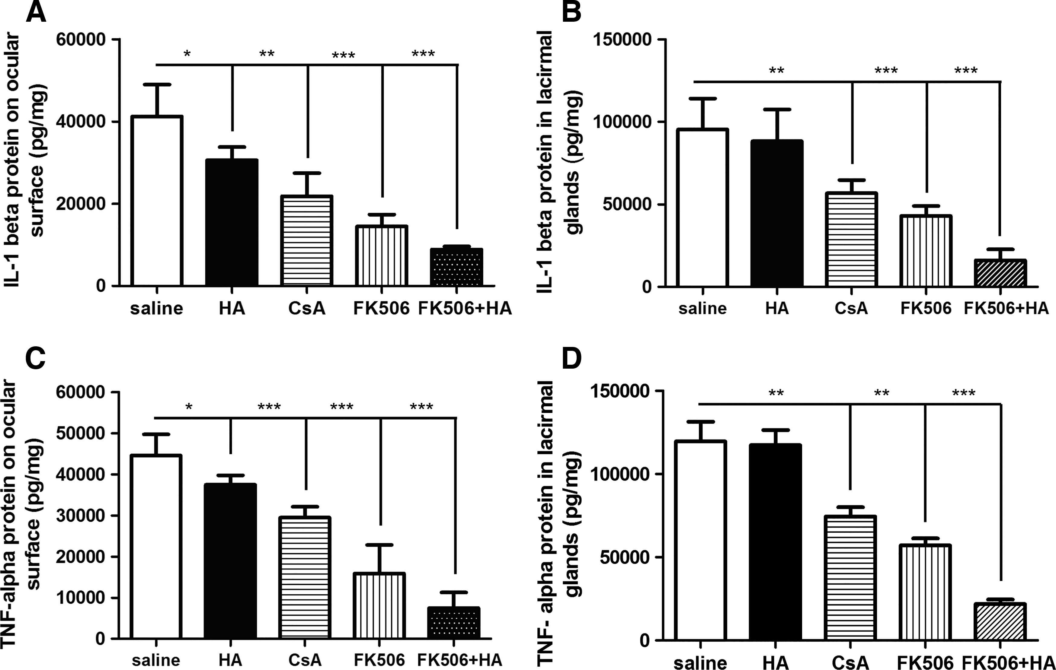
The expression of TNF-α and IL-1β proteins detected by ELISA 4 weeks after treatment. The results indicated that the levels of TNF-α (
Expression of Inflammatory Cytokines in the Keratoconjunctival Tissues and Lacrimal Glands
All data are shown as the means ± standard deviations.
HA, sodium hyaluronate; CsA, cyclosporin A.
Discussion
DED is a multifactorial ocular disease. The most common therapy is the use of lubricants, which is effective against most types of DED.1,2 Evidence has shown that inflammation is involved in the pathogenesis of the disease. 21 Anti-inflammatory drugs are beneficial for patients when irritation symptoms and inflammatory signs are present. Various anti-inflammatory treatments have been demonstrated to be useful for DED treatment. 3 Topical CsA has become an effective treatment for patients with moderate-to-severe DED.10,11 However, no single treatment successfully cures DED. Other medications have been shown to improve the quality of life in patients with DED. In the present research, we assessed the curative effects among HA, 0.05% CsA, 0.025% FK-506, and 0.025% FK-506 eye drops combined with HA eye drops in a BTX-B-induced dry eye model. We demonstrated that the topical application of 0.025% FK-506 combined with HA, 0.025% FK-506, and 0.05% CsA eye drops can elevate tear production, decrease corneal fluorescein staining scores, and reduce inflammatory cytokine secretion in both the keratoconjunctival tissues and lacrimal glands of mice with BTX-B-induced dry eye.
Several experimental dry eye models have been reported to investigate its pathogenesis and treatment and are described in the literature.22,23 The most frequently reported dry eye model in mice is created by the injection of scopolamine transdermally or subcutaneously, with or without air currents. Some new models have been used to induce dry eye, such as the BTX-B-induced dry eye model and the benzalkonium chloride eye drop-induced dry eye model.19,24,25 Among the animal models of dry eye, the BTX-B-induced mouse model of dry eye has been established to mimic non-Sjogren's disease-associated dry eye. The dry eye state can be sustained for at least 4 weeks in this model, which is a more suitable tool for investigating the pathogenesis of the chronic condition of human dry eye than other animal models. 19 In this study, topical application of FK-506+HA, FK-506, and CsA eye drops was effective in recovering the corneal fluorescein staining scores and tear production in this model. The FK-506+HA eye drops showed the earliest onset time and the best therapeutic effects, followed by the FK-506 and CsA eye drops.
We examined 5 concentrations (0.0016%, 0.0033%, 0.0125%, 0.025%, and 0.05%) of FK-506 eye drops in our preliminary experiments and found that 0.025% was the most suitable concentration. CsA eye drops (0.05%) are effective in the BTX-B-induced mouse model of dry eye, 20 and FK-506 is reported to be ∼50–100 times more potent than CsA14,15; therefore, we chose 0.0016% FK-506 (∼50 times more potent than CsA) and 0.0033% FK-506 (∼100 times more potent than CsA). However, 0.0016% FK-506 and 0.0033% FK-506 eye drops did not improve DED in the BTX-B-induced mouse model. As a result, we tested 3 other concentrations (0.0125%, 0.025%, and 0.05%) up to 0.05%, which is the only concentration widely applied in China as an eye drop. The results showed that 0.025% and 0.05% were effective in treating BTX-B-induced dry eye, and no statistically significant difference was observed between these 2 concentrations. Ultimately, 0.025% FK-506 was chosen for this experiment, whereas 0.05% CsA was reported to be an effective therapy in this model of dry eye. FK-506 was 2-fold more effective than CsA for dry eye, which is lower than the comparative potency of its immunosuppressive effects. The results may be due to 2 possible reasons. One reason may be the differences in the drug accumulation, distribution, and clearance of the 2 drugs.26,27 FK-506 is not as lipophilic as CsA and therefore does not enter the cornea as readily. Previous reports have shown that FK-506 distributes and clears more easily than CsA, meaning that there is less FK-506 accumulation in the cornea.28,29 The other reason may be the differences in the anti-inflammatory mechanisms between the 2 drugs. CsA can inhibit apoptosis by blocking the opening of the mitochondrial permeability transition pore, except as an inhibitor of calcineurin, which occurs with FK-506 administration. 30
At the molecular level, dry eye induction leads to a persistent increase in the corneal expression of inflammatory cytokines (eg, IL-1β and TNF-α).9,31 Recent studies have also demonstrated that ocular surface inflammation develops in the BTX-B-induced dry eye model. The inflammatory cytokines (eg, IL-1β and TNF-α) in the keratoconjunctival tissues and lacrimal glands were significantly increased in BTX-B-injected mice. 32 The current research demonstrated that the IL-1β and TNF-α expression levels were decreased in the keratoconjunctival tissues and lacrimal glands after the administration of FK-506+HA, FK-506, and CsA. FK-506 and CsA have been reported to decrease the expression of inflammatory cytokines by inhibiting T lymphocyte activation.33,34 However, no T lymphocytes or macrophages were identified in either the lacrimal glands or ocular surface of the BTX-B-induced mouse model. Therefore, FK-506 or CsA may decrease IL-1β and TNF-α expression in the ocular surface and the lacrimal glands via other mechanisms. In a recent report, FK-506 and CsA were shown to suppress the activation of the nuclear factor-kB (NF-κB) pathway induced by hyperosmotic stress (a key factor in the pathological process of dry eye) in a series of cell lines, including nonimmune NRK-52E cells (a rat renal tubular epithelial cell line) and LLC-PK1 cells (a porcine renal tubular cell line). 35 Our previous study showed that the anti-inflammatory effect of FK-506 was due to inhibiting the activation of NF-κB in the lacrimal glands and ocular surface epithelium and that the production of inflammatory cytokines was reduced. 18 The exact anti-inflammatory mechanism of CsA was unclear in the BTX-B-injected mice, but it is possible that it is similar to that of FK-506, based on their similar anti-inflammatory mechanisms in both lymphocytes and nonlymphocytes.
We observed some improvements, although not significant, in the tear production and corneal fluorescein staining of the HA-treated mice. This finding may indicate that the frequency of supportive eye drop application was insufficient and emphasizes the role of inflammation in DED. Interestingly, HA eye drops can reduce the expression of inflammatory cytokines in the keratoconjunctival tissues rather than in the lacrimal glands, which could be a consequence of the changes in the osmotic pressure on the ocular surface. As demonstrated previously, the tear osmotic pressure increases in DED.36,37 Hyperosmolarity alone can stimulate the expression and production of inflammatory cytokines such as IL-1β and TNF-α on the ocular surface in an in vivo animal model. 38 Therefore, the HA eye drops could relieve inflammation on the ocular surface by enhancing tear film stability, which reduces the elevated tear film osmolarity. 39 Our results showed that the treatment of DED with FK-506+HA eye drops produced the best results, treatment with FK-506 eye drops produced good results, and CsA eye drops produced the worst results. A synergistic effect may have occurred between HA and FK-506. Owing to the large number of negative charges on the molecule and the mesh structure, HA is capable of holding large quantities of water and drugs. This synergistic effect might due to 2 reasons. First, the drug-holding capacity can extend the time that FK-506 remains on the ocular surface, which can allow FK-506 to attain its full effect. Second, FK-506+HA eye drops suppress inflammation by 2 different pathways. The water-holding capacity can reduce the elevated tear film osmolarity, which can relieve inflammation in the initial stage. Moreover, FK-506 can restrain inflammation by inhibiting the activation of NF-κB.40,41 As a result, the combination of FK-506 and HA can exhibit a better anti-inflammatory effect than use of a single drug alone.
In conclusion, topical administration of 0.025% FK-506 combined with HA, 0.025% FK-506, and 0.05% CsA eye drops for 4 weeks can successfully increase tear production and reduce fluorescein staining scores. Eye drops containing 0.025% FK-506 combined with HA showed the best therapeutic effects, making it a promising therapy for treating dry eye. Future experiments should be performed to determine whether the findings from this study can be translated into a clinical benefit for patients with DED.
Footnotes
Acknowledgment
This research was partly supported by “Guangdong Scientific Program 2013” in China (Grant no.: 20130319c).
Author Disclosure Statement
No competing financial interests exist.