
Abstract
Abstract
Purpose:
The purpose of this in vitro study was to assess the potential benefits of eye drops based on hybrid cooperative complexes (HCCs) obtained from high and low molecular weight hyaluronic acid (HA).
Methods:
Rheological measurements were performed to adjust the HCC concentration toward optimal resistance to drainage from the ocular surface. The viscosity and mucoadhesion profiles of the optimized preparation were derived. Primary porcine corneal epithelial cells were used for biological studies. Cells were exposed to dehydration after being pretreated with the HCC solution, and protection from desiccation was evaluated using cell viability assays. Time-lapse experiments were carried out to evaluate the ability of the HCC preparation to promote corneal wound healing. The characterization studies were performed in comparison with a control HA solution representative of commercial HA-based products.
Results:
The HCC formulation is able to deliver twice the amount of biopolymer compared with conventional products while avoiding discomfort due to excessive viscosity. The viscosity and mucoadhesion profiles allowed the authors to predict the longer in vivo retention and, therefore, an improved HCC formulation bioavailability. The new preparation also proved superior in protecting porcine corneal epithelial cells from desiccation and in hastening corneal cell wound repair in vitro.
Conclusions:
The results suggest that the developed formulation may be a promising topical ophthalmic medical treatment.
Introduction
Hyaluronic acid (HA), also known as hyaluronan, is widely used as the active component of many artificial tear substitutes for the treatment of dry eye symptoms. Its capacity to protect corneal epithelial cells from dehydration and to promote corneal wound healing is mainly exploited. Furthermore, its viscosifying properties and mucoadhesiveness allow a reduction in the drainage rate of the preparation from the ocular surface, thus enhancing the therapy's effectiveness.1–16
Considering these HA-related beneficial effects and the high patient compliance associated with the topical treatment of eye disorders, there is a great clinical interest in high-performing HA-based eye drop preparations.
Currently available products mainly contain the linear naturally occurring form of the biopolymer, and molecular weights varying from 155 to 1,400 kDa, and concentrations in the range 0.1–0.4 wt% are encountered.17–21
Several studies have demonstrated that the biopolymer concentration is the main polymer-related parameter to adjust to maximize efficacy: the higher the biopolymer content, the higher the preparation's bioavailability, cytoprotective effect against desiccation, and healing capacity.4,17,18,21,22 However, the HA amount in eye drops is limited by the final formulation's viscosity that should not exceed 30 mPa·s to avoid discomfort for the patients.23,24
This study aimed to evaluate recently developed hybrid cooperative complexes (HCCs) of HA to set up high-performing ophthalmic formulations for topical use. Unlike currently available products, such complexes are based on both high and low molecular weight HA chains (H-HA and L-HA, respectively), which cooperatively interact during a specific patented thermal process—namely the NAHYCO™ technology—to form a HCC.25,26 The HCC solution exhibits a viscosity much lower than that of the simple mixture of the 2 components.25–27 This feature should permit an increase of the biopolymer amount in the formulation without exceeding the viscosity limit and creating discomfort. Prolonged retention and improved biological efficacy were, therefore, expected.
Furthermore, it is well established that both short and long HA chains display crucial roles at an injury site during the wound repair process of the tissues.28–31 The concurrent presence of H-HA and L-HA in HCCs could be on the basis of enhanced corneal wound repair activity compared with currently available products.
To verify the potential of HCC-based eye drops, the viscosity-optimized formulation was first identified and then in vitro investigated for mucoadhesiveness, protective effect on corneal epithelial cells from dehydration, and ability to accelerate the corneal wound healing process, compared with a conventional HA-based preparation.
Methods
Materials
HA sodium salt, lot. No. 02622 (H-HA), was kindly provided by Altergon srl (Italy). Its complete hydrodynamic characterization is reported elsewhere: it has a Mw of 1,120 ± 100 kDa and a polydispersity index Mw/Mn = 1.5 ± 0.1. 21 The hydrodynamic radius and the intrinsic viscosity values are 67 ± 7 nm and 18.7 ± 1.8 dL/g, respectively. For the preparation of hybrid cooperative complexes (HCC), the high molecular weight HA sodium salt, lot. No. 5052/1, was also kindly provided by Altergon srl (Italy). The hydrodynamic characterization provided a Mw 1500 50kDa; Mw/Mn = 1.5 – 0.1, intrinsic viscosity value 22.2 – 2.1dL/g, hydrodynamic radius 78 – 3 nm. Low molecular weight hyaluronic acid sodium salt (L-HA) was prepared by hydrolysing the H-HA powder under heterogeneous acid conditions, as reported elsewhere. 28 The purified hydrolysis product, L-HA, was characterized using the size exclusion chromatography-triple detector array equipment by Viscotek (Lab Service Analytica, Italy).32,33 The sample had a Mw equal to 110 ± 10 kDa, and a polydispersity index Mw/Mn = 1.9 ± 0.1. The hydrodynamic radius and the intrinsic viscosity were 16 ± 1 and 2.7 ± 0.1 dL/g, respectively, and were consistent with previous data.32,34,35
Mucin (from porcine stomach type II; Cat. No. M2378), Na3PO4 (Cat. No. 342483), NaH2PO4.2H2O (Cat. No. 71505), Na2HPO4.2H2O (Cat. No. 71643), EDTA (ethylenediaminetetraacetic acid disodium salt dihydrate; Cat. No. E5134), sodium hydroxymethylglycinate (Cat. No. CDS003712), and collagen (Cat. No. C9791) were all purchased from Sigma-Aldrich (Milan, Italy). Dulbecco's phosphate-buffered saline (PBS), without calcium and magnesium, was purchased from Lonza Sales Ltd., (Switzerland; Cat. No. BE17–516F). Collagenase type I (Cat. No. 17100017), dispase (Cat. No. 17105-041), DMEM/F12 (Cat. No. 11320033), fetal bovine serum (FBS; Cat. No. 16000014), and epidermal growth factor (EGF; Cat. No. PHG 0314) were purchased from Invitrogen (Milan, Italy). Gentamicin was purchased from Fisiopharma S.r.l. (Salerno, Italy). Presto Blue assay (Cat. No. A13261) was purchased from Invitrogen.
HCC preparation
The HCC complex (16g/L H-HA 1500kDa +16g/L L-HA 100kDa) was obtained following the NAHYCOTM technology procedure as described elsewhere.25–27 The H-HA/L-HA complex formation was ascertained by performing viscosity measurements according to the literature (data not shown).26,27
Rheological measurements: evaluation of viscosity and mucoadhesiveness
Rheological measurements were carried out using a Physica MCR301 oscillatory rheometer (Anton Paar, Germany) with coaxial cylinders and a Peltier temperature control. The system is described in detail elsewhere. 21
Viscosity measurements
The H-HA/L-HA complex was diluted to different concentrations in the range 0.40–0.90 wt% in the buffer used for commercial products. 21 For each solution, the dynamic viscosity was registered as a function of the shear rate in the range 1–1,000 s−1 at 35°C (50 measuring points and no time setting) and the value of zero-shear viscosity (η0, viscosity in the range of Newtonian plateau) was obtained. The dependence of η0 on HCC concentration was derived. The concentration corresponding to the target viscosity value, fixed at 24 mPa.s, was identified.
For H-HA, a 0.28 wt% solution was prepared and the flow curve was registered under the same conditions described earlier for HCC. H-HA 0.28 wt% corresponds to the viscosity-optimized H-HA-based preparation, as previously determined, and is representative of the best performing among the analyzed HA-based eye drops on the market. 21
Mucoadhesiveness measurements
Mucoadhesiveness of the samples was evaluated by means of viscosity measurements as previously described.21–23,36 In brief, the following samples were prepared:
A suspension of mucin (10 wt%) in the buffer indicated in Viscosity Measurements section. A solution of the macromolecule to test HCC or H-HA in the same buffer at the concentration corresponding to the target viscosity. A suspension containing mucin (10 wt%) and HCC or H-HA at the same concentration as in sample 2.
The procedure followed for the preparation of the solutions is described in detail elsewhere. 21 The pH and the conductivity were maintained in the range 7.0–7.6 and 12.0–14.0 mS/cm, respectively.
The dynamic viscosity of each solution was measured as a function of the shear rate (1–1,000 s−1) at 35°C.
At each value of shear rate, mucoadhesiveness of sample 2 was expressed as
where Δ(%) is the mucoadhesion index, ηmuc is the dynamic viscosity of sample 1, ηHA is the dynamic viscosity of sample 2, and ηmuc+HA is the dynamic viscosity of sample 3.
For a mucoadhesive polymer, ηmuc+HA is higher than (ηmuc + ηHA) due to the interaction occurring between the polymer and mucin.22,23,36 Δ(%) is a measure of the mucoadhesion strength.
The experiments were carried out in triplicate. The Δ(%) value at each shear rate value is reported as the mean value ± SD. A 10 wt% mucin was selected for the experiments since it allowed us to appreciate the mucoadhesion phenomenon also allowing for a good dispersion of mucin. Rheological studies were performed at 35°C since this value was reported as the temperature of the eye's surface and because many similar studies, reported in the literature, were carried out under these conditions.2,22–24
Clarity examination
HCC formulation's optical clarity was assessed by visual inspection in suitable lighting and by transmittance measurement in the wavelength range 400–800 nm using a DU 800 UV/Vis spectrophotometer (Beckman Coulter, Inc.).37,38
Cell culture and growth conditions
Primary porcine corneal epithelial cells (PCECs) were obtained by enzymatic digestion of 2 fresh porcine corneas, a gift from A.O.R.N. Antonio Cardarelli, Centre of Biotechnologies (Naples). Specifically, the corneas were washed with PBS and then immersed in a digestion solution composed of 0.06% (w/v) collagenase type I and 0.08% (w/v) dispase in the complete growth medium (DMEM/F12 containing 15%w/v FBS, 10 ng/mL EGF, and 40 μg/mL gentamicin). 14 Isolated cells were seeded and grown in complete DMEM/F12.
The epithelial cell morphology was evaluated by inverse phase microscopy. Cells were expanded to achieve the proper number to run the experiments in triplicate. In particular, cells at passage 2 and passage 3 were used for the dehydration and wound healing experiments, respectively.
Evaluation of the protective effect against dehydration
PCECs were seeded and grown to 70% confluence in 24-multiwell plates (seeded cells/well: 5 × 10 4 ). The medium was then replaced with the HCC- and the H-HA-based preparations and with the same after 1:3, 1:10, and 1:30 dilutions. Specifically, H-HA 2.8 wt% in PBS was prepared and then diluted to the optimal concentration (0.28 wt%) (1:10 dilution) in the cell culture medium. The same procedure was followed for the HCC preparation: a 10-fold concentrated HCC solution in PBS was diluted to 0.55 wt% in cell culture medium. For both the positive (CTR+) and the negative (CTR−) control, the medium was replaced with PBS not containing HA and diluted 1:10 in cell culture medium. Cells were incubated under cell culture conditions for 2 h. Untreated controls (CTR−) and cells treated with the HA samples were then exposed to dehydration. Next, the medium was removed and the multiwells were incubated at 37°C without the lid until the effect of the applied stress on cells was evidenced (i.e., morphological changes) in the CTR−.
The cells in the positive control were not exposed to desiccation but kept under normal culture conditions throughout the experiment. Images of cells were acquired using a LEICA microscope (Model: DMi1; Microsystem CMS GmbH) with a 10 × objective and a Nikon Coolpix4500 camera (30 × final magnification). Cell viability was then evaluated using the Presto Blue assay (Cat. No. A13261; Invitrogen, Gibco) according to manufacturer's instructions. Cell viability (%) was calculated with respect to the positive control (100% viability) and normalized with respect to CTR−. The experiment was performed in triplicate. In each replicate, the formulations were also analyzed in triplicate, thus 9 samples were analyzed for each formulation. Results of the replicates were consistent. Results from 1 replicate are reported as the mean value ± SD. A Student's t-test was performed for comparison between groups. P values <0.05 accounted for significant differences.
Evaluation of the effect on corneal wound healing
Cells were seeded in a 12-well plate precoated with collagen at a cell density of 1.8 × 10 5 cells/well (4 × 10 4 cell/cm 2 ) until complete confluence was reached. Scratch wounds were created mechanically with a sterile pipette tip (Ø = 0.1 mm). The effect of the HA formulations on the rate of corneal repair was tested by incubating the scratched monolayers of porcine corneal epithelial cells with 0.55 wt% HCC and 0.28 wt% H-HA in DMEM/F12 1.5% FBS. Wound repair was monitored and analyzed using a time-lapse video microscopy station (OkoLab, Naples, Italy), equipped with the custom-tailored software OKO-Vision 4.3 software.25,39 Immediately after the scratch, the multiwell was placed into the station and several fields of view were selected for each well. Images of the selected areas were acquired by using an AxioVision200 Microscope equipped with CCD-gray-camera (ORCA ER; Hamamatsu, Photonics, Japan) and a 10 × objective every 30 min. A quantitative analysis of the rate of wound closure was thereby obtained. At each time interval, the percentage of repaired area, the wound closure (%), was calculated as [(area t0 – area t)/(area t0) × 100] by the software. Each sample was analyzed in triplicate. For each replicate, a minimum of 5 fields of view were used for deriving the overall averaged curves of wound closure (%) as a function of time, thus assuring the statistical significance of the experiment. Each field of view (∼1 × 10 6 μm 2 ) represents 5% of the total scratched area (∼20 × 10 6 μm 2 ) of each well. Fields of view showing similar scratch width were selected for the analysis.
Results
HCCs: identification of the viscosity-optimized formulation
The zero-shear viscosity of HCC solutions was studied as a function of the HCC concentration to identify the viscosity-optimized formulation. Results are shown in Fig. 1. η0 exponentially increased with the HCC concentration. The optimal viscosity value was set at 24.2 mPa.s.21,23,24 Based on the curve in Fig. 1, the complex concentration corresponding to the “ideal zero-shear viscosity” was 0.55 wt%. Such formulation was selected for further investigation.
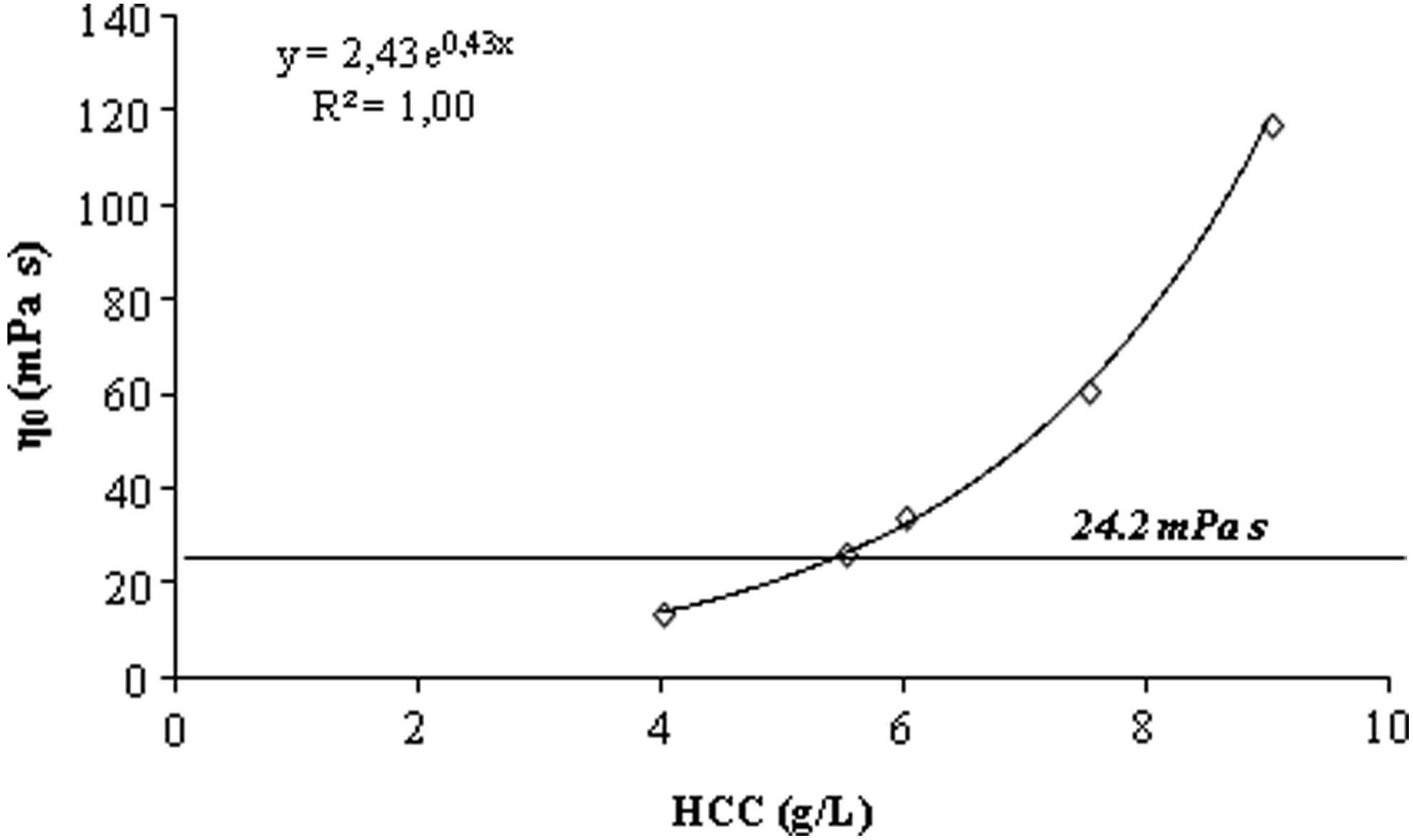
Zero-shear viscosity as a function of HCC concentration. The η0 value selected as the optimal viscosity is evidenced. HCCs, hybrid cooperative complexes.
Evaluation of the viscosity and mucoadhesion profiles of the optimized formulation
The dynamic viscosity profiles of the optimized HCC preparation and the conventional H-HA 0.28 wt% solution are shown in Fig. 2a. Although the formulations exhibit the same (optimized) η0, the dynamic viscosity varies differently with the shear rate. In particular, both solutions present a pseudoplastic behavior, but for the HCC-based formulation, viscosity decreases less with the shear rate.
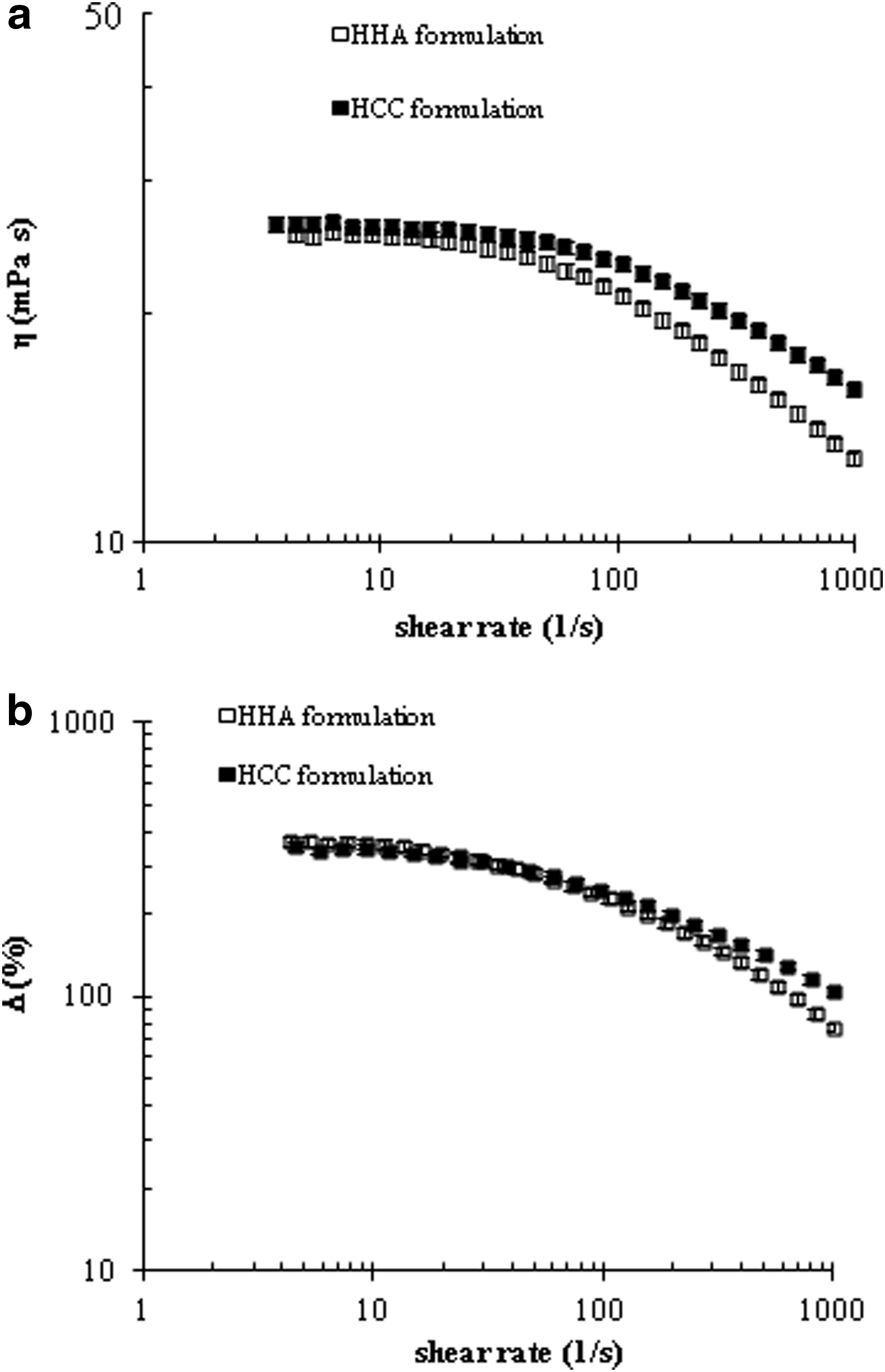
The mucoadhesion profiles are shown in Fig. 2b. The mucoadhesiveness decreases with shear rate for both the formulations. The mucoadhesion indexes were similar in a wide range of shear rate; however, at shear values >400 s−1, the HCC–mucin interaction proved slightly but significantly stronger.
Clarity examination
The HCC 0.55 wt% formulation appeared transparent at visual inspection. The appearance of the preparation with respect to distilled water is provided in the Supplementary Fig. S1 (Supplementary Data are available online at www.liebertpub.com/jop). The percentage transmittance of the formulation in the range 400–800 nm, registered with respect to distilled water, is also reported (Supplementary Fig. S1). The formulation exhibited percentage transmittance values higher than 95% in the whole visible range of wavelength, thus indicating a high transparency of the formulation.
In vitro evaluation of corneal epithelium protection against dehydration
HCC-based formulation was evaluated for the capacity to protect PCECs from dehydration with respect to H-HA 0.28 wt%. Experimental results are shown in Figs. 3 and 4. Optical microscope images of PCECs captured at the end of the desiccation experiment are shown in Fig. 3. In particular, images of cells exposed to desiccation under no protective conditions (CTR), or exposed to the same stress after being previously treated with the HA formulations (not diluted and after 30-fold dilution), and of cells that were not exposed to any stress (CTR+) are shown. The results of the quantitative analysis of cell availability are shown in Fig. 4. The images in Fig. 3a and b indicate cell mortality with respect to CTR+ as well as a nontypical morphology for cells stressed under no protective conditions (CTR−). Regardless of the specific formulation used, when treated with the undiluted HA preparations before dehydration, cells show their typical morphology (Fig. 3c, d.) The quantitative data confirmed that the applied stress was responsible for about 50% of cell mortality in the CTR− compared with the CTR+ (Fig. 4). When cell viability was preserved by the HA-based formulations, 80–100% survival rates were estimated. The same result was obtained when the biopolymer concentration was lowered to one-third (data not shown). HCC formulation proved effective in preserving cell morphology (data not shown) and viability (Fig. 4) even after 1:10 dilution. Under the same dilution conditions, effects of the stress became evident for cells treated with H-HA. As shown in Fig. 4, the survival rate is still significantly higher than that for CTR− (about 65% cell viability), but lower than the rate for preincubation with HCCs. When the biopolymer concentration was lowered to 1/30th of the original, no protective effect could be observed for H-HA 0.28 wt%, whereas a cell survival rate significantly higher than the control was still observed for the HCC 0.55 wt% formulation (Fig. 4).

Optical microscope images of PCECs under normal culture conditions
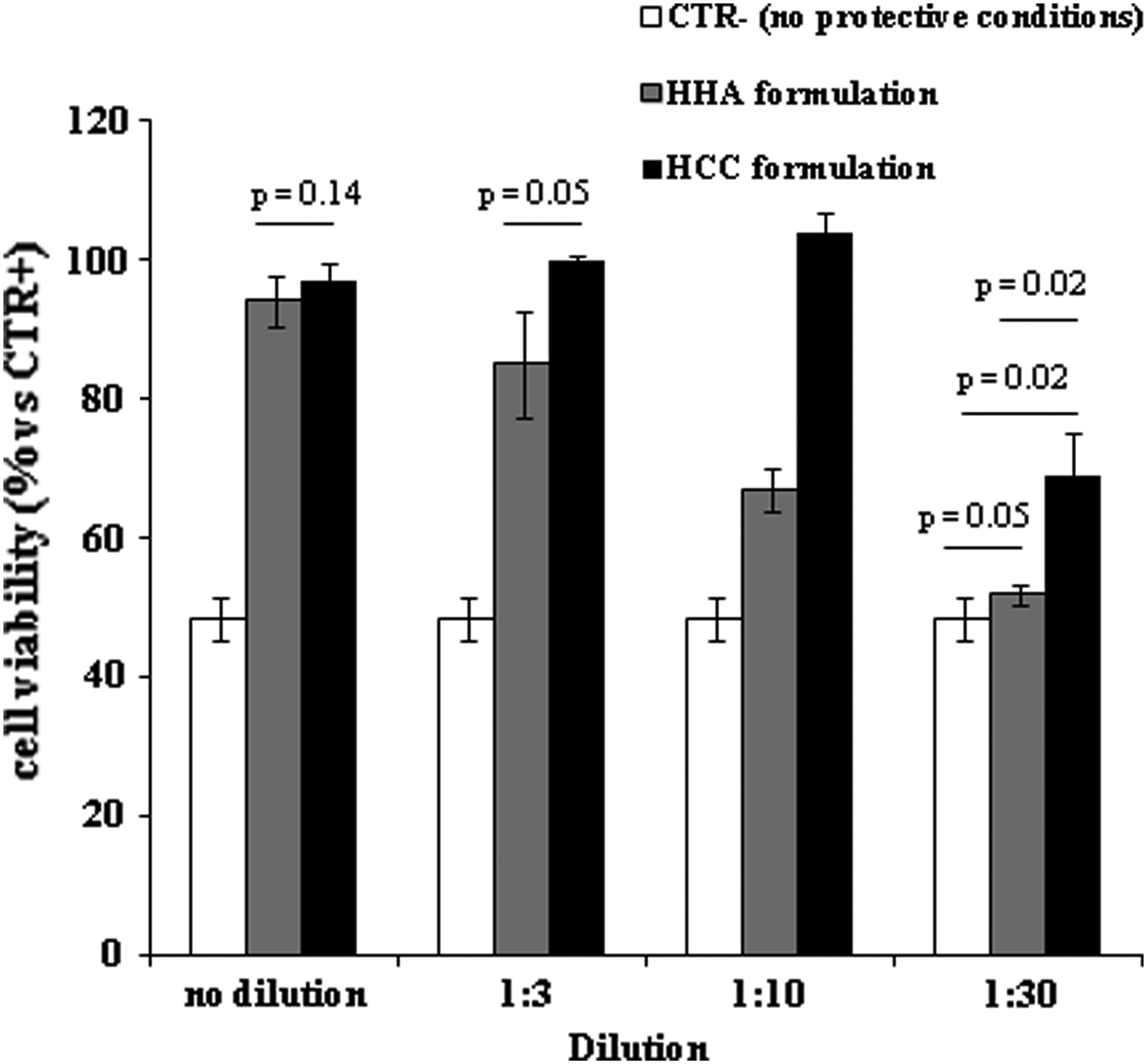
Quantification of the protective effect from dehydration displayed by the HCC- and the H-HA-based preparations. Viability (%) with respect to CTR+ of PCECs dehydrated under no protective conditions (CTR−) and for PCECs exposed to the same stress after treatment with the formulations, differently diluted.
In vitro corneal wound healing
Time-lapse experiments were performed on PCECs to investigate the corneal reparative activity of the HCC formulation compared with the conventional HA-based product. The results are shown in Fig. 5. Specifically, the percentage of wound closure is reported as a function of time in Fig. 5a. The curves indicate a faster repair in the presence of the HCC formulation than in the presence of the conventional HA formulation. In particular, as indicated in Fig. 5b, in the presence of HCCs, 50% wound repair was reached at 2.9 ± 0.5 h, whereas the same result was registered at 4.2 ± 0.1 and 5.3 ± 0.2 h for the H-HA formulation and for the control, respectively. The wound closure in the presence of HCCs proceeded faster until the complete repair was reached, and the differences in the reparative rate increased. Actually, in the presence of HCCs, 80% wound closure was accomplished after 4.5 ± 0.5 h of incubation, whereas 7.2 ± 0.2 h and 10.6 ± 0.7 h were needed to reach the same extent of repair in the presence of H-HA and the control, respectively.
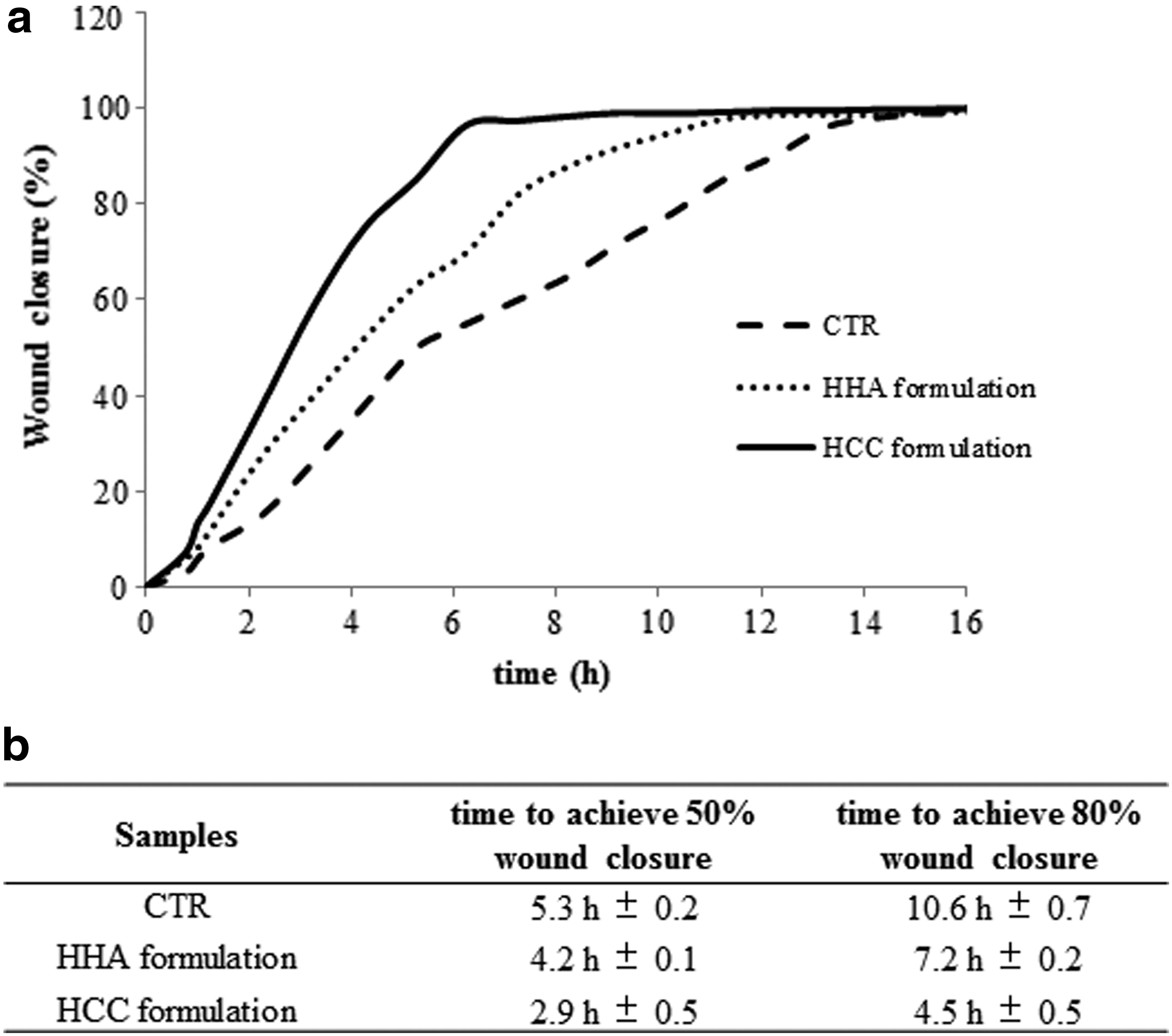
In vitro wound healing capacity of the formulas.
Discussion
In this work, a HCC of H-HA and L-HA was used for the development of a novel HA-based formulation intended for topical ophthalmic use, expecting improvements in efficacy with respect to conventional formulations on the market. Eye drops' efficacy increases with formulation's viscosity; however, viscosity should not exceed 30 mPa.s to avoid foreign body sensation and blurred vision.23,24 In this study, the optimal viscosity value was set at 24.2 mPa.s: this value is the zero-shear viscosity of a commercial already clinically exploited product, and is close to the upper limit for the intended application.21,23,24 The viscosity studies performed allowed to identify HCC 0.55 wt% as the “optimum” (Fig. 1). This result indicates that, with respect to conventional products, the use of the hybrid complex allows an increase, specifically a doubling of the total biopolymer content in the formulation also including a low molecular weight component, while maintaining optimal viscosity for the intended application. The optimized HCC preparation was characterized with respect to the conventional H-HA 0.28 wt% solution, representative of the products currently available on the market.
The viscosity and mucoadhesion profiles were compared to fully evaluate in vitro the relative potential of the formulations to be retained in the precorneal area. The data obtained (Fig. 2) suggest a similar resistance to drainage at rest (similar viscosity and mucoadhesiveness at low shear rates), but affected by blinking (high shear rates), the HCC-based formulation outperforms the H-HA-based formulation, both in terms of viscosity and capacity to interact with mucin. The slighter reduction of HCC viscosity with increasing shear rates (Fig. 2a) is consistent with previous findings demonstrating that shorter HA chains maintain viscosity at high shear rates better than longer chains.21,40,41 The decrease of formulations' mucoadhesiveness with the shear rate (Fig. 2b) confirmed the trend reported in the literature.21,23,36 The stronger HCC–mucin interaction registered under high shear conditions (Fig. 2b) is consistent with previous studies on the dependence of HA mucoadhesiveness on molecular weight and concentration demonstrating that regardless of the molecular weight, the biopolymer concentration effect is predominant at high shear rate values. Based on the rheological data shown in Fig. 2, a prolonged retention on the corneal surface and, therefore, a higher efficacy can be predicted for the HCC formulation.
To evaluate the potential effectiveness of the HCC-based formulation in the treatment of eye dryness disorders, its capacity to preserve the viability of PCECs during desiccation trials was evaluated in vitro and compared with H-HA 0.28 wt%. The solutions were also tested after dilution, allowing for the fact that eye drops are diluted in the lachrymal fluid immediately after instillation and that further dilution occurs when the solution is steadily drained from the ocular surface by blinking. Results for H-HA 0.28 wt% (Figs. 3 and 4) are in agreement with data previously obtained in our laboratory. 21 The survival rate for cells preincubated with the HCC solution (Fig. 4) is similar to that for cells treated with comparable concentrations of linear HA (about 500 kDa Mw) in a similar experiment. 21 This suggests that the better performance of the HCC formulation compared with the H-HA formulation (Figs. 3 and 4) can be ascribed to the higher biopolymer concentration increasing the water retaining capacity.17,42,43 Based on the results of the desiccation trial, in the case of in vivo equal drainage (dilution) rates, the efficacy of the HCC-based formulation is expected to last longer than that of conventional H-HA. Moreover, since the viscosity and mucoadhesion profiles indicate that HCC is likely to be retained longer on the ocular surface, the efficacy of the protection from desiccation should be further improved.
The time-lapse experiments demonstrated the HCC's higher in vitro corneal reparative activity (Fig. 5). The corneal healing rate observed for the H-HA solution with respect to the control is in agreement with literature data.5,7 The higher rate of wound closure observed for the HCC formulation is expected to be related to the synergistic effect of H-HA and L-HA. 28 Such an effect has already been demonstrated when HCC was used for the wound healing of keratinocytes, alone and in coculture with fibroblasts. 25 Furthermore, despite the same viscosity, the higher total HA amount in the HCC than in the H-HA formulation is also expected to contribute to the enhanced wound healing ability. Data obtained suggested that HCC-based eye drops display strong potential in the treatment of scratches and abrasions to the cornea that represent the most common eye injuries.
Finally, the authors want to mention that the safety of HCC-based formulations has already been proved in vivo. 44 Other preparations, based on HCCs obtained by the patented technology employed in this study, are currently used as class III devices in other fields of applications (e.g., aesthetic medicine). 26
Conclusions
HCCs were used for the first time to set up a formulation intended for topical ophthalmic use. The HCC-based optimized formulation contains a twofold higher biopolymer amount than commercialized products, and also has a low molecular weight component, while not exceeding the viscosity limits for the specific application. Compared with a conventional HA-based product, the rheological and biological in vitro characterization of the HCC-based product predicted longer retention time on the ocular surface (higher bioavailability), enhanced cytoprotective effect from dehydration, and improvements in corneal wound repair. The outcomes suggest the newly developed formulation as a promising topical medication for the treatment of dry eye disorders and, generally, for the treatment of corneal injuries and could represent a solid basis prompting further in vivo studies to definitely prove the effectiveness of the preparation.
Footnotes
Author Disclosure Statement
No competing financial interests exist.
References
Supplementary Material
Please find the following supplemental material available below.
For Open Access articles published under a Creative Commons License, all supplemental material carries the same license as the article it is associated with.
For non-Open Access articles published, all supplemental material carries a non-exclusive license, and permission requests for re-use of supplemental material or any part of supplemental material shall be sent directly to the copyright owner as specified in the copyright notice associated with the article.