
Abstract
Abstract
Purpose:
To investigate the anti-(lymph)angiogenic and anti-inflammatory effects of albendazole and to study whether these effects are additive with bevacizumab therapy in a murine corneal suture model.
Methods:
Corneal neovascularization (NV) and lymphangiogenesis (LY) were compared in a corneal suture model after administration of a subconjunctival injection of albendazole, bevacizumab, dexamethasone, or phosphate-buffered saline (PBS). Immunohistochemical staining and analysis were performed in each group. Real-time polymerase chain reaction (RT-PCR) was performed to quantify the expression of inflammatory cytokines (tumor necrosis factor [TNF]-alpha and interleukin-6), vascular endothelial growth factor (VEGF)-A, VEGF-C, vascular endothelial growth factor receptor (VEGFR)-2, and VEGFR-3. To evaluate the additive effect of albendazole, corneal NV and LY were also analyzed in a combined group of albendazole and bevacizumab therapy and the additive effect was compared with that in the group of double dose of bevacizumab.
Results:
The albendazole group showed less NV and less LY compared with the PBS control group (P < 0.01). When albendazole was combined with bevacizumab therapy, a significant decrease in NV and LY was seen compared with bevacizumab treatment alone, and with albendazole alone (all P values <0.05). The combination group showed better antilymphangiogenesis effect than the group of double dose bevacizumab. The albendazole-treated group showed reduced expression of VEGF-A, VEGF-C, TNF-alpha, and VEGFR-2 compared with corneas from the PBS group (P value <0.05 in all respective comparisons).
Conclusion:
Albendazole significantly decreased NV and LY in the cornea. This beneficial effect is additively enhanced when combined with bevacizumab treatment.
Introduction
Corneal transparency is required for good vision, and corneal avascularity is important for maintaining corneal transparency.1,2
Multiple pathological conditions lead to the development of angiogenesis and lymphangiogenesis in the cornea, which decreases visual acuity.3–5
In pathologic (lymph)angiogenesis, the vascular endothelial growth factor (VEGF) family of molecules is essential for growth of blood and lymphatic vessels into cornea.6–8 Angiogenic responses to VEGF-A are mediated by vascular endothelial growth factor receptor-1 (VEGFR-1, Flt-1) and VEGFR-2 (KDR). VEGF-C binds VEGFR-2 and VEGFR-3 and promotes lymphangiogenesis.1,9
To prevent pathologic angiogenesis, numerous drugs have been developed as an antiangiogenesis strategy. As 1 antiangiogenesis strategy, repurposing of drugs has been tried with chloroquine, propranolol, diclofenac, itraconazole, and clarithromycin.10–14
Repurposing of anthelmintic drugs for cancer therapy is currently being studied. 15 Anthelmintic drugs such as mebendazole and albendazole have shown anticancer and antiangiogenesis activity in a variety of diseases.16–18
Albendazole, methyl [5-(propylthio)-1-H-benzimidazol-2Yl] carbamate, is a benzimidazole derivative broad-spectrum anthelmintic drug. 16 It has been explored as a potential inhibitor of VEGF, hypoxia inducible factor 1-α, and tumor angiogenesis over the past few years.17,19 The antitumor effect of albendazole is related to its inhibition of tubulin polymerization and cell cycle arrest.17,19 In combination with chemotherapeutics, albendazole shows superior anti-VEGF activity in a xenograft model of cancer.17,19 In light of its antiangiogenesis properties coupled with VEGF inhibition, we hypothesized that albendazole could be repurposed in the cornea as a new treatment for pathologic angiogenesis.
The aim of this study was to evaluate the impact of albendazole on corneal neovascularization (NV) and lymphangiogenesis (LY) in murine corneal suture models.
Methods
The experiments were performed in accordance with guidelines of the Association for Research in Vision and Ophthalmology and were approved by the Institutional Animal Care and Use Committee of St. Vincent's Hospital.
Experimental corneal suture model and subconjunctival injection
In single treatment comparison, 40 eyes of 20 mice (BALB/c) were used. In combination treatment comparison, 36 eyes of 18 mice were used.
Under surgical microscope, 2 corneal sutures (10–0 nylon) were placed between the corneal center and the limbus at the 12 and 6 o'clock positions with a bite length of ∼200 μm and with a depth of near full corneal thickness. To decrease interprocedure variation, 1 surgeon (Y.K.C.) placed sutures in all mice of the suture model.
After making sutures in corneas, 4 test materials (subconjunctival injection, 10 μL) were individually injected into each appropriate group on the day of suture, and twice weekly (1st and 4th day of the week) thereafter until 3 weeks postsuture: albendazole (20 mg/mL, 8 eyes of 4 mice), bevacizumab (Avastin®, 25 mg/mL, 8 eyes of 4 mice), glucocorticoid (dexamethasone sodium phosphate, 5 mg/mL, 8 eyes of 4 mice), and phosphate-buffered saline (PBS) (8 eyes of 4 mice). Using the tip of a 0.5-inch 33-gauge needle with a 45°bevel on a 10 μL gas-tight syringe (Hamilton, Reno, NV), each material was injected into the subconjunctival space, respectively.
After harvesting, NV and LY were compared among groups to assess the antiangiogenic and antilymphangiogenic effects of the treatments.
Immunohistochemical staining
NV and LY were measured in the corneas. After harvesting corneas, immunohistochemical staining for CD31 and LYVE-1 was performed as previously described and is as follows.20,21
Fresh harvested corneas were dissected, rinsed in PBS for 30 min, and fixed in 100% acetone (Sigma, St. Louis, MO) for 20 min. After washing in PBST (0.1% Tween®20/PBS), nonspecific binding was blocked with 3% bovine serum albumin (BSA)/PBS for 3 nights at 4°C. Incubation with 1:500 fluorescein isothiocyanate-conjugated monoclonal antimouse CD31 antibody (558738; BD Biosciences, San Jose, CA) and 1:200 rabbit anti-LYVE-1 (ab14917; Abcam, Inc., Cambridge, MA) in 3% BSA/PBS at 4°C overnight was followed by incubation with 1:1,000 goat antirabbit antibody-Alexa Fluor®546 (A11071; Life Technologies, Carlsbad, CA) for 1 h with subsequent washes in PBST at room temperature. Corneas were mounted with an antifading agent (VECTASHIELD®; Vector Laboratories, Burlingame, CA).
Fluorescent microscopic examination
After immunohistochemical staining and flat mounting of the cornea, images of the corneal vasculature were captured and quantified using a camera attached to a fluorescent microscope (OLYMPUS BX51, Tokyo, Japan) as previously described and are as follows.20,21
NV and LY were quantified by setting a threshold level of fluorescence above which only vessels were captured and processed using Image J (National Institutes of Health, Bathesda, MD). The total corneal area was outlined using the innermost vessel of the limbal (rim of the cornea) arcade. The areas of NV and LY were calculated as follows: NV (%) = (NV area of cornea/total cornea area) × 100; LY (%) = (LY area of cornea/total cornea area) × 100.
Quantitative real-time polymerase chain reaction analysis of gene expression in mouse cornea
After harvesting, the expression of VEGF-A, VEGF-C, VEGFR-2, VEGFR-3, TNF-alpha, and interleukin (IL)-6 in each group was analyzed using real-time polymerase chain reaction (RT-PCR) as previously described.22–24
Total RNA was purified with an RNeasy Mini Kit (Qiagen, Hilden, Germany). Complementary DNA synthesis and thermocycling were performed. Published primer sequences for mouse GAPDH, 25 mouse VEGF-A, 26 mouseVEGF-C, 26 mouse VEGFR-2, 26 mouse VEGFR-3, 26 mouse TNF-alpha, 27 and mouse IL-628 were used. Each gene expression level was analyzed through the Ct method, using GAPDH expression as an internal control. The relative expression level of each sample is expressed as a fold change compared with the normal control (PBS).
Comparison of each group and combination group
Single treatment comparison
The 4 groups analyzed included albendazole (20 mg/mL, 8 eyes of 4 mice), bevacizumab (Avastin, 25 mg/mL, 8 eyes of 4 mice), glucocorticoid (dexamethasone sodium phosphate, 5 mg/mL, 8 eyes of 4 mice), and PBS (8 eyes of 4 mice).
The areas of NV and LY were analyzed and compared.
Combination treatment comparison
To compare combination treatment, after making sutures in mice corneas, the test materials (subconjunctival injection, 10 μL) were individually injected subconjunctivally into each appropriate group on the day of suture, and twice weekly (1st and 4th day of the week) thereafter until 3 weeks postsuture. Then the bevacizumab only group (25 mg/mL, 10 eyes of 5 mice, 10 μL), albendazole only group (20 mg/mL, 8 eyes of 4 mice, 10 μL), combination group (10 eyes of 5 mice) of bevacizumab (25 mg/mL, 10 μL) and albendazole (20 mg/mL, 10 μL), and bevacizumab double dose group (25 mg/mL, 20 μL) were compared.
Statistical analysis
Statistical analysis was performed using SPSS 12.0 (Chicago, IL). Postoperative NV and LY of each group were compared with those of the corresponding control group using ANOVA and post hoc test. P values <0.05 were considered statistically significant.
Results
Single treatment comparison
Neovascularization
As shown in Fig. 1, the albendazole [mean ± standard error of the mean (SEM), 13.47% ± 2.54%], bevacizumab (8.03% ± 1.68%), and dexamethasone (12.51% ± 2.12%) groups demonstrated less corneal NV compared with the PBS group (22.94% ± 1.04%) (P = 0.017, P < 0.01, P = 0.011, respectively).
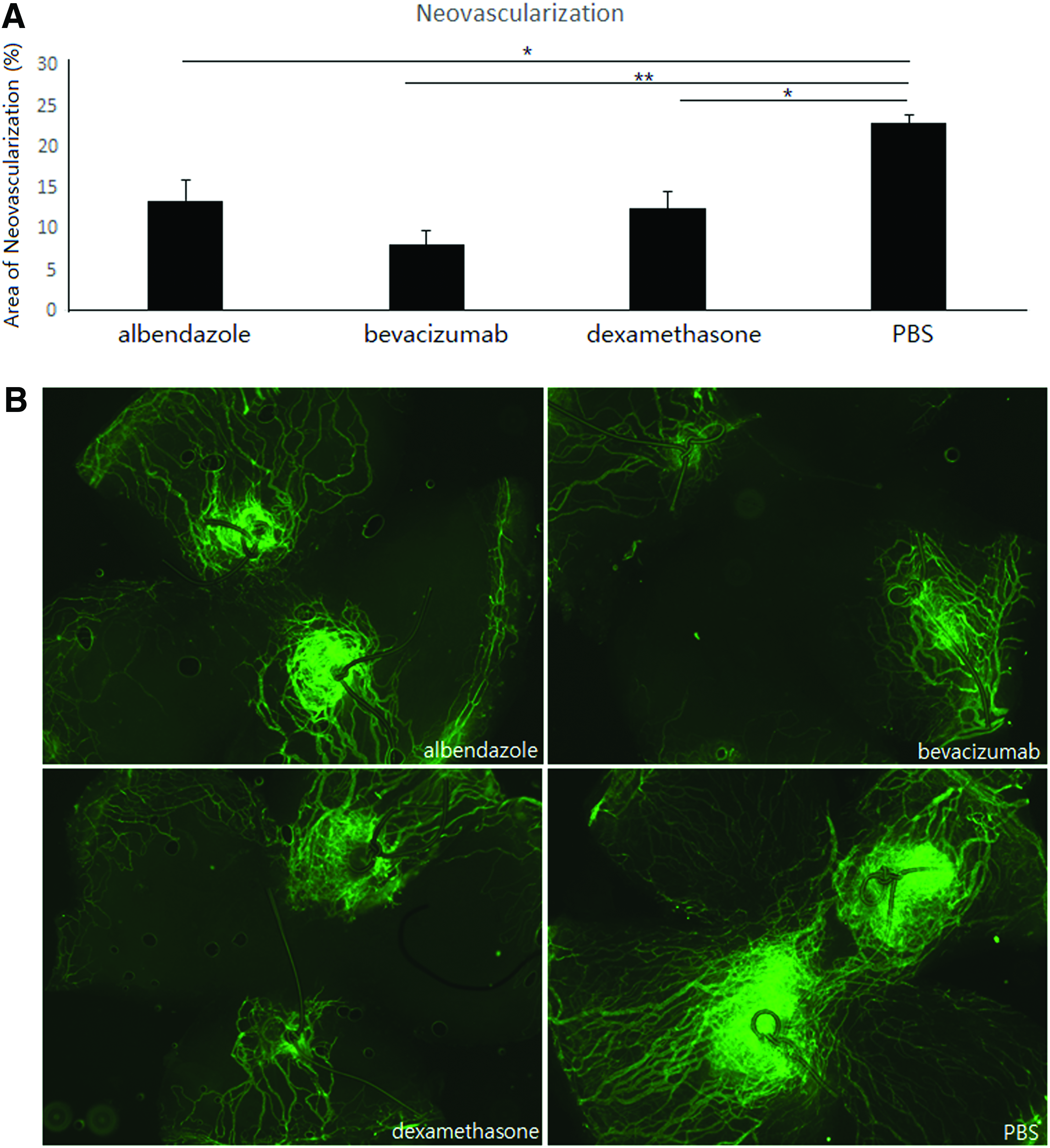
Neovascularization (NV).
Among albendazole, bevacizumab, and dexamethasone groups, there was no significant difference in NV (P = 0.183).
Lymphangiogenesis
As shown in Fig. 2, the albendazole (mean ± SEM, 5.65% ± 1.22%), bevacizumab (4.88% ± 1.44%), and dexamethasone (4.79% ± 1.61%) groups demonstrated less corneal LY compared with the PBS group (11.81% ± 0.71%) (P = 0.019, P < 0.01, P < 0.01, respectively).
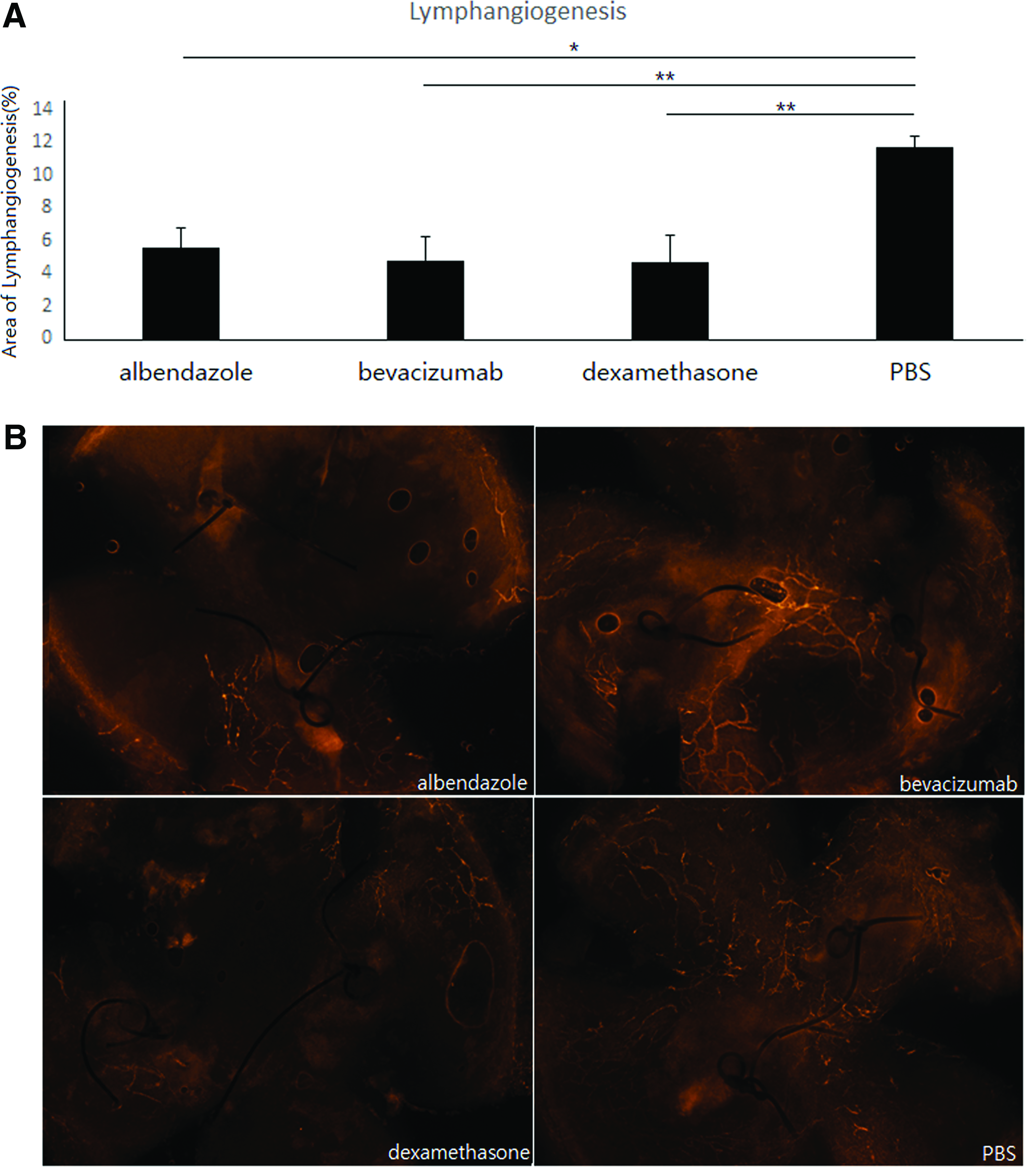
Lymphangiogenesis (LY).
Among albendazole, bevacizumab, and dexamethasone groups, there was no significant difference in LY.
Real-time polymerase chain reaction
Figure 3 shows the mRNA expression levels of VEGF-A, VEGF-C, TNF-alpha, IL-6, VEGFR-2, and VEGFR-3 in each group in suture model. mRNA expression ratios were normalized by GAPDH (PBS group = 1.0). A P value represents statistical significance compared with PBS group.
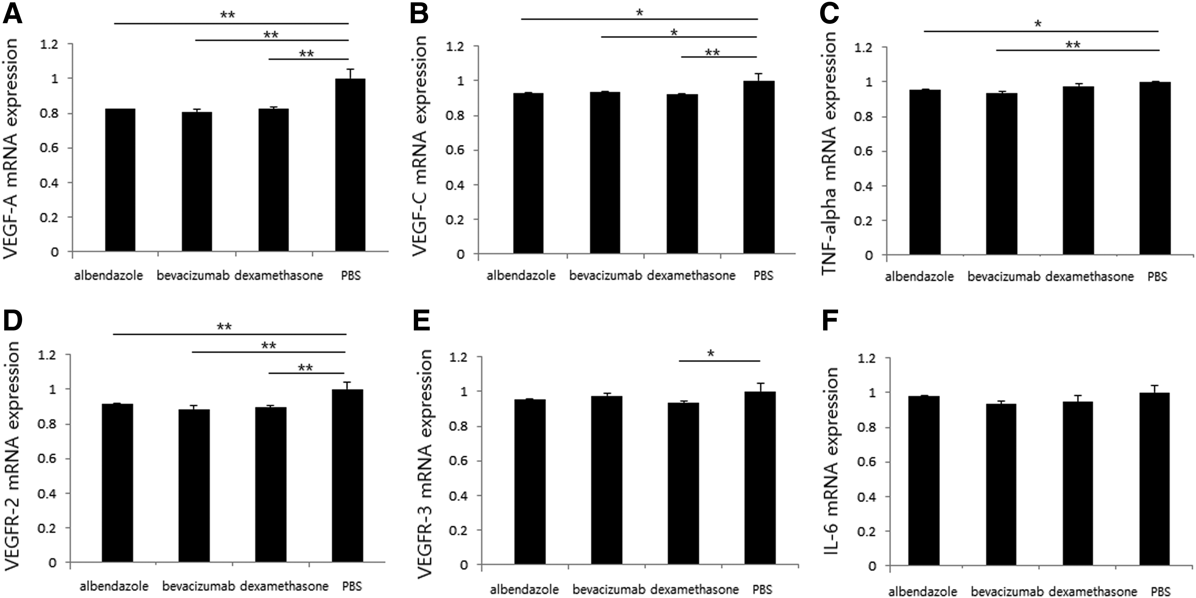
Comparison at 3 weeks postsuture of mRNA expression in corneas of mice treated with albendazole, bevacizumab, dexamethasone, or PBS. mRNA expression ratios were normalized to GAPDH (PBS group = 1.0).
VEGF-A: All 3 groups, albendazole, bevacizumab, and dexamethasone, showed a significant reduction in VEGF-A expression compared with that of the PBS group (P < 0.01, P < 0.01, P < 0.01, respectively). There was no significant difference in VEGF-A level among the albendazole, bevacizumab, and dexamethasone groups (P > 0.05) (Fig. 3A).
VEGF-C: All 3 groups, albendazole, bevacizumab, and dexamethasone showed a significant reduction in VEGF-C expression compared with that of the PBS group (P = 0.02, P = 0.016, P < 0.01, respectively). There was no difference in VEGF-C level among the 3 treatment groups, albendazole, bevacizumab, and dexamethasone (P > 0.05) (Fig. 3B).
TNF-alpha: TNF-alpha expression was significantly lower in the bevacizumab group (P < 0.01) and albendazole group (P = 0.035) than in the PBS group (Fig. 3C).
VEGFR-2: All 3 groups, albendazole, bevacizumab, and dexamethasone, showed significant reduction in VEGFR-2 expression compared with that of the PBS group (P < 0.01, P < 0.01, P < 0.01, respectively). There was no significant difference in VEGFR-2 expression among albendazole, bevacizumab, and dexamethasone groups (P > 0.05) (Fig. 3D).
VEGFR-3: There was no significant difference in VEGFR-3 expression among the albendazole, bevacizumab, and dexamethasone groups (P > 0.05) (Fig. 3E).
The dexamethasone group showed reduced VEGFR-3 expression compared with that of the PBS group (P = 0.049).
IL-6: There was no significant difference in IL-6 level among the albendazole, bevacizumab, dexamethasone, and PBS groups (Fig. 3F).
Combination treatment comparison
The combination effect of albendazole and bevacizumab was evaluated.
Neovascularization
The combination group of bevacizumab with albendazole (mean ± SEM, 7.29% ± 0.65%) showed less NV than the bevacizumab only group (9.64% ± 0.71%) and albendazole only group (12.39% ± 1.81%) (P = 0.026, P = 0.011, respectively). Bevacizumab double dose group (8.46% − 0.98%) did not show a significant antiangiogenesis effect than bevacizumab only group. Bevacizumab double dose group did not show a significant antiangiogenesis effect than combination group of bevacizumab and albendazole (all P values >0.05) (Fig. 4).
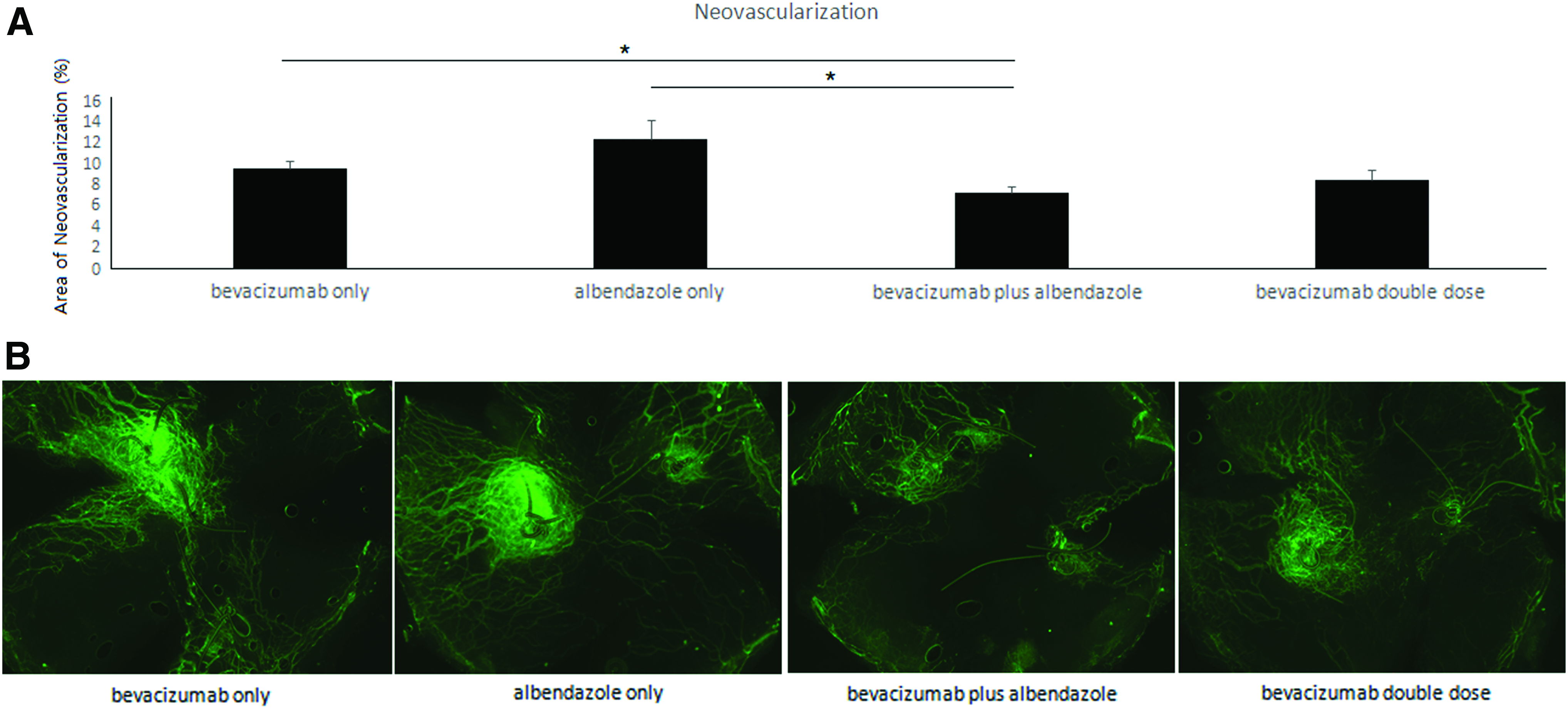
Neovascularization (NV).
Lymphangiogenesis
The combination group of bevacizumab and albendazole (mean ± SEM, 2.77% ± 0.23%) showed less LY than bevacizumab only group (4.30% ± 0.67%) and albendazole only group (5.94% ± 0.92%) (P = 0.047, P = 0.002, respectively). The combination group of bevacizumab and albendazole also showed less LY than the bevacizumab double dose group (4.41% ± 0.75%) (P = 0.037) (Fig. 5).
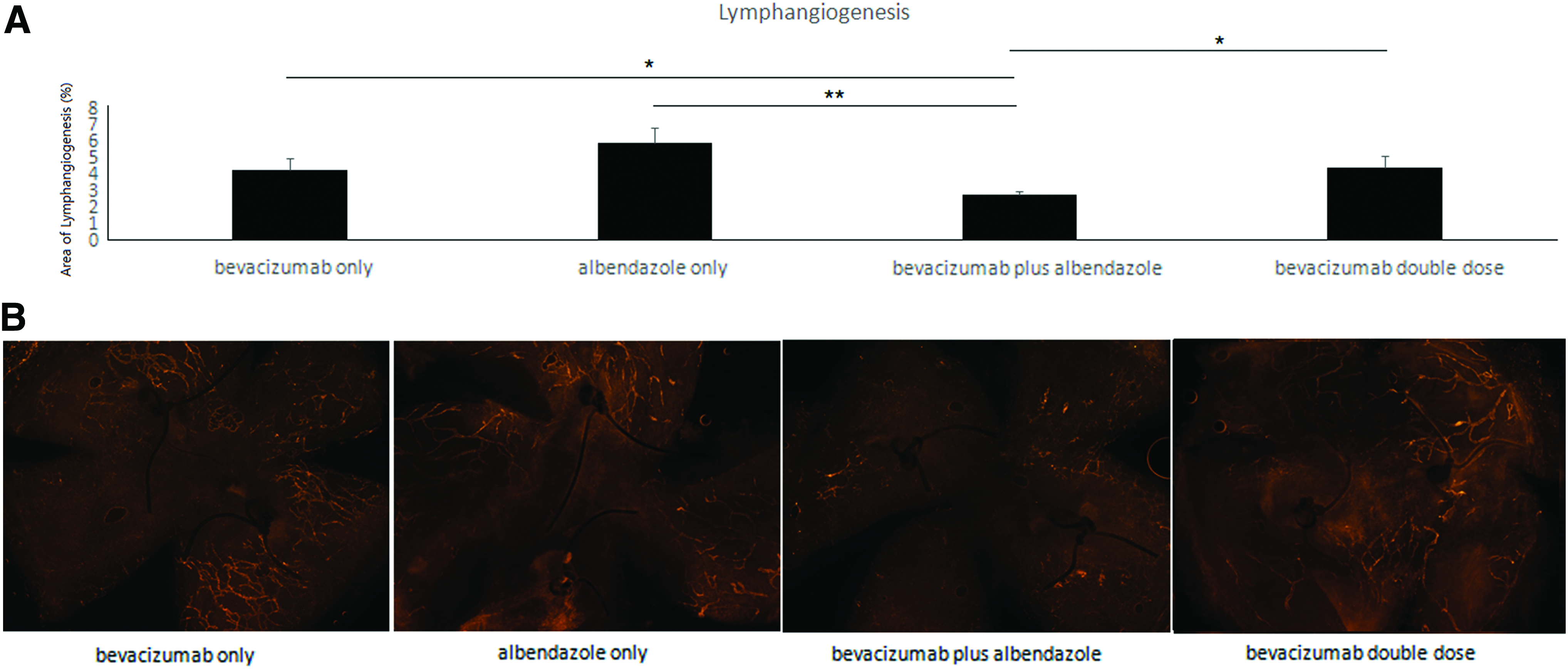
Lymphangiogenesis (LY).
Discussion
Normal cornea is free of blood and lymphatic vessels. 1 Pathologic blood and lymphatic vessels in the cornea are related to diverse ocular diseases and result in decreased visual acuity.2,3,6
To decrease pathologic angiogenesis, modulation of (lymph)angiogenesis has been tried widely.4,7,29
VEGF and VEGFRs are the central regulation of angiogenesis and lymphangiogenesis. VEGFs and VEGFRs are the main molecular target of antiangiogenic therapy, such as soluble VEGFRs, VEGF trap, decoy receptors, and inhibitors of tyrosine kinase receptor.3,5,9 Among recent trials, in an attempt to regulate pathologic angiogenesis, repurposing drug therapy is currently being studied.
Our study is also a trial of repurposing drug therapy.10–15
We have previously tried to repurpose clarithromycin,13,24 itraconazole, 23 and timolol 30 as antiangiogenesis therapy on cornea. Clarithromycin, a well-known antibacterial drug, showed significant antilymphangiogenic and anti-inflammatory effects in corneas as a topical therapy. 24 As a topical therapy, itraconazole, an antifungal drug, showed antiangiogenic and antilymphangiogenic effects in corneal suture and penetrating keratoplasty models. 23 As a topical drug, timolol maleate, an antiglaucoma beta-blocker, showed antiangiogenic and antilymphangiogenic effects in the cornea. 30
Repurposing approved drugs with antiangiogenic effects is a wise and promising strategy to offer more effective options to patients with ocular disease. Repurposing drugs allow substantial advantages of faster and safer preclinical and clinical validation protocols. The potential benefits of repurposing drugs as topical therapy include established efficiency and safety.
Albendazole, methyl [5-(propylthio)-1-H-benzimidazol-2Yl] carbamate, was originally an anthelmintic drug.16,17
We investigated its effect on corneal anti-(lymph)angiogenesis in this study.
In single treatment groups, albendazole showed less NV and LY than the PBS group.
However, the albendazole group did not demonstrate better antiangiogenic effects than the pre-existing commercially available anti-inflammatory and antiangiogenesis agents, dexamethasone and bevacizumab (Avastin).
We then tried combination treatment, with albendazole plus bevacizumab.
In a comparison of combination treatment, albendazole was found to have a significant additive effect when combined with bevacizumab. The combination group showed better effects in decreasing NV and LY than bevacizumab alone as follows. The combination group of bevacizumab and albendazole (mean ± SEM, 7.29% ± 0.65%) showed less NV than the bevacizumab only group (9.64% ± 0.71%, P = 0.026). The combination group (2.77% ± 0.23%) also showed less LY than the bevacizumab only group (4.30% ± 0.67%, P = 0.047).
Although bevacizumab double dose group did not show a significant effect than bevacizumab only group neither in NV nor LY (P > 0.05), combination group showed significant better effect than bevacizumab only group in both NV and LY.
In LY, combination group even showed better anti-LY effect than bevacizumab double dose group (P = 0.037).
These results shown in our study (Figs. 4 and 5) can cautiously suggest that albendazole has additive effect when combined with bevacizumab in anti-(lymph)angiogenesis.
We tried 2-step experiments, first single treatment and second combination treatment. In both experiments, bevacizumab and albendazole were tested and their antiangiogenesis effect in 2 different step experiments was not significantly different. With bevacizumab treatment, NV in single treatment experiment (mean ± SEM, 8.03% ± 1.68%) and combined treatment experiment (9.64% ± 0.71%) was not statistically different (P > 0.05). LY of bevacizumab group in single treatment experiment (4.88% ± 1.44%) and combined treatment experiment (4.30% ± 0.67%) was not statistically different (P > 0.05).
With albendazole treatment, NV in single treatment experiment (mean ± SEM, 13.47% ± 2.54%) and combined treatment experiment (12.39% ± 1.81%) was not statistically different (P > 0.05). LY of albendazole group in single treatment experiment (5.65% ± 1.22%) and combined treatment experiment (5.94% ± 0.92%) was not statistically different (P > 0.05).
Our RT-PCR result showed less VEGF-A and VEGF-C expression in the albendazole group than the PBS group (P < 0.05). Although VEGFR-2 expression decreased in response to albendazole treatment, VEGFR-3 expression did not decrease in response to albendazole treatment. However, albendazole did decrease LY and NV compared with the control PBS group. We suspect that antiangiogenesis induced by albendazole is related to the VEGF-A–VEGFR-2 pathway.2,31–33 LY may have been medicated by VEGF-C–VEGFR-2, or VEGF-A, which directly induce lymphangiogenesis through VEGFR-2.2,31–33
Although albendazole was known to have poor aqueous solubility, 17 albendazole delivered to the subconjunctival space showed a significant effect to decrease NV and LY in the cornea.
Our study identified the novel possibility that albendazole can be used as an antiangiogenesis treatment in the cornea. Especially, the additive effect when combined with bevacizumab was significant on the cornea.
Footnotes
Authors' Contributions
Y.K.C. designed the study, performed the experiment, and wrote the article. E.Y.S. assisted in the molecular work of the experiment. H.U. taught all the procedures of animal and molecular experiments of the study. B.A. designed the study.
Author Disclosure Statement
No competing financial interests exist.