
Abstract
Micelles have been studied in the targeting of drug substances to different tissues as a nano-sized delivery system for many years. Sustained drug release, ease of production, increased solubility, and bioavailability of drugs with low water solubility are the most important superiorites of micellar carriers. These advantages paved the way for the use of micelles as a drug delivery system in the ocular tissues. The unique anatomical structure of the eye as well as its natural barriers and physiology affect ocular bioavailability of the drugs negatively. Conventional dosage forms can only reach the anterior segment of the eye and are used for the treatment of diseases of this segment. In the treatment of posterior segment diseases, conventional dosage forms are administered sclerally, via an intravitreal injection, or systemically. However, ocular irritation, low patient compliance, and high side effects are also observed. Micellar ocular drug delivery systems have significant promise for the treatment of ocular diseases. The potential of micellar systems ocular drug delivery has been demonstrated by in vivo animal experiments and clinical studies, and they are continuing extensively. In this review, the recent research studies, in which the positive outcomes of micelles for ocular targeting of drugs for both anterior and posterior segment diseases as well as glaucoma has been demonstrated by in vitro, ex vivo, or in vivo studies, are highlighted.
Introduction
The eye is a unique organ that differs from other parts of the body due to its complex anatomy, physiology, and its natural effective barriers. Delivery of drug molecules at therapeutic levels to the targeted tissues of the eye with minimum side effects is crucial in drug development for use in treating anterior and posterior segment diseases where conventional dosage forms can only be effective to a certain level. Therefore, the development of drug carriers for ocular administration is a challenging but noteworthy part of pharmaceutical research.
The eye is anatomically divided into two parts: an anterior segment, which is the front one-third of the eye, and the posterior segment, which is the back two-third of the eye. 1 Its natural barriers include tear film, cornea, conjunctiva, sclera, blood-aqueous humor or iris-ciliary body, lens and blood-retina barriers, which limit the penetration of drug substances to the targeted ocular tissues. 2 However, the blood-retinal barrier affects the penetration of drugs to the posterior segment of the eye; other barriers mainly affect the delivery of drugs to its anterior segment.2–4 Thus, the limitation of these barriers should be overcome to improve the ocular delivery of drugs and to provide therapeutic drug concentrations in the targeted tissues of the eye.
Topical drug delivery is considered the first choice for the treatment of ocular diseases. However, in most cases, conventional dosage forms could be inefficient to achieve effective drug concentrations in the target tissues.5,6 To overcome this situation, drugs are generally administered via systemic route at high doses, but serious systemic side effects can be observed. 2 Thus, different topical drug delivery strategies have been developed to increase the penetration of drugs to the target tissue, bypassing the barriers of the eye.2,4
In recent decades, the potential of nanocarriers for ocular drug delivery has been widely investigated and it is hoped that nanotechnology-based delivery systems improve the ocular delivery of drugs. Different types of nanocarriers, including nanoparticles, colloidal nanocarriers, vesicular nanocarriers, and dendrimers, have been developed to increase ocular permeability of drugs, providing improved drug delivery and decreased side effects.7–14
The 29 (2) issue of the Journal of Ocular Pharmacology and Therapeutics, which was published in 2013 by the guest editor of Uday Kompella, addressed the ocular-targeted nanocarriers in different strategies. 15 Molokhia et al. reviewed the ocular devices such as implants and inserts developed for use in the treatment of anterior ocular diseases, 16 whereas Cholkar et al. examined the anterior ocular drug delivery systems for pharmaceutical formulation development. 17 In the same journal, Rodriguez et al. described lipid nanoparticular systems that were developed for use in the treatment of retinal diseases whose clinical or preclinical studies are continuing. 18 Besides, Kambhampati et al. examined only dendrimers as the ocular drug delivery system according to the targeted diseases 19 and Novack provided updated information on commercial products developed as an ocular drug delivery system. 20
In recent years, micellar drug delivery systems have been investigated to deliver drugs for both anterior and posterior segment diseases due to numerous surperiorities of micelles in ocular drug delivery. In 2012, Pepic et al. reviewed ocular polymeric micelles in terms of their physicochemical properties, ability to pass ocular barriers, ocular toxicity, patient compliance, and industrial applicability. 21 In the same year, Cholkar et al. classified micelles according to the treatment of anterior and posterior segment targets. 22 In 2014, Vaishya et al. examined the research studies in which the structure of ocular micelles was argued about. 23 In 2017, Mandal et al. described polymeric micelles only by classifying them according to preparation methods; they also addressed micelle-based hydrogels for ocular delivery. 24
In this review, we mainly focus on the recent research studies on the development of micelles as ocular drug delivery systems. The characteristics of the micellar carriers are highlighted, the research studies based on ocular application of micellar carriers are summarized, and an overview of the potential of micelles for the treatment of anterior and posterior segment diseases of the eye is provided.
Anatomy and physiological barriers of eye—ocular drug administration routes
The eye that is embryologically an extension of the central nervous system is one of the complex and unique organs in the human body. 25 The eyeball, which is also knowns as “bulbus oculi,” is located in the orbita and orbital bones protect it. 26 There are three main layers, namely tunica fibrosa oculi (corneal-scleral layer), tunica vasculosa oculi (vascular layer), and tunica interna oculi (neural layer) 26 and two main segments are divided by the lens, anterior and posterior segments. The anterior segment includes the cornea, conjunctiva, pupil, aqueous humor iris, and ciliary body. The sclera, choroid, retinal pigment epithelium, retina, and vitreous humor are in the posterior segment. 27 The anatomy and segments of the eye are shown in Fig. 1.
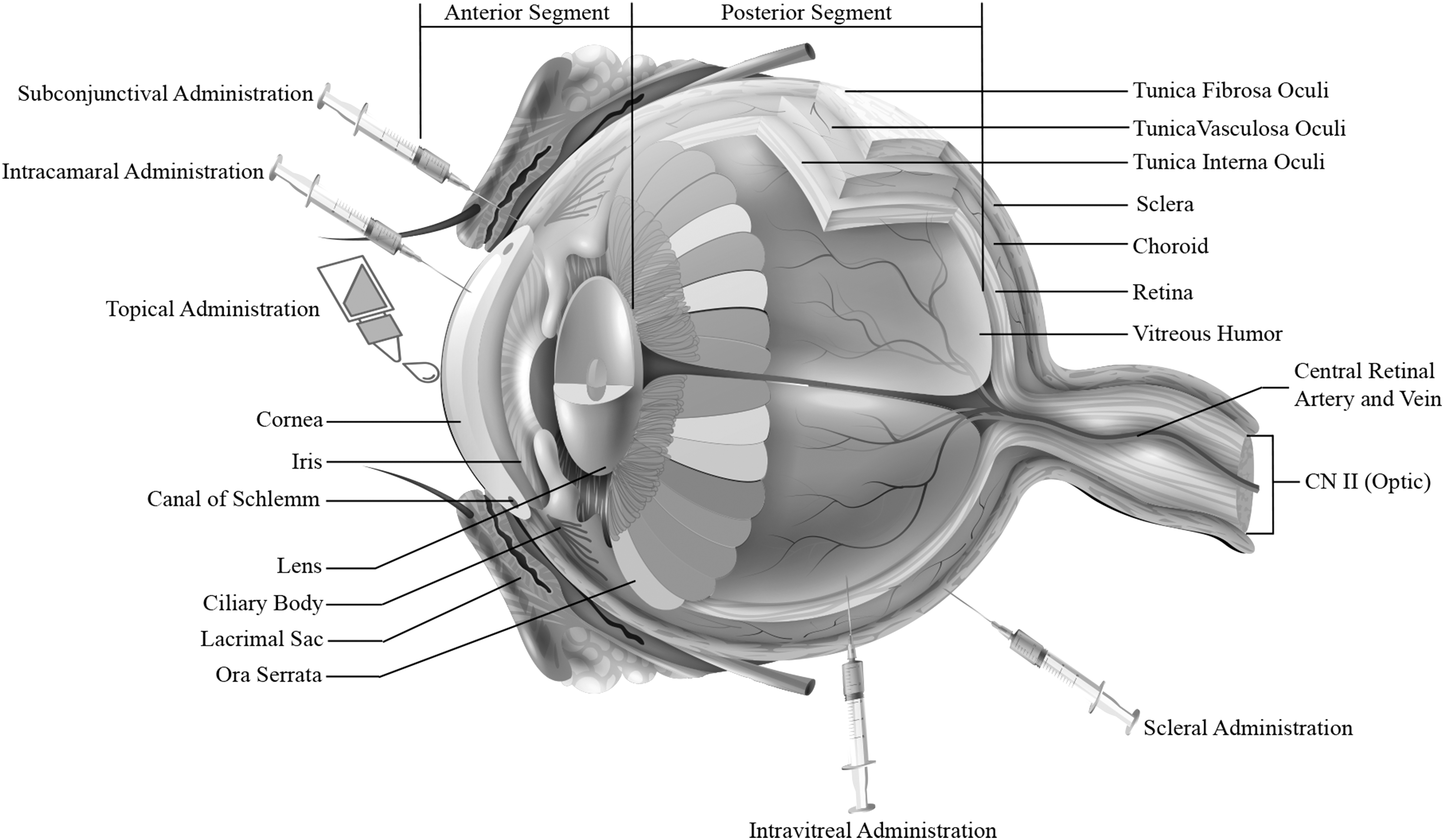
Anatomy of the eye and ocular drug administration routes.
Tunica fibrosa oculi is the fibrous layer of the eye. It does not contain blood and lymph vessels. 28 It consists of cornea, conjunctiva, and sclera. The cornea is the thick and transparent part of the eye.29,30 The cornea consists of five layers, from front to back: corneal epithelium, bowman's membrane, stroma, Descemet's membrane, and endothelial layer.31,32 The conjunctiva is a mucosal membrane where the corneal epithelium continues. It envelops the anterior part of the sclera. The sclera consists of collagen fibers and five layers: Tenon's capsule, episclera, scleral spur, limbus, and posterior sclera.30,33,34 Tunica vasculosa oculi or uvea is the middle vascularized layer. This quite thick layer contains the choroid, ciliary body, and iris. The choroid is a tissue-containing dense capillary of this layer. The ciliary body provides the secretion of the aqueous humor filling the anterior segment. At the same time, it provides ocular accommodation.28,31 Tunica interna oculi is the inner layer where the light is transformed into a neural impulse and transmitted to the brain.35,36
The eye has complex anatomy and physiology as well as effective natural barriers that limit the ocular absorption of drugs and bioavailability. These barriers are tear film, cornea, conjunctiva, sclera, blood-aqueous humor or iris-ciliary body, lens and blood-retina barriers. The blood-retinal barrier affects the penetration of drugs to the posterior segment, whereas other barriers affect the anterior segment.2–4
Different drug delivery routes have been developed to increase the penetration of drugs to the target tissue, bypassing the barriers. Drugs are mainly administered to the eye in three different routes. These are topical, periocular, and systemic administrations. Tear film and cornea are barriers for topical drugs.2–4,37 Most of the ophthalmic drugs are applied topically. The tear film is the most important barrier for these topical drugs.5,6 It is responsible for the removal of drugs by the nasolacrimal drainage and participation in the systemic circulation.3,38–40 The cornea acts as a barrier for hydrophilic drugs due to tight junctions of the corneal epithelium. On the other hand, the corneal stroma is hydrophilic due to collagen fibers and acts as a barrier for lipophilic drugs.2,4
In systemic administration, drugs are administered in high doses and the risk of systemic side effects increases.2,37,41 As an alternative to them, periocular administration is used in four different routes: intracameral, subconjunctival, scleral, and intravitreal. However, these applications and methods have disadvantages, such as limited delivery; being used in the administration of drugs that do not pass through the cornea or target posterior segment; and problems for patient compliance, which are mainly based on the injection procedure and cause problems for patient compliance.3,4,6,37 Conjunctiva, blood-aqueous humor, and blood-retina barriers also cause the elimination of drugs because of systemic circulation.2,4,42,43 The lens acts as a barrier for drugs through active transport diffusion mechanisms.2,42
Micelles
Micelles are nanocarriers consisting essentially of a hydrophobic core and a hydrophilic shell, consisting of amphiphilic copolymers or surfactants. The hydrophobic core is an alternative for targeting hydrophobic drugs, in particular.7,8 The major feature that distinguishes micelles from other drug delivery systems is that they apply not only to lipophilic drugs but also to hydrophilic drugs. In some special cases, such as targeting hydrophilic drugs to hydrophobic environments, amphiphilic copolymers or surfactants may be misaligned to form “reverse micelles.” 23 The other important difference of the micelles is that they can remain stable in the circulation system for a long time. 43 Thus, they are eliminated more slowly than other drug delivery systems. Also, they passively accumulate in the inflammation area and tumors that exhibit enhanced permeability and retention effect due to their nano sizes.44–46 Because of this feature, micelles provide advantages, especially in anti-inflammatory treatment.46,47
Stability problems of micelles can be solved more easily than other drug delivery systems. It is important that the lyophilization process can be applied or the pH and molarity range where the micelle can remain stable can be easily adjusted by adding suitable buffers. Polyethylene glycol ( ) chain can be added to the polymers/surfactants to form the hydrophilic shell of the micelles. It is known that PEGylated micelles can remain stable for a longer period and can easily accumulate, especially in the tumor or inflamed area.48,49 However, the most important point to be considered is the risk of being activated by the accelerated blood clearance system (ABC) by repeated application of PEGylated nanocarriers. Blood clearance of PEGylated nanocarriers that are administrated after ABC is activated increases, because these PEGylated nanocarriers accumulate in the spleen and liver.50,51
The other advantages than as drug delivery systems are that they are easily prepared, the desired particle size can be adjusted, and they can be easily targeted to tissue by adding a mucoadhesive, thermosensitive, or pH-sensitive agent into the formulation.46,47, 52,53
Micelles are used as a drug delivery system for drug targeting to different tissues. Different types of cancer derivatives,54–57 nasal,58,59 transdermal,60,61 and ocular21–24 targeting are examples of micellar systems studied so far.
Formation and preparation methods of micelles
Critical micelle concentration (CMC) is the most important parameter in the formation of micellar structures. Amphiphilic copolymers or surfactants, which are their components, are present in aqueous media separately at their low concentrations. However, the concentrations of a surfactant or amphiphilic copolymer molecules increase the interface and the system reaches saturation level. If the molecules are still added to the system after saturation, the aggregates that form micelles come into being by themselves. This concentration at which micelles are formed is called the CMC. The low CMC value of micelles as a drug delivery system provides a great advantage. Micelles with low CMC values cannot easily decompose to monomers in the systemic circulation and can maintain their stability for a long time. 9 The structure and formation mechanisms of micelles are shown in Fig. 2.

Structure and formation mechanisms of micelles.
Different techniques are used for preparation of micelles, including the direct dissolution method, thin-film hydration method, lyophilization procedure, dialysis process, and oil-in-water emulsion (O/W) method. 24 The direct dissolution method is the simplest micelle preparation method known and is based on the principle that the drug and polymer/surfactant form micelles by themselves. To form micelles, the polymer/surfactant is present in water in an amount of CMC or greater. The main disadvantage of this method is that the amount of drug loaded is low. In the thin-film hydration, which is a very simple and fast preparation method, a solution of the drug and polymer/surfactant is formed in a volatile organic solvent. The organic solvent is then evaporated to give a thin film around the flask. This film is mixed by hydrating with an aqueous phase. In the lyophilization method where large-scale preparation can be done, the drug and the polymer/surfactant must be resistant to lyophilization. In the organic solvent that is suitable for lyophilization, the drug and polymer/surfactant are dissolved; then, this solution is lyophilized by mixing with water. In the dialysis method, the prepared solution of the drug and polymer/surfactant in the water-miscible organic solvent is filled into dialysis bags. The organic solvent in the dialysis bag placed in water is replaced by water over time. The biggest disadvantages of this method are that it is difficult to remove all micelles from the dialysis bag after preparation and is suitable only for laboratory-scale preparation. In the O/W emulsion method, after the solution of the drug is prepared in the organic solvent that is immiscible with water, this solution is mixed with water until a homogeneous emulsion is obtained. The organic solvent is then evaporated. The polymer/surfactant is soluble in the aqueous phase or the organic solvent.
Characteristic features of micelles
Increasing the penetration to the target tissue and high efficacy of the micelle depends on some of its characteristic features. These features are their particle size and its polydispersity index (PDI), surface charge, encapsulation efficiency, drug release behavior, and physical stability. Depending on the target tissue, different features of micelles also become important. For instance, pH and osmolarity of micellar carriers are important characteristic features for the ocular micellar system,9,62,63 which is one of the main topic of this review.
The transport of nano-sized delivery systems to the target tissues by passing both dynamic and static barriers of the eye depends on their size. Nanocarriers with a particle size of less than 400 nm are known to have higher penetration into polarized epithelial cells. 64 For this reason, the particle size and PDI values of micelles are considered very important characteristic features. Studies have shown that micelles less than 100 nm can cross corneal barriers. 21 On the other hand, it is known that the scleral pore has a size between 20 and 80 nm. 60 Therefore, those should be considered to develop a micellar ocular drug delivery system.
The physicochemical properties of the micelles that can remain stable with electrostatic and/or steric stabilization depend on the surface charge. On the other hand, surface charge affects the biological properties of micelles. Electrostatic interactions (e.g., hydrogen bonds or ionic) with the target tissue, cell, or membrane are directly dependent on surface charge of the micellar carrier. 21 For instance, it has been shown that negatively charged micelles can bond better with negatively charged mucin than neutral or positively charged micelles. 65
Molecular properties of drug and drug-core interactions, polymer/surfactant used, and preparation method are factors affecting drug release from micelles. 9 The target segment of ocular micelles changes the properties of the desired drug release. For example, micelles targeted to the anterior segment are expected to release faster than micelles targeted to the posterior segment because of the nasolacrimal drainage. 21
One of the biggest factors that determines the success of the produced micelles as a drug delivery system is the stability of the micelles. Micelles that come into contact with many cells and proteins after administration and are exposed to physiological pH and tonicity undergo some changes. 39 The most important thing in these inevitable changes is that the micelles do not release the drug in their core quickly. Micelle stability is examined by thermodynamic and kinetic stability tests. Thermodynamic stability is directly related to the CMC value. Micelles are thermodynamically stable if the amount of polymer/surfactant in the aqueous medium is above the CMC value. However, below the CMC value, it tends to decompose to unimers. Kinetic stability defines the behavior of micelles in the aqueous solution over time.9,48
There are different methods applied to increase the stability of the micelles. Lyophilization is the most preferred method. Micelles with stability problems in the dispersion medium can be lyophilized and have a longer shelf life. Another method to increase the stability of the micelles is to minimize the effect of physiological pH, tonicity, cells, and proteins that cause changes to the structure. For this purpose, the dispersion medium can be adjusted to the pH and molarity where the micelle can remain stable.48,49 The most important issue to be considered here is that the adjustments to be made in pH and molarity to ensure the stability of the micelle do not go far beyond the physiological limits of the target tissue. Another method to increase stability is the addition of PEG chain to polymers/surfactants. This PEGylation process increases the stability of the micelles. However, it should be considered that the ABC system is active in long-term use.50,51
A drug that can be applied to the eye must have a similar pH (6.5–7.6) and osmolarity (300 mOsm/kg) to the eye. 62 Although there are exceptions to this, 66 pH and osmolarity buffers are added to commercial products for this purpose. The pH and osmolarity values of the micelles should be examined in terms of ocular applicability.
Structures of micelles
Micelles are classified into three types based on their structures as surfactant micelles, polymeric micelles, and poly-ion complex micelles. 24
Surfactant micelles
Surface-active agents, commonly known as surfactants, are amphiphilic molecules consisting of a hydrophilic (polar) head and lipophilic (nonpolar) tail, and they are known to reduce the interfacial surface energy. 67 The main use of surfactants in pharmaceutical science is to increase the solubility or permeability of the components. The usage of surfactants, which are also widely used in ocular drugs, varies depending on their physicochemical structure. Usage purposes of the ocular drugs are that they can be listed as solubilizers, emulsifiers, wetting agents, stabilizers, antimicrobial agents, charge inducers, gelling agents, and corneal permeation enhancers. 68 Surfactants are generally classified according to their surface loads and are divided into four groups: non-ionic, anionic, cationic, and zwitterionic.
One of the controlled drug release system micelles produced with surfactants that tend to form aggregates on or above the CMC value equilibrates with molecular bonds such as hydrogen bonding, electrostatic interactions, and vander waals forces. On the other hand, the ionic forces in the environment, temperature, pH, and total amount of surfactant in the environment affect the dimensions and stability of the micelles produced. 69 Surfactants used in ocular micelle preparation not only provide controlled drug release but also enhance the aqueous solubility of drugs and their penetration into the ocular tissues. As penetration is enhanced, they increase the corneal permeability of the drugs and allow the drugs to reach deeper tissues by overcoming the corneal barriers. Thus, the ocular bioavailability of the drug is increased. 68
There are some theories for surfactants to increase ocular penetration. One of them is that they increase the permeability of the cell membrane by reducing the expression of P-glycoprotein, which is responsible to reduce the absorption and bioavailability of drugs in cornea and RPE.70,71 Another theory is that they increase the transcellular permeation by changing the physical properties of the lipid bilayer membrane of the corneal epithelium. However, the integrity of the lipid bilayer is very important. If penetration is enhanced in high doses, the physical changes caused can result in the removal of phospholipids from the cell membrane, dissolution of the cell membrane, and serious toxic effects. 72 Other theories are that surfactants increase the direct intake of drugs into the cell by endocytosis73,74 and decrease the nasolacrimal drainage by affecting the droplet size in liquid dosage form. 72
Brij 35/78/98, cremophor (EL, RH40), polyoxyl-40- stearate, tyloxapol, octoxynol-40, Solutol HS 15, polaxemer 407 (Pluronic F127), and
Micellar Ophthalmic Drug Products that Contain Surfactants
CsA, cyclosporine A.
Polymeric micelles
Polymeric micelles consist of block copolymers. The polymer is any large molecule substance that contains certain repeating units. They are substances in which one or more atoms or groups of atoms form long chains by nonpolar bonding. It is structurally divided into two groups, synthetics and biopolymers. 81 Hydrophobic or ionic bonds between polymer monomers are effective. 72 Particle sizes are generally between 10 and 100 nm. 21 Since the CMC values of polymeric micelles are lower than the micelles produced with a surfactant, their stability is higher.82,83 Besides, prolonged contact time with tissues due to long stability, low toxicity, and good biological distribution are among the other advantages of polymeric micelles.84,85
Polymeric micelles provide ocular bioavailability by increasing paracellular or transcellular drug delivery. Permeation enhancement mechanism is one of the theories considered for surfactant micelles. It is believed that by creating physical changes in the structure of the lipid bilayer, they facilitate the separation of phospholipids from the cell membrane, thereby facilitating the passage of the drug through the formed spaces.70,86
Polymers such as polyethylene glycol, poly(glycolide), poly(ɛ-caprolactone), poly(lactide-co-glycolide), polypropylene oxide, and poly-L-lysine are predominantly used for preparation of micelles in the ocular drugs. 23
Poly-ionic micelles
Poly-ionic micelles consist of electrostatic interactions between poly-ion copolymers and opposite-charged ionic drugs. 10 They are generally preferred as alternative nanosystems for antisense oligonucleotide and gene delivery. 23 PEG, which is a neutral block, is frequently used as a stabilizing agent in the production of poly-ionic micelle. 87 These micelles become more stable and provide an alternative for ocular drug targeting. 23
Ocular diseases and disorders
Ocular diseases and disorders are phenomena that affect vision and eye health. 1 They can be diagnosed at any age. 88 Although the disease or disorder can develop directly depending on the eye, it can also be a secondary one. If it is secondary, there may be primary diseases such as immunodeficiency, neurological problems, or idiopathic problems. The eye is a unique organ due to its anatomy and physiology. It is also common for an existing disease to easily affect other tissues of the eye and impairment of eye integrity. For this reason, finding the main reason for the disease and making an accurate diagnosis is very important for treatment.
Diseases of the eye are divided into two groups: ocular and extraocular. Ocular diseases are seen in the eye tissues, whereas extraocular diseases are related to the orbita and the muscle tissue. 83 These ocular diseases can be treated by medicine, surgery, or devices. Many parameters that affect the success of the treatment in medical treatment are mentioned in section “2.2. ocular barriers and ocular drug administration routes.” The fact is that the drug administration route may be changed because of the target tissue that is in the anterior or posterior segment. On the other hand, the type carrier or delivery system used in the ocular drug delivery systems, which is one of the innovations in medical treatment, varies according to the segment where the target tissue is located. Table 2 shows anterior and posterior ocular diseases.25,83
Glaucoma affects both the anterior and posterior segments of the eye.
Micellar drug delivery systems for treatment of ocular diseases
Conventional dosage forms administered to the eye generally target the anterior segment of eye and have little effect on a possible disease in the posterior segment.3–6 On the other hand, low bioavailability due to ocular barriers and therefore high dosing frequency and low patient compliance are disadvantages of this dosage form. The fact that gels and ointments cause blurred vision is another factor that decreases patient compliance. To prevent all these negativities, ocular drug delivery systems targeting specific tissues are being developed.
Micro- and nanoparticles, 7 microemulsions, 7 nanosuspensions, 7 solid lipid nanoparticles, 11 liposomes, 12 cubosomes, 7 dendrimers, 7 niosomes, 7 discosomes, 7 hydro gelling system, 10 ocular lens/inserts/implants,8,13,14 and micelles 9 have been developed as ocular drug delivery systems. Micelles are one of the drug delivery systems that have been preferred for ocular drug targeting for many years and are examples that have been approved by clinical studies.75–80 Its main advantages are that they increase the solubility of poorly soluble drugs in water and sustain drug release because of the hydrophobic core. 9 Another important advantage is that they increase the ocular bioavailability of the drugs, because they have high permeations to the ocular tissues, thus reducing the daily drug frequency and toxicity due to the drug dose. 7 Other advantages include ease of production and reproducibility, the ability to produce the desired quantity at one time, no discomfort when used, a clear aqueous solution of the final form that increases patient compliance, and adjustable particle sizes. 9
If we compare these advantages with other ocular drug delivery systems, we can see that they provide superiority to microparticles in terms of particle size. Since microparticles cannot pass through the ocular barriers, it is known that ocular permeations are lower than nanocarriers.21,43,60 Although nanoparticles are similar in size to micelles, low drug loading capacity creates an important disadvantage against micelles. 43 On the other hand, having toxic effects for dendrimers makes micelles superior. 89 Sustained drug release of ocular inserts, implants, or contact lenses has been demonstrated by many studies, and they also have approved products. However, these products have difficulties in application and cause inflammation.8,90
The main limitation of micellar carriers is that amphiphilic polymers/surfactants used in their production have been shown to probably cause ocular irritation. 72 Another disadvantage of micelles is their scale-up problems in production. 52 The applicability of the lyophilization method, which is suitable for large-scale productions, has been demonstrated in a few studies.91–93
Micelles for the anterior segment diseases
Although conventional ocular dosage forms are used for the anterior segment, low bioavailability has led to anterior tissues targeting when developing drug delivery systems. Micellar drug delivery system studies have been performed for the treatment of diseases such as keratitis, conjunctivitis, chemical burns, dry eye syndrome, anterior uveitis, and corneal neovascularization. 94
The studies with micelles have been continuing both intensively and successfully in recent years. In vivo studies of cyclosporine A (CsA) loaded octoxynol-40 micelles developed by Mandal et al. showed statistical improvement in conjunctiva after 5 days of single- and multi-dose administration. Subsequently, phase III studies of micelles, which have been clinically studied, have been finalized and the product has been approved by the FDA for treatment of dry eye disease signs and symptoms. 72 Dexamethasone (DEX) is an active ingredient that is used in ocular inflammations and is an eye drops solution. Alami-Milani et al. have developed DEX loaded Poly(ɛ-caprolactone)-b-poly(ethylene glycol)-b-poly(ɛ-caprolactone) (PCL-PEG-PCL) micelles. Corneal permeation of sustained-release micelles was found to be 2-fold higher than commercial eye drops. 95
The studies regarding micellar ocular delivery systems targeting drugs to the anterior segment of the eye published in the past 6 years are given in Table 3.
Micellar Ocular Drug Delivery System Studies Targeting to the Anterior Segment
5DUP3, 5-(dodec-1-ynyl)-uracil phosphoramidite 3; DEX, dexamethasone; DSPE-PEG, 1,2-distearoyl-sn-glycero-3-phosphoethanolamine-N-[amino(polyethylene glycol)]; mPEG-b-PLGA, Methoxy poly(ethylene glycol)-b-poly(lactide-co-glycolide); mPEG-PCL, methoxy poly(ethylene glycol)-block-poly(ɛ-caprolactone); mPEG-PLA, methoxy poly(ethylene glycol)-b-poly(
Micelles for the posterior segment diseases
It is difficult to reach the posterior segment bypassing the ocular tissues through a conventional dosage form. Therefore, different drug delivery routes have been found, such as scleral, intravitreal, and systemic. However, side effects such as ocular irritation and systemic toxicity may be seen in these applications. Considering all these conditions, drug targeting to the posterior segment becomes very important.
The micellar drug delivery system has been studied so far for neovascularization, macular degeneration, diabetic macular edema, posterior inflammations, and posterior uveitis. 94 Vaishya et al. developed DEX loaded MPEG-PCL micelles for use in the treatment of posterior uveitis. The permeation of conjunctival cells and sclera of the micelles with sustained drug release was 2 and 2.5 times higher than the DEX suspension, respectively. 128
The studies regarding micellar ocular delivery systems targeting drugs to the posterior segment published in the past 6 years are given in Table 4.
Micellar Ocular Drug Delivery System Studies Targeting the Posterior Segment
Cholesterol-PEG, cholesterol- poly(ethylene glycol); CSO-SA, chitosan oligosaccharide-stearic acid; CSO-VV-SA, chitosan oligosaccharide-valylvaline-stearic acid; INU-EDA-RA, inulin-ethylendiamine-retinoic acid; MPEG-b-PAE, methyl ether poly(ethylene glycol)-b-poly(b-amino ester); mPEG-PCL, methoxy poly(ethylene glycol)-block-poly(ɛ-caprolactone).
Micelles for the glaucoma
Glaucoma, a disease characterized by an increase in intraocular pressure (IOP), ultimate damage to the optic nerve, fiber layer, and ganglion cells, is the second leading cause of irreversible blindness after cataract.135,136 It is a disease affecting both the anterior and posterior segments of the eye.
Many active ingredients with different mechanisms of action are used in the medical treatment of glaucoma. Latanoprost and timolol are examples of drugs with different mechanisms that are frequently used in treatment. Latanoprost, the prostaglandin F2α analog, is one of the active substances used in glaucoma treatment by increasing the flow of aqueous humor 137 ; timolol, the beta-blocker, inhibits aqueous humor production 138 ; and both reduce IOP. No product on the market combines these two drugs with different mechanisms of action. Xu et al. produced contact lenses nano-decorated with latanoprost and timolol loaded mPEG-PLA micelles in their study. It was observed that the micelle loaded contact lenses reached the highest release value in the 120–144 h interval by sustained release. In pharmacokinetic studies, latanoprost was detected for 96 h and timolol was detected for 120 h in tear fluids; bioavailability improved 7.3 times and 2.2 times, respectively. In vivo pharmacodynamic studies showed that a continuous decrease in IOP was detected for 168 h in rabbits with high IOP. The relative pharmacological availability of CLs-M was 9.8 times as high as the eye drops. 139
All micellar ocular drug delivery system studies for the treatment of glaucoma in the past 6 years are given in Table 5.
Micellar Ocular Drug Delivery System Studies for the Treatment of Glaucoma
IOP, intraocular pressure; mPEG-PCL, methoxy poly(ethylene glycol)-block-poly(ɛ-caprolactone); mPEG-PLA, methoxy poly(ethylene glycol)-b-poly(
Micelles for the myopia
Myopia is a refractive disorder in which the eye does not refract light properly. Light does not focus correctly so images are not clear. Li et al. developed pirenzepine loaded mPEG-PDLLA micelles. The aqueous solution of micelles that has a 280 nm particle size increased corneal permeability by increasing the partition coefficient. In vivo absorption results showed that bioavailability was increased. 143
Conclusion
Micelles as ocular drug delivery carriers have significant promise for the treatment of both anterior and posterior segment diseases as well as glaucoma treatment due to their numerous superiorities such as the increasing of aqueous solubility of drugs, providing sustained drug release, and improving ocular permeation of the drugs. In this review, we have highlighted recent research studies that draw on the positive outcomes of micelles as an ocular drug delivery system.
When all the studies were examined, it was seen that the research studies are mostly focused on the treatment of ocular inflammation and dry eye in anterior segment diseases and the treatment of macular degeneration in posterior segment diseases. This fact draws attention that DEX is a frequently studied active drug molecule as a micellar drug delivery system. The data indicated that its micellar carriers showed higher permeation to both anterior and posterior eye segments compared with its commercial eye drops. These results suggest that DEX loaded micelles may successfully pass clinical trials and might become commercial products in the future. In addition, micelles drug delivery systems of CsA have also been studied in many research studies. Cequa, a cyclosporine loaded micelle product that has been the subject of many studies, is highly promising for new studies in this field.
Developments in glaucoma, a disease affecting both segments, suggest that drugs with different mechanisms of action may be loaded in the same micelles. This development, which makes combined treatment possible, promises great hope for the treatment of the disease.
On the other hand, the development of micellar drug delivery systems in the treatment of myopia, which is not a disease but an eye disorder, paves the way for medical treatment in permanent eye disorders. If medical treatment can be performed in such diseases that can be treated by using glasses or surgical procedures under normal conditions, the quality of life of the people will increase significantly.
Future perspective
The development of new drugs instead of the existing molecules is a long and challenging process. Modifying existing drug molecules is a wiser approach to solve all these problems. The drug delivery systems developed for this purpose aim at the administration of any drug to the target tissue in as low doses as possible. Thus, systemic absorption of drugs can be prevented, their toxicity is reduced, and their absorption and bioavailability are increased. In the development of drug delivery systems, a drug's solubility and solubilization, lipophilicity, permeability, gastrointestinal metabolism, degree of ionization, stability in biological fluids, protein binding, systemic pharmacokinetics, and pharmacodynamics properties play a very important role because they affect the pharmacokinetics of that molecule. 144 Although biological membranes are generally hydrophilic, most of the drugs currently in treatment are lipophilic.145,146 Therefore, the absorption and bioavailability of lipophilic drugs are very low. Drug delivery systems, among other advantages, first improve the aqueous solubility of these lipophilic drugs and increase the absorption.
Micellar drug delivery systems have always been considered promising nanocarriers in ocular targeting, from the moment they were discovered until today. Ophthalmic drug products that contain surfactants to solubilize the drug, which in principle are micellar formulations that have been approved, are available (Table 1). An eye drop, named TJ Cyporin, produced by Taejoon Pharm contains cyclosporin A, which is also in a micellar form. However, polymeric micellar carriers are not yet approved as commercial products; the promising data from recent research studies indicate possible opportunities for ocular delivery of micellar carriers. Having higher ocular permeation compared with conventional dosage forms and suitable particle sizes that are capable of crossing the ocular barriers are the biggest data supporting this opinion. Also, the declaration of some of the used polymers/surfactants as reliable excipients by the FDA,147,148 ease of production, adjustable stability, and particle size are other factors that favor micelles to be targeted in ocular targeting.
Having high ocular permeation and in vivo efficacy, not causing ocular toxicity are, undoubtedly, very important to develop drug delivery systems and give an idea about whether that system will turn into a commercial product. However, while talking about the future perspective in the development of these systems, only experimental studies are not convincing. The presence of commercial products that are approved or whose phase studies are in progress distinguishes ocular targeted micellar drug delivery systems from other systems.
Footnotes
Author Disclosure Statement
No competing financial interests exist.
Funding Information
No funding was received for this article.