
Abstract
Purpose:
Cataracts are the leading cause of blindness worldwide, resulting in over 30 million surgeries each year. These cases are expected to double within the next 10 years. About 25% of all patients develop secondary cataracts or posterior capsule opacification (PCO) postsurgery. PCO is a vision impairment disorder that develops from myofibroblasts migration and contraction that deforms the capsule surrounding the lens. Currently, Nd:YAG laser therapy is used to treat PCO; however, laser is not available worldwide and adverse side effects may arise. Thus, there is a considerable unmet need for more efficacious and convenient preventive treatments for PCO. Our work focuses on engineering an innovative, prophylactic sustained release platform for DNA-based nanocarriers to further reduce the incidence of PCO.
Methods:
Novel, optically clear, self-assembled poly(
Results:
The 29 (w/v)% polymer hydrogels with the 3DNA nanocarriers presented over 80% of light transmittance, soft mechanical properties (<350 Pa), and sustained release for 1 month.
Conclusions:
In this work, we show for the first time that the hydrophobic PLGA-PEG-PLGA hydrogels can be used as platforms for sustained delivery of nucleic acid-based nanocarriers. This work demonstrates that polymeric formulations can be used for the extended delivery of ocular therapeutics and other macromolecules to treat a variety of ocular conditions.
Introduction
Poor drug bioavailability continues to be a major concern associated with ocular therapy. The physiology and anatomy of the eye make it a protected organ, constraining the absorption of therapeutically relevant concentrations of drugs. One of the greatest challenges in ophthalmology is to provide minimally invasive, targeted, and sustained therapy to a particular area within the eye.1,2 This can be done by extending the residence time and duration of drug release, at the tissue region of interest. The majority of ophthalmic therapeutics are administered either topically (solutions and suspensions) or intravenously (systemically). Topical administration is common for disorders in the anterior portion of the eye, but these therapies are limited in their bioavailability due to tear turnover, nasolacrimal drainage, metabolic degradation, and nonproductive adsorption/absorption. In most topical applications, only 1%–7% of the administered drug reaches therapeutic site, and the rest is systemically drained.1,2 Systemic administration is generally used to treat disorders in the posterior portion of the eye 3 ; and the therapeutic effect is greatly impeded by the blood–ocular barrier and liver metabolism. 4
Tissue barriers within the eye have been effectively bypassed using novel, colloidal drug delivery systems, for example, nano/microparticles, nanosuspensions, liposomes, and nanohydrogels.2,5 The molecular architecture of these systems can be engineered for enhanced bioavailability of the targeted drug at a local site, improved permeability across biological membranes, improved drug stability, and sustained release of peptides and proteins.5,6 Noting the need for biocompatibility, FDA approved degradable polymers such as poly(
Injectable, in-situ forming, biodegradable PLGA-PEG-PLGA hydrogels are versatile and tailorable, allowing them to be used for a wide variety of applications. These gels can be used in combination with other natural and synthetic polymers to design more efficient matrices for the extended and controlled release of drugs. 9 Different drugs and targeting moieties can be incorporated into the system, allowing for the design of more specific and effective drug release platforms. For instance, these injectable systems can be used for a variety of ocular applications, including but not limited to, macular degeneration and secondary cataracts or posterior capsule opacification (PCO). The autocatalytic reactions of the polymer provide controllable drug release through bulk or surface erosion. 8 Therefore, these gels have become more popular for the controlled and extended delivery of therapeutic cells, growth factors, and other therapeutic peptide/protein injections.
Hydrophobic PLGA-PEG triblock copolymer depots are widely used as platforms for the delivery of small, hydrophobic drugs such as dexamethasone10,11 and hydrocortisone butyrate
12
to treat posterior ocular disorders. Recently, the release of negatively charged, hydrophilic drugs, proteins, peptides, and nucleic acid conjugates from these hydrophobic matrices has been analyzed. It has been determined that the release profile can be controlled and extended using polycations such as poly(ethyleneimine) (PEI) or poly(
Secondary cataracts or PCO is a prominent vision impairment disorder that affects about 25% of patients after primary cataract surgery. PCO is characterized by myofibroblast (differentiated Myo/Nog cells) migration and contraction onto the lens capsule. Myo/Nog cells are a subpopulation of cells present on the lens capsule that differentiate into myofibroblasts as a result of wounding.15,16 These cells deposit fibers and wrinkle the capsule, preventing light from reaching the retina (Fig. 1). The wrinkling of the retina causes light diffraction which gives rise to PCO. 16 To prevent PCO, Myo/Nog cells must be specifically targeted by a monoclonal antibody (mAb) called G8 and depleted. This depletion can be achieved by using cytotoxin doxorubicin intercalated within DNA-based nanocarriers (3DNA®; Genisphere, LLC, Drive Hatfield, PA) that have the G8 mAb on their surface. Myo/Nog cells undergo receptor-mediated endocytosis (via G8 mAb) to internalize the nanocarriers into acidic compartments that enhance doxorubicin release, which induces apoptosis. 16 The 3DNA nanocarriers are built from DNA to provide a biodegradable, nontoxic, and nonimmunogenic platform17–19 engineered for specific targeting and delivery of therapeutic payloads.19–24
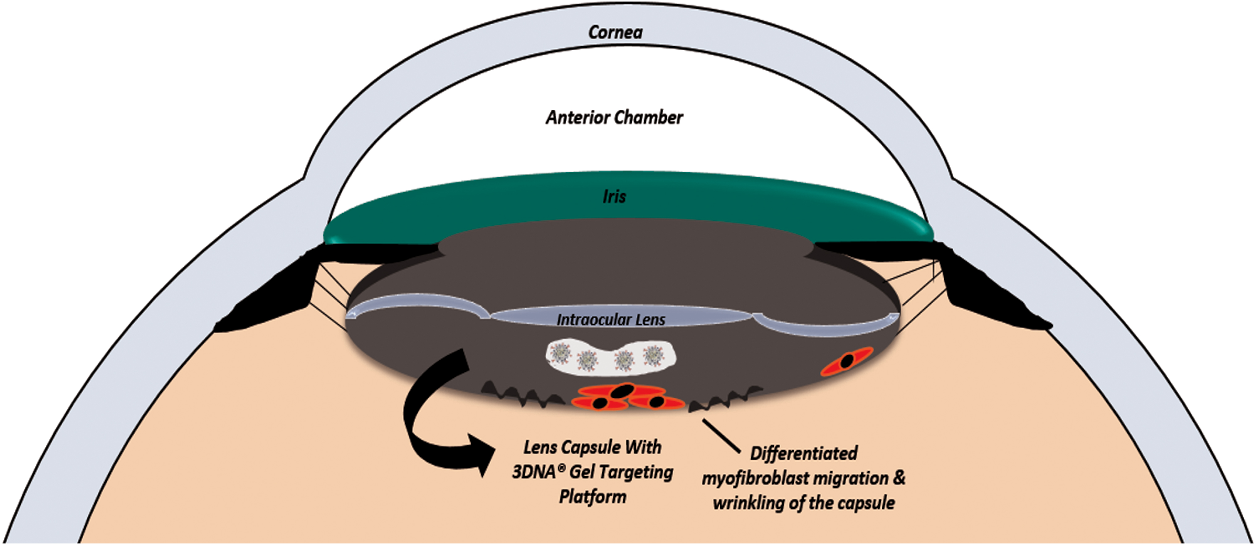
Proposed prophylactic solution for secondary cataracts. DOX-loaded 3DNA® nanocarriers embedded in injectable PLGA-PEG-PLGA hydrogel systems are being designed as a prophylactic treatment for PCO. It can be safely administered during cataract surgery. Approximately, 60 to 100 μL of the sustained delivery formulation can be injected behind the intraocular lens. PCO, posterior capsule opacification; PLGA-PEG, poly(
In preparation and continuation of Gerhart et al.'s in vitro and in vivo work,15,16 we are focusing on the development of a prophylactic, polymer-based formulation for the sustained release of 3DNA nanocarriers to reduce the incidence of PCO and protect the lens capsule from cells migrating from the ciliary processes.15,25 Approximately, between 60 and 100 μL of the drug-loaded 3DNA nanocarriers/PLGA-PEG-PLGA gel, systems can be administered during cataract surgery, after the intraocular lens has been successfully implanted and the filling gel is aspirated (Fig. 1).
In this work, we show for the first time the release of G8 mAb functionalized 3DNA nanocarriers loaded with doxorubicin (3DNA:DOX:G8) from optically clear, self-assembled PLGA-PEG-PLGA hydrogels over the period of 1 month. This study is a proof of concept that sustained release of the drug-loaded DNA-based nanocarriers may further reduce the incidence of PCO.
15
These drug delivery platforms need to meet the following criteria to be injected into the lens capsule, underneath the intraocular lens, at the time of cataract surgery: gelation temperature (GT) should occur at the physiological temperature of the human lens capsule (34°C −35°C),
26
allow over 80% of light transmittance at visible wavelengths (455–700 nm)27,28 and have a modulus of <350 Pa to prevent any damage to surrounding ocular tissue.29,30 Based on our previous polymer characterization study,
9
the triblock copolymers that best met such criteria had the following parameters: lactic acid to glycolic acid (
Methods
Materials
Triblock copolymer, PLGA-PEG-PLGA, with
3DNA nanocarrier synthesis and formulation
3DNA nanocarriers were manufactured as previously described.18,31 In brief, pairs of 7 unique single-stranded DNA oligonucleotides are hybridized and crosslinked to make 5 unique monomers which are hybridized and crosslinked in a step-wise process (Fig. 2A). The resulting 2-layer structure is double-stranded in all regions except the periphery, consists of ∼3,000 DNA bases, has a Mw of 1,000 kDa, and has a zeta potential of −30 mV (Fig. 2B). Alexa Flour 647®-labeled oligos were crosslinked to peripheral 5′ regions of the 2-layer 3DNA, and oligos for hybridization with antibody-oligo conjugates were crosslinked to peripheral 3′ regions. Doxorubicin (Sigma-Aldrich, St. Louis, MO) was intercalated into the double-stranded regions of 3DNA® by incubating at room temperature at a molecular ratio of ∼500:1 DOX:3DNA. The G8 mAb was conjugated to a single-stranded DNA oligonucleotide via amine-to-sulfhydryl attachment using a heterobifunctional crosslinker (LC-SMCC; Pierce). The antibody-oligonucleotide conjugate was hybridized to 3DNA or 3DNA:DOX by incubating at 37°C at a molecular ratio of 24:1 conjugate:3DNA, yielding ∼4 G8 mAbs per nanocarrier (Fig. 2C). To determine the particle size of the 3DNA nanocarriers, dynamic light scattering was used (Malvern Zetasizer Nano-ZS). A 60 nm latex standard was used to confirm if the instrument was working appropriately. The 3DNA formulations used in this study had a diameter of 74 ± 2.6 nm, while the 3DNA:G8 formulations had a diameter of 82 ± 1.6 nm.
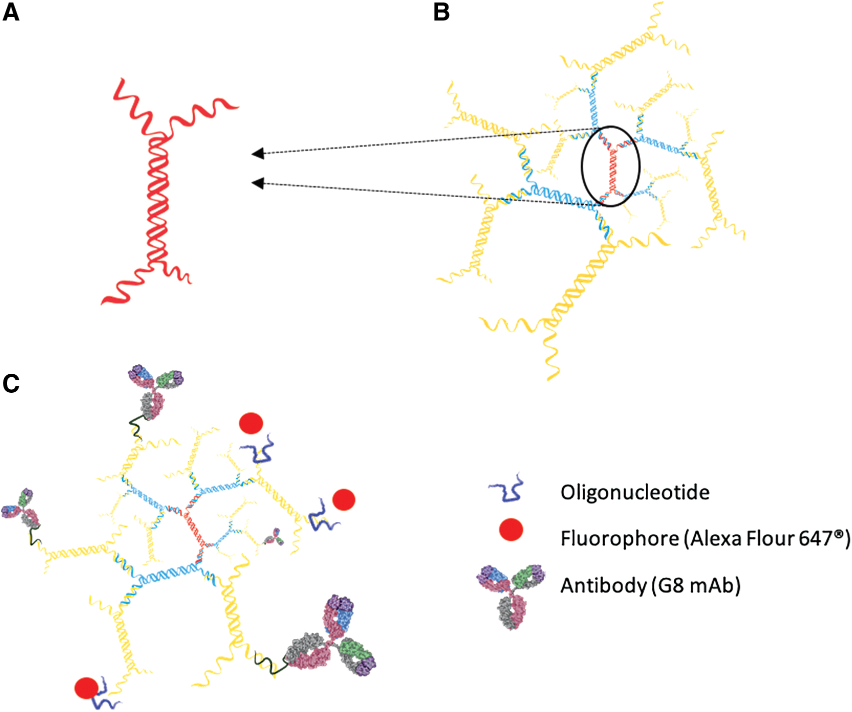
3DNA Nanocarriers with monoclonal G8 antibody and fluorophore.
Formulating self-assembled, injectable hydrogels
Solutions consisting of 29 (w/v)% polymer in PBS were mixed on a rotator for 24 h until the polymer completely dissolved. They were stored at 5°C for an additional 24 h minimum before testing. To ensure maximum DNA loading, 100 μL aliquots were freeze dried and reconstituted in PBS with either 3DNA, 3DNA:DOX, 3DNA:G8, or 3DNA:DOX:G8. Hydrogels in PBS only and with 3DNA:G8 were used for cryo-SEM (Phenom™; Pure Microscope). A 20 μL aliquot of gels at 35°C were quenched using liquid nitrogen and fixed on a flat metal disk. SEM images were taken at the conditions of 5 kV.
Sol-gel phase transition measurement
The sol-gel transition, or gelation temperature (GT), is the point at which the solution transforms into a physical hydrogel. It is one of the most important parameters in self-assembled systems that are used as potential injectable drug delivery vehicles.32,33 The GT can be tailored to a desired application by varying the following triblock copolymer properties:
The sol (flow) or gel (no-flow) phase transition temperatures of the triblock copolymers at 29 (w/v)% in PBS were determined by the vial-inverting method, 38 with a temperature increment of 2°C per step. A total of 0.5 mL of polymer and 3DNA solution was added into a 2 mL vial and immersed in a water bath. The temperature was adjusted between 25°C and 50°C and the vials were allowed to equilibrate for 20 min at each temperature. If no flow was observed within 30 s of inverting the vial, the triblock copolymer was considered a gel. The polymer hydrogel formulations that were studied are shown in Table 1.
Polymer Solutions and 3DNA Formulations
29 (w/v)% polymer solution concentration.
PBS, phosphate-buffered saline.
Light transmittance
Transmittance of 29 (w/v)% PLGA-PEG-PLGA hydrogels with the 3DNA reagents was measured on a 96-well plate at 35°C (ocular temperature) using a UV-spectrophotometer (SpectraMax M3; Molecular Devices) (sample size: 100 μL). The wavelength range was between 400 and 700 nm.
Dynamic rheological measurements
Rheology was used to confirm the ability of the different triblock copolymers to self-assemble into a hydrogel. The viscoelastic properties of the hydrogels were investigated using a Discovery Hybrid Rheometer (DHR) (TA Instruments, New Castle, DE). A strain-controlled temperature ramp was performed with a sample quantity of 100 μL, between 2 flat plates of diameter 20 mm with the gap between the plates set to 0.3 mm. The heating rate was 1°C/min and the strain was 3%, at a frequency of 1.0 Hz. Oscillatory dynamic rheology as a function of temperature was used to analyze the mechanical modulus of the gel. The shear storage modulus (G′) represents the elastic behavior, or the energy stored in the hydrogel, during deformation. G′ as a function of temperature should be near or close to zero at temperatures below the critical GT. At this point, the hydrogel solutions behave as Newtonian fluids with low viscosity, suggesting good injectability. With increasing temperature, G′ increases because a bridged micelle network starts to form until it reaches its maximum strength point/maximum storage modulus.39,40
Release of 3DNA nanocarriers
PLGA-PEG-PLGA solutions [29 (w/v)% in PBS] were used for release studies. A 100 μL aliquot of each 3DNA reagent was added to freeze-dried hydrogels at the aforementioned concentration (Table 1). All formulations were prepared at room temperature. The release was conducted in microfluidic devices mimicking the anatomical shape of the anterior portion of the eye (anterior and posterior chambers) and the physiological aqueous humor flow rate (2.4 ± 0.6 μL/min 41 ) (n = 3 per formulation). Although the lens capsule does not come in direct contact with fluid, fluid flow must be considered. 41 The lens capsule is the membrane that encloses the lens. This membrane is permeable to water, glucose, and small molecules that diffuse freely. As shown in Fig. 3A, the aqueous humor is secreted by the ciliary body, flows between the lens and the iris, passes through the pupil into the anterior chamber, and exists through the trabecular meshwork, located at the base of the cornea.41,42 Figures 3A and B show the microfluidic device, which was designed to replicate the anatomical shape and physiological fluid flow rate of the eye. Each device has 2 inlet and outlet flowlines. Each inlet line has a rate of 1.2 μL/min, making the total flow rate 2.4 μL/min. The anatomical shape, the physiological volumes, and the flow rate of the lens capsule are well represented on our microfluidic devices. The formulation was pipetted onto the posterior portion of device at room temperature. These were brought to 35°C where the formulations turned into nanogels and were maintained as nanogels for the entirety of the experiment. It is hypothesized that the point of gelation, the 3DNA reagents sit outside the micelles interacting with PEG chains and water molecules, similar to the case of HA arrangement at the time of gelation. 43
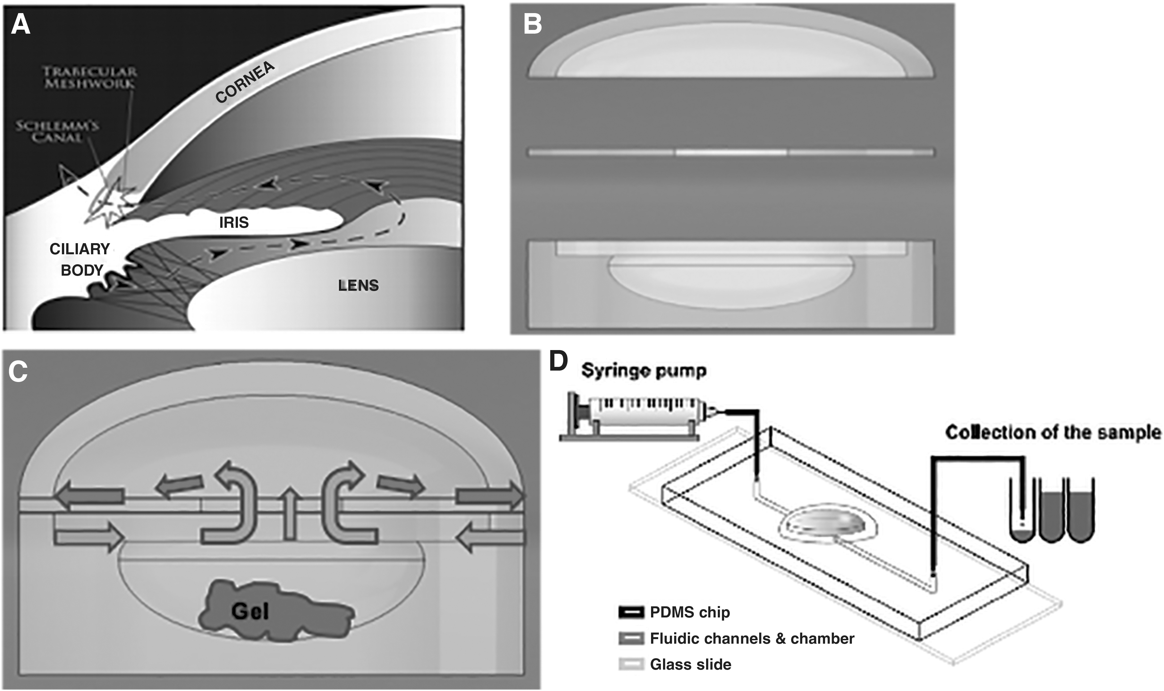
Microfluidic device mimicking the lens capsule shape and fluid flow rate.
A flow pump (Model 220; kd Scientific, Inc.) set at 1.2 μL/min was used to ensure continuous release media throughout the experiment (Fig. 3D). The release samples were collected at different time points. A 150 μL aliquot of each release sample was added to a black, 96-well plate (Corning®) and stored at 4°C overnight. 3DNA concentration in release media was tested via UV-Spectrophotometry (SpectraMax M3; Molecular Devices) of Alexa Fluor 647, with excitation and emission wavelength of 642 and 670 nm, respectively. Statistical analysis of particle size and time (due to degradation) versus fractional release percentage was performed using a 2-way ANOVA (R Studio); P values <0.05 was considered statistically significant. This article emphasizes the statistical significance between particle size and fractional release percentage.
Results
Sol-gel phase transition
The GT of the different formulations are presented on Table 2. All systems with 3DNA nanocarriers showed a lower GT compared to systems with no DNA. These self-assembled hydrogels go through 2 stages: transparent gel stage and opaque gel stage. Table 2 also shows the temperature at which the gels became opaque.
Phase Transition Temperature of the Different Hydrogels Formulations
Light transmittance
Figure 4 illustrates the light transmittance properties of the 29 (w/v)% PLGA-PEG-PLGA copolymers with different 3DNA nanocarrier formulations at 35°C. Only systems with 3DNA:DOX:G8 and control gels in PBS allowed for >80% light transmittance across the visible wavelength (455–700 nm). Wavelengths lower than 455 nm, including UV, were filtered out because of the scattering by the gel structure, or absorption from the solvent or gelator—in this case PLGA. 44 The ability of these hydrogels to filter out UV wavelengths is essential for preserving healthy eye conditions. Moreover, hydrogels with 3DNA, 3DNA:DOX:G8, and gels in PBS only allowed for over 80% light transmittance between 540 and 700 nm wavelengths. The rest of the hydrogel formulations presented <80% light transmittance.
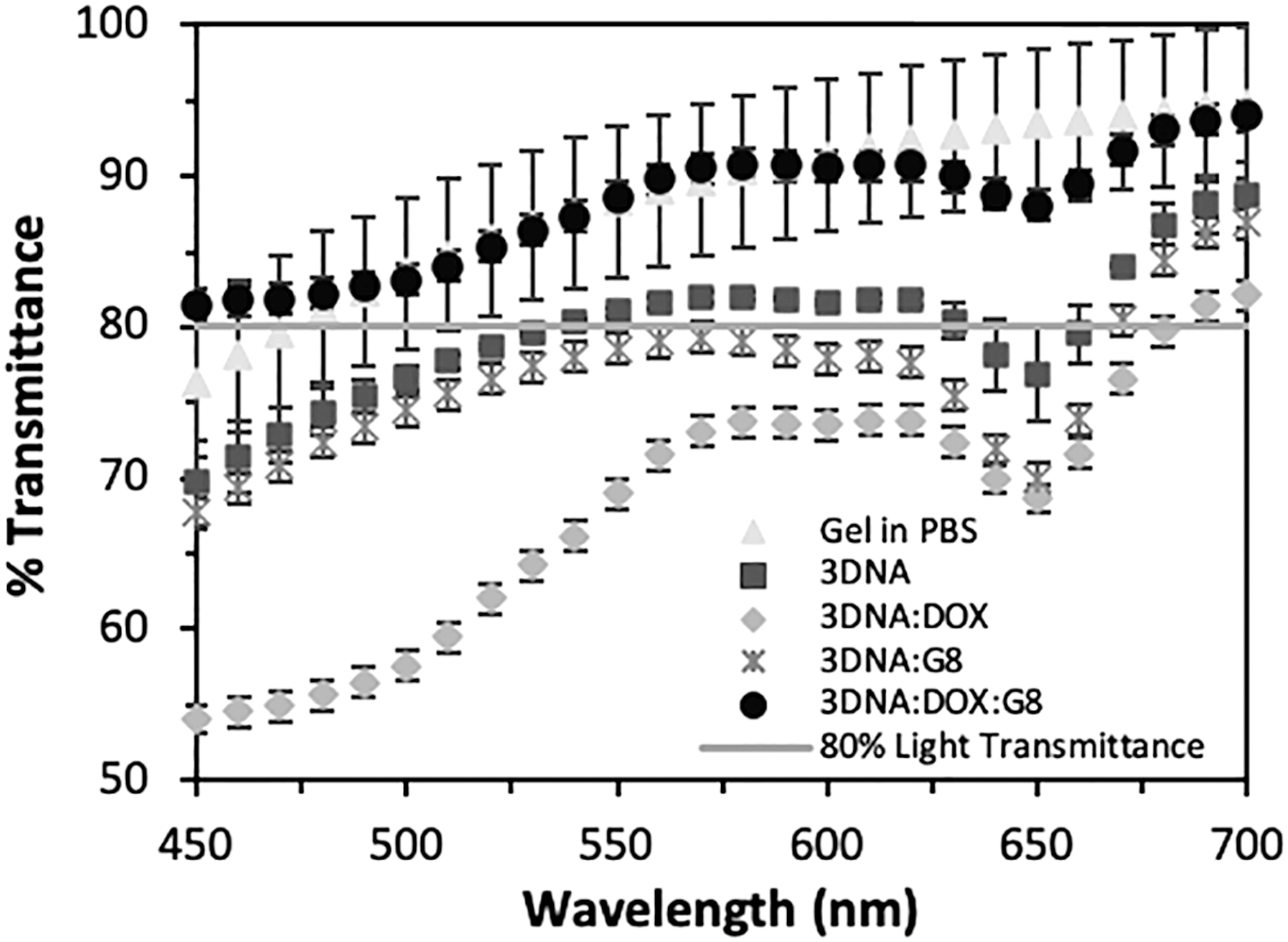
Light transmittance percentage (%) of hydrogels with 3DNA reagents. Polymer hydrogels in PBS only and with 3DNA:DOX:G8 presented over 80% over the wavelength spectrum of interest (455–700 nm). The third formulation that presented light transmittance above 80% between 540 and 700 nm is gels with 3DNA. Gels with 3DNA:G8 and 3DNA:DOX presented <80% light transmittance. These differences can be attributed to the microscopic structure formed by each of the different 3DNA reagents at the time of gelation. DOX, doxorubicin; PBS, phosphate-buffered saline.
Hydrogel mechanical properties
Figure 5 and Table 3 show the storage modulus of the polymer hydrogels with the presence of the 3DNA reagents. The addition of the 3DNA complexes make the hydrogels softer overall, compared to gels in PBS only. Hydrogels with 3DNA:Dox show a higher modulus than hydrogels with 3DNA only, while hydrogels with 3DNA and 3DNA:G8 show a higher modulus than hydrogels with the complex of interest, 3DNA:Dox:G8. The G8 antibody makes the overall 3DNA complex more hydrophilic, entrapping more water at the gelation point (Fig. 8B). Having hydrogels with modulus <350 Pa is deformable material and will be suitable for intraocular implantation.29,30
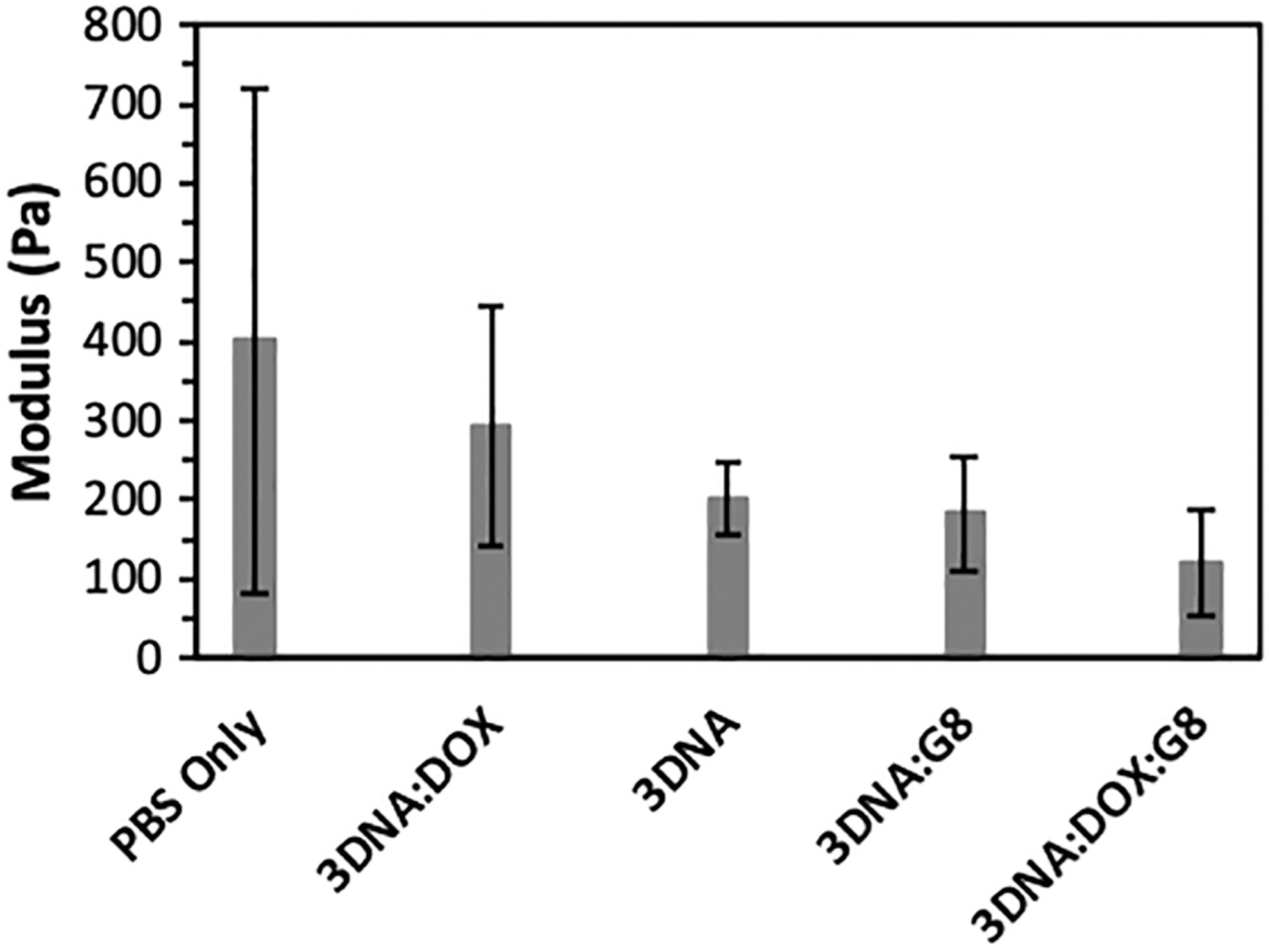
Modulus values of polymer formulations. Gels in PBS only show the highest modulus, 401 Pa. Adding the 3DNA complexes lower the modulus linearly, with 3DNA®:DOX:G8 presenting the lowest modulus 120 Pa. The presence of the G8 mAb further lowers the modulus due to its hydrophilicity. mAb, monoclonal antibody.
Normalized Peak Modulus Values (%) in Decreasing Order of Gel Stiffness
Modulus values have been normalized with respect to gels in PBS only, which are considered 100% stiff gels for the purpose of data analysis.
Table 3 shows the moduli values as well as the normalized values of the polymer formulations. The data were normalized with respect to gels in PBS only, as 100% stiff gels. The stiffness decreases linearly. For instance, 3DNA®:DOX showed 73% less stiffness compared to gels in PBS only. Gels with 3DNA are 50% less stiff, and gels with the G8 mAb are 45% to 30% more deformable (lower stiffness). Having the G8 mAb present in the formulation increases the softness (larger pores, more water accessibility) of the overall polymer hydrogel structure by >50%, as observed on Fig. 8B.
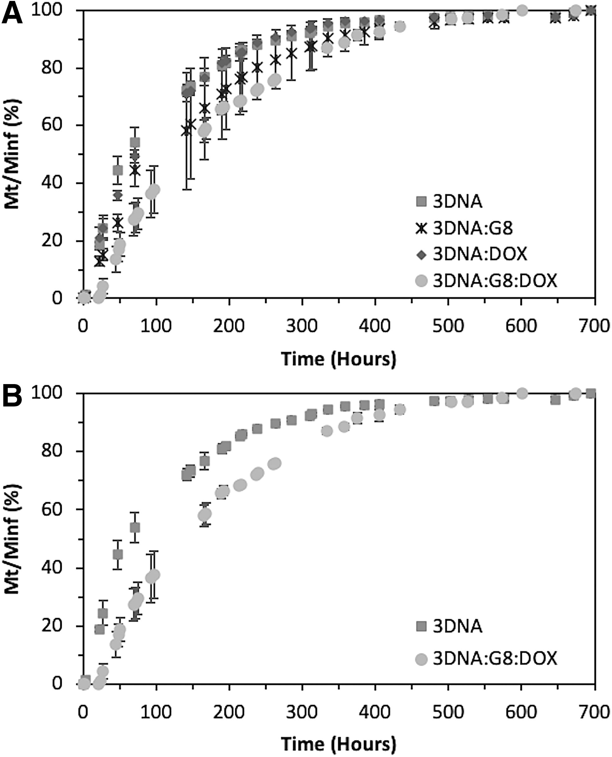
Release of 3DNA complexes
Figure 6 shows the release profile of 3DNA reagents from the optically clear, self-assembled polymers at a concentration of 29 (w/v)%. The release profile is mainly dominated by the size of the different 3DNA complexes in the formulation. 3DNA nanocarriers without the G8 mAb have a diameter size of 74 ± 2.6 nm, while 3DNA nanocarriers hybridized with the G8 mAb-oligo conjugate have a diameter size of 82 ± 1.6 nm. The release profile between 3DNA and 3DNA:DOX is not statistically significant, regardless of the presence of DOX. However, there is a statistically significant difference in the release profile of the reagents with the G8 mAb (Fig. 6B), with P value <0.05 (P = 1.28E-4), and ∼87% of the total mass of 3DNA:DOX:G8 reagents loaded were released in the period of 1 month. This is indicative that the particle size diameter difference plays an important role in determining the release profile of the 3DNA reagents.
Release profile of 3DNA nanocarriers.
Discussion
Phase transition
PLGA-PEG-PLGA triblock copolymers in aqueous media show a spontaneous, chemical free, and temperature-dependent self-assembly process that enables the formation of 3-dimensional structures at the sol-gel transition or GT. 45 GT is triggered by the hydrophobic effect as temperature increases, also known as a reverse thermal gelation mechanism. This mechanism is characteristic of the triblock copolymer theoretical self-assembly process, in which flower-shaped micelles form, aggregate, and bridge into a hydrogel.33,46 An optimal balance of hydrophobicity and hydrophilicity plays a key role in gelation, which could be easily tuned by alterations in the amphiphile's composition or the solution formulation. 47 The GT of PLGA-PEG-PLGA systems is also tunable by altering the compositional elements of the triblock copolymer such as Mw, LA/GA ratio, PEG Mw, PLGA Mw, block chain ratios (PLGA/PEG ratio), and solution concentration among others. These triblock copolymers regularly become a hydrogel in aqueous solvents as the triblock copolymers become more hydrophobic. 48 However, in this research, the triblock copolymer's compositional elements are constant ensuring less variability in the GT as well as the diminishment of critical effects induced by small amounts of additives (other homopolymers, different salts, and so on). 49 Administration of drug-loaded 3DNA nanocarriers as a sustained delivery formulation may reduce the incidence of PCO even further and protect the lens from cells migrating from the ciliary processes.15,25
Based on our observations, as the 3DNA nanocarriers were added to the polymer solution the GT decreased compared to systems in PBS only. 3DNA nanocarriers tend to be more hydrophilic because the DNA backbone is negatively charged, the G8 antibody is hydrophilic, and DOX is embedded within the central double-stranded regions, preventing further hydrophobic interactions with the PLGA hydrophobic pockets. Hydrophobic interactions and hydrogen bonding are the driving forces between hydrophobic polymer entities and DNA.50–52
An increase in entropy due to water release from the interface between 2 hydrophobic regions allows the hydrophobic interactions to take place while hydrogen bonding occurs between the carbonyl and ether groups of PLGA-PEG-PLGA. Cosolutes, in this case, the 3DNA nanocarriers, play an important role in the change of GT and opacification of the hydrogel. At room temperature, both the cosolute and the polymeric system are soluble in aqueous solvents. However, as temperature increases, the 3DNA structures favor water relative to the PLGA-PEG-PLGA system. In theory, phosphate groups act as kosmotropic elements in nonionic solutions. In other words, phosphate groups bring order to the solution (water-making structures) that dramatically reduces the GT due to enhanced dehydration of the polymer as temperature increases. This effectiveness follows the Hofmeister ion series, in which anions are more effective than cations in polymeric solutions.49,53,54 The change in hydration regions/shells interferes in the way the polymer interacts with water and itself. The destabilizing effect of ions in solution reduces the interactions required to form a polymer network, hence speeding up the gelation process.
Moreover, the gel window of these self-assembled hydrogels is composed of 2 regions, transparent gels (closer to GT) and opaque gels (above GT). Temperatures higher than the GT lead to a more significant (stronger) hydrophobic effect, coarsening the micelle network. 55 When the micelle cluster size falls in the range of the visible wavelength spectrum of interest (455–500 nm), the gel becomes opaque to the eye. This happens because visible wavelengths at the size of the micellar cluster or smaller are either absorbed or scattered. 48
Light transmittance
Drug delivery systems that are implanted in the lens capsule or in the vitreous humor are not required to be fully transparent, since the adult/elderly lens filters out short blue visible light (400–500 nm).27,28 Figure 4 shows the light transmittance percentage of the different hydrogel formulations. Samples with >80% transmittance (3DNA®:DOX:G8 and gels in PBS only) indicate that these hydrogels meet the optical clarity criteria.
In general, hydrogels and other soft materials like glasses are disordered microscopically, but behave like solids macroscopically (gel transition or jamming transition). One of the most significant factors for transparency is the microstructure that includes micelle size, micellar boundaries (amorphous regions between adjacent micelles), crystallization of micelles structure, pore size, and least contamination.56–59 It has been hypothesized that the polymer concentration affects micellar morphology,60,61 which indeed influences light transmittance. At high enough concentrations, the self-assembled nanocomplexes can order themselves in much the same way as lyotropic liquid crystals do. 61 An isotropic micellar lattice or homogeneous liquid crystal theoretical structure, that is, cubic lattice, generally makes hydrogels more transparent.49,62,63 It has been shown that the interactions between PEG-PLGA-PEG and DNA do not have a drastic effect on the DNA shape and charge. Hence, light transmittance is not affected by the 3DNA nanocarriers. The loosely interactive triblock copolymer shows limited effect on DNA double helix structure, forming slightly compacted, yet, flexible polymer/DNA complexes—unlike cationic polymers which usually condense DNA into smaller particles (50–200 nm).50,64,65 Light transmittance can also depend on the gel's final geometry. 66 As specified in the methods section, our in vitro testing sample is 100 μL, which will be held the same for our future in vivo work. However, after cataract surgery, the gel may acquire a slab geometry, which can allow for improved light transmittance.
Modulus
Hydrogels with a modulus <350 Pa are deformable and suitable for intraocular implantation.29,30 Soft hydrogels prevent further damage to surrounding ocular tissue postintervention. Figure 5 shows that the modulus of hydrogels with all different 3DNA nanocarriers decreased linearly, compared to hydrogels in PBS only. Although all hydrogels have the same polymer concentration [29 (w/v)%], adding 3DNA nanocarriers modifies the overall hydrogel mechanical properties. In this case, adding the G8 mAb antibody causes significant differences in the gel mechanical properties.
Hydrogels with 3DNA:DOX showed a 73% decrease is softness compared to gels in PBS only. However, 3DNA:DOX reagents presented a higher modulus (294 ± 46 Pa) than gels with 3DNA (201 ± 46 Pa). This change can be attributed to the subtle changes of the DNA double helix structures (partial unwinding) produced by the intercalation of doxorubicin as well as some partial leaking of doxorubicin. Small hydrophobic molecules may help enhance the hydrophobic interactions as these are encapsulated in the hydrophobic micellar cores. As a consequence, the hydrogel becomes stiffer compared to hydrophilic molecules. 67 Hydrogels with 3DNA only presented lower modulus (201 ± 46 Pa) compared to hydrogels in PBS only (401 ± 318 Pa) and 3DNA:DOX (294 ± 46 Pa). This can be attributed to a higher entrapment of water due to the hydrophilicity of DNA. Furthermore, adding the G8 mAb to the system makes the hydrogels more deformable, up to 45% and 30% less stiff as in the case of 3DNA:G8 and 3DNA:DOX:G8, respectively.
For the system of preference (3DNA:DOX:G8), the presence of the G8 mAb plays a bigger role in modifying the overall architecture of the hydrogel (Fig. 8B), than the potential leaking of DOX or the minimal unwinding of the double helix structure of DNA.
Dynamic release
In this work, 3DNA nanocarriers released from the self-assembled hydrogel are tracked using Alexa Fluor 647, as explained in the Methods section. Doxorubicin is not measured since it is specifically intercalated in the double helix regions of the 3DNA nanocarriers. Doxorubicin is released from the 3DNA nanocarriers after internalization into the acidic compartments of Myo/Nog cells. 16
PLGA and its derivatives have been extensively studied as drug delivery carriers for proteins, drugs, and other macromolecules such as RNA, peptides, and DNA. 68 Their high biocompatibility and controlled biodegradability make them efficient drug delivery carriers. PLGA-based delivery platforms start degrading upon immediate contact with water through hydrolysis of the ester bonds. Hydrolysis creates acids, which further catalyze the degradation process. This autocatalytic phenomenon is known to cause heterogeneous degradation (bulk erosion) inside PLGA-based matrices. 69 Release of DNA from self-association polymers such as PLGA-PEG-PLGA tends to rely on diffusion, disruption of weak interactions, and the degradation rate of the polymer. 65 The release of the 3DNA nanocarriers is strongly coupled to diffusion through water-filled pores and the degradation rate of the polymer matrix, as previously reported.65,69,70
Hydrophilic drugs can draw more water into the matrix, enhancing pore formation and hydrolysis (Fig. 8B). Moreover, the rate of release is influenced by the diameter size of the 3DNA nanocarriers, polymer degradation/erosion, and possible polymer/3DNA interactions. 3DNA formulations used in this study had a diameter of 74 ± 2.6 nm, while the 3DNA:G8 formulations had a diameter of 82 ± 1.6 nm. Figures 6A and B show that the release profile of the complex of interest (3DNA:DOX:G8) presents a slower release rate compared with the controls: 3DNA; 3DNA:DOX; and 3DNA:G8. The release profile of 3DNA and 3DNA:DOX is not statistically significant since both complexes have the same size, regardless of the fact that there is DOX present. Doxorubicin is a small, hydrophobic, cytotoxic drug that has been widely used for chemotherapy. It has been known for its ability to kill cancerous cells for solid and liquid tumors as well as its strong side effects. 71 When mixed with DNA, doxorubicin shows a preferential intercalation into the G-T complexes of DNA through hydrogen binding. 72 The intercalation may cause subtle/partial unwinding of the DNA double helix structure.71,72 These subtle structural changes may restrict the nanocarrier from diffusing out of the hydrogel, as in the case of 3DNA:DOX:G8 (Fig. 6B).
Furthermore, Figs. 6B and 7 show the fractional release profile (Fig. 6B) and cumulative mass release profile (Fig. 7) of 3DNA and 3DNA:DOX:G8 with diameter size of 74 ± 2.6 and 82 ± 1.6 nm, respectively. The presence of the G8 mAb makes the overall complex ∼10% larger in diameter. As observed in Fig. 7, the size difference reduces the release rate of the nanocarriers. In other words, the bigger the nanocarrier, the slower the release rate. The overall release rate is controlled by the size difference of the DNA-based reagents, diffusion through water-filled pores, and polymer degradation/erosion. 73 Drug release also depends on the gel's final geometry. 74 As specified in the Methods section, our in-vitro testing sample is 100 μL, which will be held the same for our future in-vivo work. However, after cataract surgery, the gel may acquire a slab geometry. The drug rate is dependent upon surface area, which can be determined in future in-vivo studies.

Cumulative mass release profile of 3DNA nanocarriers. Cumulative release profile shows that the release rate greatly depends on the size of the different 3DNA reagents, as it can generate more 3DNA/polymer interactions. The bigger the overall diameter, the slower the release rate.

SEM images of hydrogels in PBS only
For in-vivo work, it is important to keep in mind the anatomical shape of the lens capsule with regard to the nanocarriers diffusing in and out of the capsule. The lens capsule is an active membrane that allows for passive diffusion of metabolic substrates and waste through the interwoven fibrous network. No evidence has been found for pores on the lens capsule. 75 It selectively filters molecules based on their size and charge. Intermediate size molecules diffuse through the lens capsule at a rate that is directly correlated to their Mw. Negatively charged particles have significantly lower diffusion rates because the overall charge of the lens capsule is also negative. 76 We do not present a concern that the nanocarriers will diffuse out of the lens capsule since both the lens capsule and the 3DNA are negatively charged.
Conclusion
In this study, we show, for the first time, sustained release of doxorubicin-loaded 3DNA nanocarriers targeted with a mAb (G8 mAb) specific to Myo/Nog cells for 1 month. These nanocarriers were released from optically clear self-assembled PLGA-PEG-PLGA hydrogels that are well known for releasing small, hydrophobic drugs. This study is a proof of concept that a sustained release formulation of a 3DNA therapy is possible to make, and has favorable characteristics that may further reduce the incidence of PCO, as proposed by Gerhart et al.15,16 The 3DNA gel formulations of interest (3DNA:DOX:G8) met the specific criteria for injections into the lens capsule: gelation at 35°C, strength modulus <350 Pa, and over 80% light transmittance over the required spectrum (455–700 nm). This work shows promising results that hydrophobic matrices such as PLGA-PEG-PLGA can be used as platforms for minimally invasive local treatments and prolonged delivery of proteins, peptides, and nucleic acid structures.
Footnotes
Acknowledgment
We thank Genisphere, LLC, our collaborator, for facilitating their 3DNA® nanocarriers that were specifically synthesized for this project. This work was supported by funding from the Cooper Foundation.
Author Disclosure Statement
This work is a subject of patent. The patent pending number: 62/479,716. M.E.B., L.L.O., and George-Weinstein Mindy have financial interest in this patent.