
Abstract
Purpose:
Benzalkonium Chloride (BAK) is reported to have the potential to damage the cornea. We developed a composition with broad-spectrum antimicrobial activity without preservatives by combining trometamol, boric acid, and ethylenediaminetetraacetic acid (TBE). This study aimed at evaluating the corneal damage caused by TBE and comparing it with that caused by BAK.
Methods:
SV40-immortalized human corneal epithelial cell line (HCE-T) was treated with BAK or TBE, and the cell viability was measured. The exposure time that caused 50% cell death (CDT50) was calculated. Transepithelial electrical resistance (TEER) was measured before and after treatment with BAK or TBE. Occludin was detected with immunostaining and Western blotting after treatment with BAK or TBE. The effect of BAK or TBE on membrane-associated mucins was evaluated with rose bengal (RB) staining.
Results:
In the BAK group, cell viability decreased in a dose-dependent manner. The viability of the TBE group was significantly greater than that of the BAK group. The CDT50 of the TBE group is greater than that of the BAK groups. In the BAK groups, the recovery of TEER was delayed in a dose-dependent manner, whereas in the TBE group, the recovery occurred earlier. Localization of occludin was disrupted, and the amount of occludin was significantly reduced among the cells exposed to BAK. The area stained with RB in the BAK groups increased, whereas that in the TBE group did not increase.
Conclusion:
These results suggest that the application of TBE would be useful for developing preservative-free ophthalmic preparations that offer both sufficient safety and antimicrobial activity.
Introduction
Preservatives are a major component of multidose eye drops. By providing a level of antimicrobial activity in an eye drop, preservatives prevent ocular infections caused by microorganism contamination and extend the shelf life by avoiding biodegradation and maintaining drug potency. 1 Benzalkonium Chloride (BAK), a quaternary ammonium compound, is commonly used in ophthalmic solutions because of its broad antibacterial spectrum. The mechanism of its antibacterial action involves the destruction of the microbial cell membrane. BAK is a type of cationic surfactant that can affect cell membrane permeability, enable intracellular substances to flow out, and cause death of the microorganism. 2 However, both in vitro and in vivo studies have shown that BAK may have a toxic effect on the conjunctiva and the cornea.3–5
In clinical practice, it has been observed that the ophthalmic solution containing BAK can cause keratoconjunctive epithelium disorder and aggravation of disease. BAC is used at a concentration of 10–200 μg/mL in ophthalmic preparations. 6 Long-term BAK exposure in a glaucoma patient could worsen the condition. 7 BAK may cause or aggravate dry eye disease and allergic conjunctivitis because of its toxic and proinflammatory effects. 8 Therefore, BAK-free or preservative-free ophthalmic preparation may be a better choice for patients with glaucoma or dry eye syndrome. 9 Although unit dose vials that are discarded after a single use do not require a preservative to prevent microbial growth, they involve the issues of high manufacturing cost and difficulty in opening for elderly patients.
We developed a composition with broad-spectrum antimicrobial activity without preservatives by combining trometamol, boric acid, and ethylenediaminetetraacetic acid (TBE). 9 TBE inhibits the growth of bacteria and fungi, and the antimicrobial activity is synergistically improved by combining the 3 components (Fig. 1 and Table 1). 9 Although TBE may be useful for the development of preservative-free ophthalmic preparations, the safety of TBE remains unclear. This study aimed at evaluating the safety of TBE by comparing the toxicities of TBE and BAK in human corneal epithelial cells.
Scanning electron micrograph of Escherichia coli
Minimum Inhibitory Concentration of Each Component Solution and Mixed Solution
Reprinted with permission from Takizawa et al.
Methods
Samples
BAK (Maruishi Pharmaceutical Co., Ltd., Osaka, Japan) was dissolved in phosphate buffer saline (PBS) (FUJIFILM Wako Pure Chemical Corporation, Osaka, Japan) to adjust to a pH of 7.0 at the following concentrations: 200, 50, 30, 20, and 10 μg/mL. TBE was prepared to 1 mg/mL trometamol (Kanto Chemical Co., Inc., Tokyo, Japan), 10 mg/mL boric acid (Kanto Chemical Co., Inc.), and 1 mg/mL ethylenediaminetetraacetic (EDTA) (Nagase ChemteX Co., Ltd., Osaka, Japan) in PBS, and the pH was adjusted to 7.0.
Cell culture
SV40-immortalized human corneal epithelial cell line (HCE-T: RCB2280) was procured from the RIKEN BRC, Japan. HCE-Ts were cultured in a supplemented hormonal epithelial medium that was Dulbecco's modified Eagle's medium/Ham's F12 (DMEM/F-12; Thermo Fisher Scientific, Inc., MA) containing 5% heat-inactivated fetal bovine serum (FBS; Thermo Fisher Scientific, Inc.), 5 μg/mL insulin (Thermo Fisher Scientific, Inc.), 10 ng/mL epidermal growth factor (Thermo Fisher Scientific, Inc.), 0.5% dimethyl sulfoxide (FUJIFILM Wako Pure Chemical Corporation), 100 units/mL penicillin, and 100 μg/mL streptomycin (Thermo Fisher Scientific, Inc.). HCE-Ts were maintained at 37°C in 5% CO2, and the growth medium was replaced every other day.
Normal human corneal epithelial cell (HCEC) was purchased from Kurabo Industries Ltd. (Japan). The HCECs were placed on cell culture inserts (Corning, Inc., NY) and grown by using the Oculife Complete Medium Kit (Kurabo Industries Ltd.) to achieve confluence at 37°C in 5% CO2, and the medium was replaced every other day. After reaching confluence, the culture medium of the bottom was changed to Oculife Complete Medium with 1.15 mM CaCl2 (Nacalai Tesque, Inc., Kyoto, Japan), 20 IU/mL retinol palmitate (DMS, Inc., Tokyo, Japan), and 1% FBS. The medium level was lowered to the bottom of the membrane, and the culture medium in a culture insert was removed completely to expose the cells to air. It was reported that air exposure condition (airlifting) promoted mucin expression.10,11 During airlifting, the growth medium was replaced every day.
Cell viability assay
To determine the cytotoxic effects of the samples, the viability of HCE-Ts was assessed by using a Cell Counting Kit-8 (CCK-8; Dojindo Laboratories, Inc., Tokyo, Japan). The HCE-Ts were seeded in a 96-well plate at 2.0 × 10⁴ cells/well and incubated for 24 h. After removing the culture medium, the cells were treated with 100 μL of PBS, TBE, or BAK solutions (10, 20, or 30 μg/mL) for 1, 2, 3, 5, 7, 10, 15, 20, or 30 min. The samples were removed by washing with PBS, and the cells were incubated with 100 μL of serum-free culture medium for 24 h. Ten microliters of CCK-8 reagent was added to each well, and the cells were incubated at 37°C for 2 h. The absorbance was measured at 450 nm by using a microplate reader (Molecular Devices Japan, Inc., Tokyo, Japan).
The mean absorbance value corresponding to the control cells (PBS treated cells) was taken as 100%, and the cell viability of cells treated with each sample was calculated. The exposure time that caused 50% cell death (CDT50) was calculated from the quadratic formula based on the survival rate.
Immunocytochemistry
The HCE-Ts were seeded in a 24-well plate at 5.0 × 10⁴ cells/well and incubated for 72 h. After removing the culture medium, the cells were treated with 300 μL of PBS, TBE, or BAK solutions (10 or 30 μg/mL) for 10 min. The cells were rinsed with PBS and fixed with incubation in chilled methanol (stored at −20°C) for 15 min.
The cells were blocked with 1% bovine serum albumin (Sigma-Aldrich Corp., MO) in PBS for 15 min at room temperature. Incubation with primary antibodies (mouse anti-Occludin monoclonal IgG; Thermo Fisher Scientific, Inc.) was performed in a blocking solution at 4°C overnight. The cells were washed thrice with PBS for 5 min at room temperature and then incubated with secondary antibody (goat anti-mouse polyclonal IgG-conjugated Alexa Flour 488; Thermo Fisher Scientific, Inc.) in a blocking solution at 4°C overnight. Excess antibodies were removed by washing thrice with PBS for 5 min at room temperature. The cells were mounted on a coverslip with VECTASHIELD mounting Medium with DAPI (Vector Laboratories, Inc., CA) and observed with a fluorescence microscope (Olympus Corp., Tokyo, Japan).
Western blotting
The HCE-Ts were seeded in a 6-well plate at 2.0 × 10⁵ cells/well and incubated for 48 h. After removing the culture medium, the cells were treated with 1 mL of PBS, TBE, or BAK solutions (10 or 30 μg/mL) for 10 min. The cells were rinsed with PBS twice and then collected by scraping into 1 mL of PBS. Cells were pelleted by centrifugation at 1,500 g for 5 min. After removing the supernatant, cells were suspended in 50 μL RIPA buffer (Nacalai Tesque, Inc.) and lysed on ice for 30 min. The samples were cleared by centrifugation at 15,000 g for 15 min. The protein concentration was determined by the BCA protein assay (Thermo Fisher Scientific, Inc.).
Approximately 10 μg of total cell protein was prepared in NuPAGE LDS sample buffer (Thermo Fisher Scientific, Inc.) supplemented with 50 mM dithiothreitol (Thermo Fisher Scientific, Inc.) and then boiled at 95°C for 5 min. Samples were subjected to electrophoresis (4%–12% Bis-Tris Plus Gels; Thermo Fisher Scientific, Inc.). The separated proteins were transferred to PDVF membranes (Bio-Rad Laboratories, Inc., CA). The membranes were incubated in 5% skim milk (FUJIFILM Wako Pure Chemical Corporation) in TBS-T (blocking buffer) for 30 min at room temperature. After blocking, incubation with primary antibodies (mouse anti-Occludin monoclonal IgG; Thermo Fisher Scientific, Inc.) was performed in blocking buffer at 4°C overnight.
Membranes were washed thrice with TBS-T for 10 min at room temperature and then incubated with secondary antibody (horse anti-mouse IgG polyclonal IgG-conjugated horseradish peroxidase, Cell Signaling Technology, Inc., MA) in blocking buffer for 3 h at room temperature. Besides this, membranes for detection of beta-actin were incubated with antibody (mouse anti-beta-actin polyclonal IgG-conjugated horseradish peroxidase; Cell Signaling Technology, Inc.) in blocking buffer for 3 h at room temperature. The membranes were washed thrice with TBS-T for 10 min at room temperature. Antibody–antigen complexes were detected by using the Pierce ECL-Plus kit (Thermo Fisher Scientific, Inc.) and the Amersham ECL Prime (Cytiva, Inc., MA).
Measurement of band intensities by densitometry was performed with Amersham Imager 600 (Cytiva, Inc.), and calculations were made by using beta-actin as loading control by employing Amersham Imager 600 software.
TEER measurement
Transepithelial electrical resistance (TEER) was determined by using a Millicell-ERS Voltohmmeter (Merck Millipore Corp., MA). The HCE-Ts were seeded in cell culture inserts at 5.0 × 10⁴ cells/well and incubated for 96 h. The culture medium was replaced with fresh medium; thereafter, the cells were incubated for 1 h. After removing the culture medium in the top chamber, the cells were treated with 100 μL of PBS, TBE, or BAK solutions (10, 50, or 200 μg/mL) for 1 min. The samples were removed by washing with culture medium, and they were then replaced with fresh culture medium.
The TEER was measured before the treatment (Pre), and at 0, 15, 30, 45, 60, 90, and 120 min after the treatment. The TEER of the insert without the cells was measured as the control to determine the intrinsic resistance of the filter, and it was subtracted from all the readings. The TEER relative rates of change against Pre were calculated.
Rose bengal assay
The HCECs were seeded in cell culture inserts at 1.0 × 10⁵ cells/well and incubated for 72 h under normal culture conditions. Then, the cell culture condition was changed to airlifting, and the HCECs were cultured for 7 days in this condition. After rinsing the cells with PBS, the cells were treated with 100 μL of PBS, TBE, or BAK solutions (5, 10, or 50 μg/mL) for 5 min. The samples were removed by washing with PBS, and the cells were incubated with 0.1% RB (Sigma-Aldrich) in PBS for 5 min. The RB solutions were aspirated, and the cells were assessed for the extent of dye penetration with a microscope (Olympus, Tokyo, Japan). Pictures were taken with an AxioCam camera (Carl Zeiss Meditec GmbH), and the images were processed further for dye penetration quantification by using the ImageJ software (NIH, MD).
Statistical analysis
Cell viabilities of the TBE group (n = 6) and the BAK groups (each group n = 6) were compared by using Dunnett test. The values of the TEER relative rate of change were compared between the TBE group (n = 4) and BAK groups (each group n = 4) by using Dunnett test. The band intensities of TBE group and BAK groups were compared by using Dunnett test. The areas that excluded RB were compared between the TBE group (n = 4) and the BAK groups (each group n = 4) by using Dunnett test. Data are presented as the mean ± standard error values. All the statistical analyses were performed with JMP.
Results
Cell viability assays
To investigate the toxic effect of BAK and TBE, the HCE-T cells were treated with BAK or TBE, cell viability was measured by using a CCK-8 assay, and CDT50 was calculated (Fig. 2 and Table 2). The cell viability decreased in a dose- and time-dependent manner after treatment with BAK, whereas no obvious decline in cell viability after treatment with TBE was observed (Fig. 2). The CDT50 values of the group treated with 10, 20, and 30 μg/mL BAK were 5.12, 2.41, and <1.00, respectively, whereas that of the TBE group was >30.00 (Table 2).
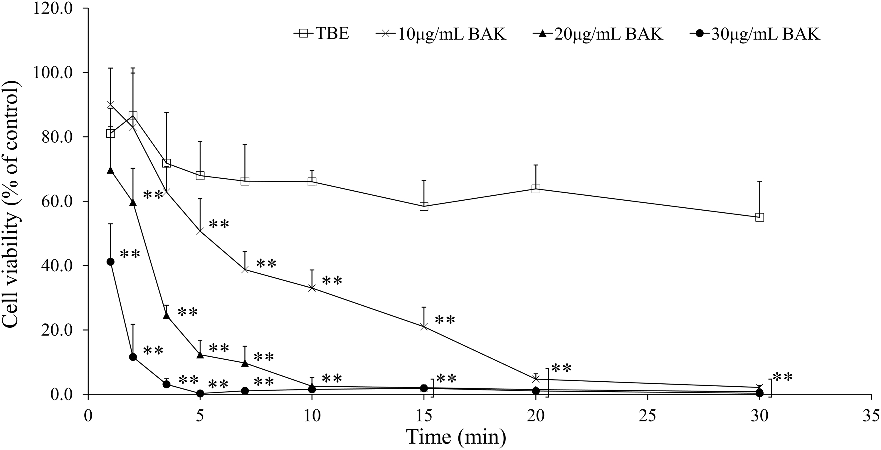
Effect of TBE and BAK solutions on the cell viability. HCE-Ts were exposed to TBE (□) or BAK solutions diluted at different concentrations: 10 μg/mL ( × ), 20 μg/mL (▴), and 30 μg/mL (•). Cell viability was measured by using CCK-8, and the data were normalized to control (PBS treated) value (100%). All the data are expressed as the mean ± standard error (n = 6) values. **P < 0.01 versus TBE. TBE, trometamol, boric and ethylenediaminetetraacetic acid;
CDT50 of TBE and BAK Solutions
TBE, trometamol, boric and ethylenediaminetetraacetic acid;
TEER measurement
The effect of exposure to BAK or TBE on barrier function of the HCE-T cell monolayer was assessed with TEER measurement. In all the treated groups, transient TEER decreases were detected just after treatment, and those in the BAK treatment groups increased in a dose-dependent manner (Fig. 3). The recovery of TEER in the BAK group was delayed in a dose-dependent manner, and the TEER value of the 200 μg/mL BAK-treated group did not show a tendency to recover within 120 min after treatment (Fig. 3). In the TBE group, TEER recovery was faster than that in the BAK groups (Fig. 3).
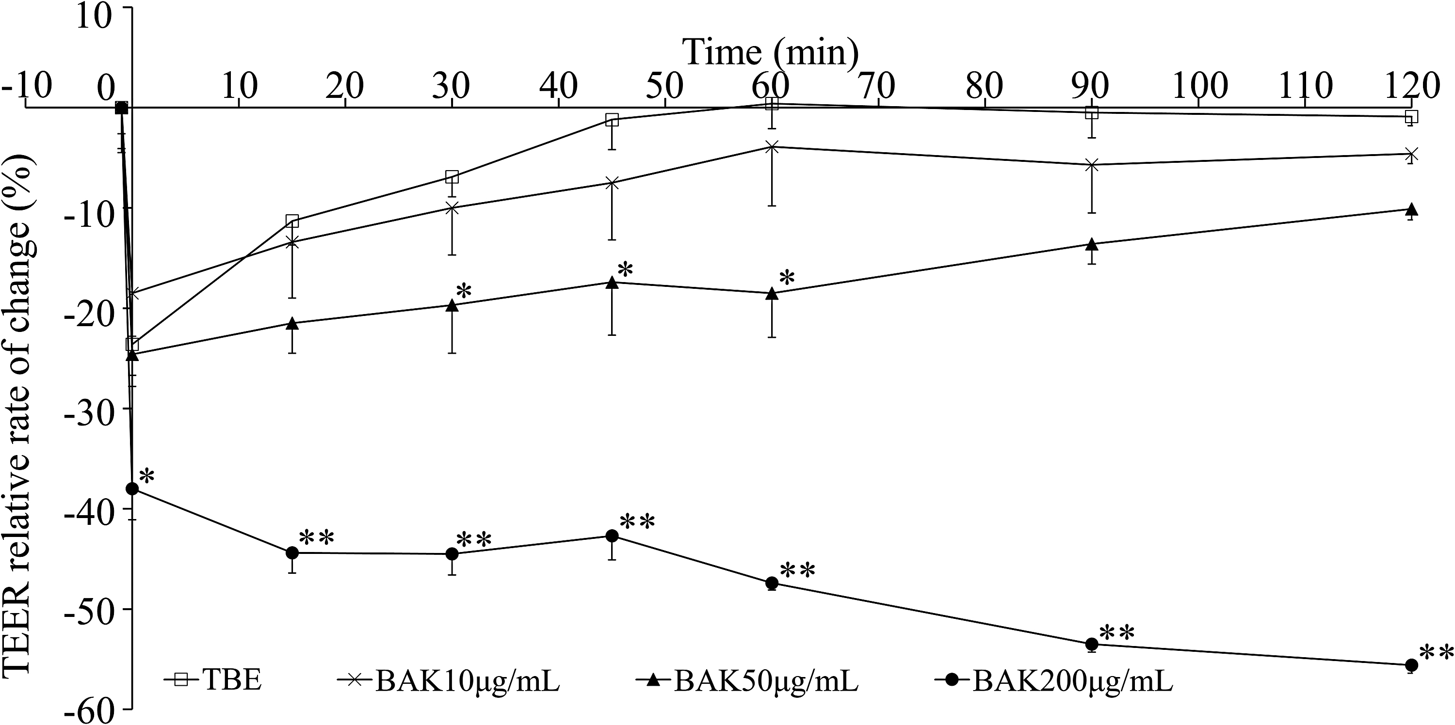
Changes in TEER induced by treatment with TBE or BAK solutions. The HCE-Ts were exposed to TBE (□) or BAK solutions diluted at different concentrations: 10 μg/mL ( × ), 50 μg/mL (▴), and 200 μg/mL (•). TEER values were measured before treatment (Pre) and at 0, 15, 30, 45, 60, 90, and 120 min after the treatment of samples. The TEER relative rates of change against Pre were calculated. All the data are expressed as mean ± standard error values (n = 4). *P < 0.05 versus TBE. **P < 0.01 versus TBE.
Localization of occludin after exposure to BAK and TBE
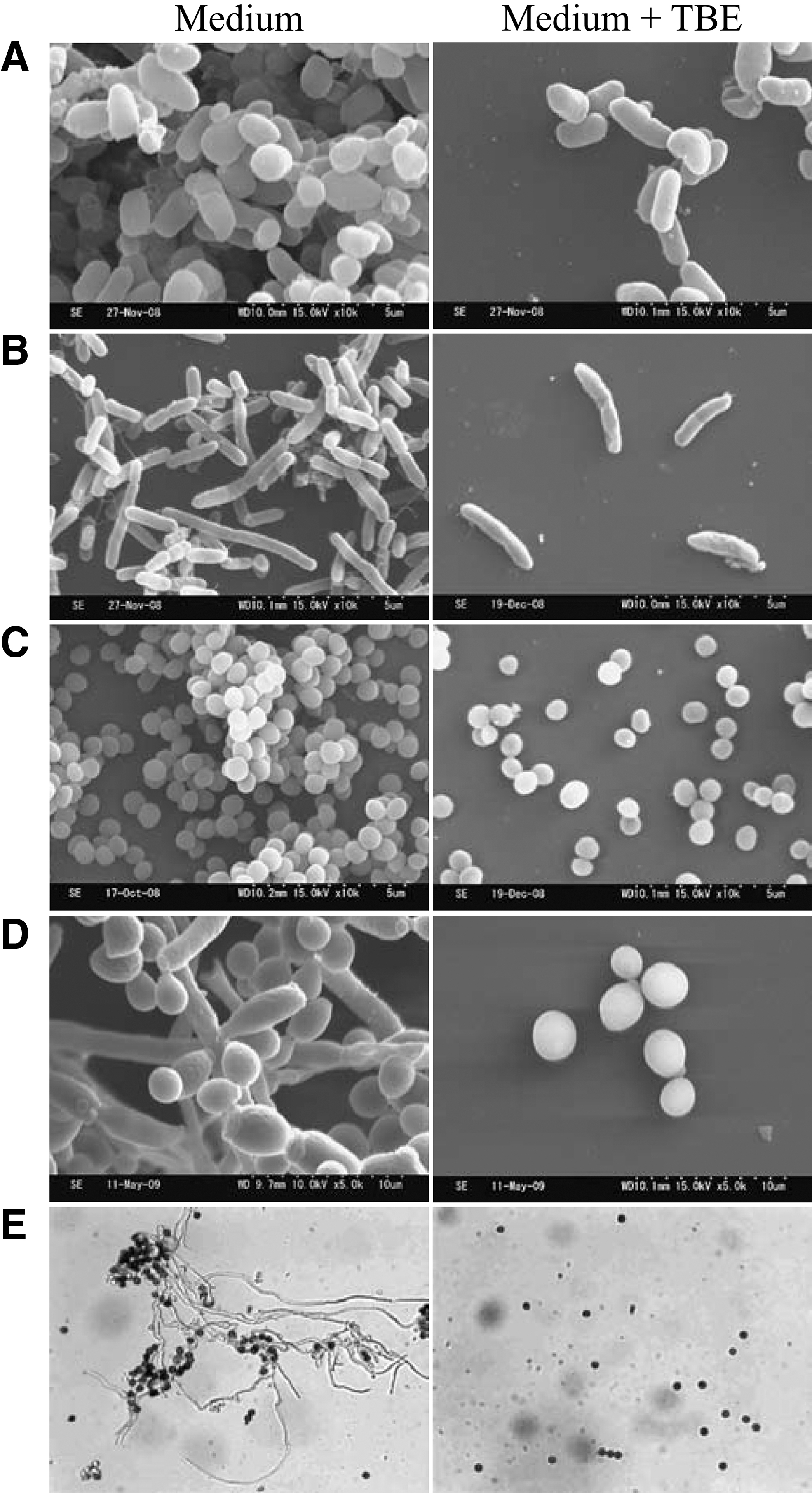
Figure 4A–D shows that occludin localizes at the region of the cell–cell junction. In the PBS group, as a negative control, occludin localization was present on the cell membrane (Fig. 4A). There was no difference in the localization of occludin between the cells exposed to PBS and those exposed to 10 μg/mL BAK or TBE (Fig. 4B, C). The disruption of occludin localization was observed among cells exposed to 30 μg/mL BAK (Fig. 4D).

Microscopic images of immunofluorescence analysis of occluding localization in HCE-Ts
Western blotting of occludin
The effect of exposure to TBE or BAK on occludin expression in HCE-Ts was analyzed by Western blotting. The band intensities of occludin in the groups exposed to 10 μg/mL BAK or TBE did not change compared with those of occludin in the PBS group as a negative control. In contrast, the band intensities of occludin in the group exposed to 30 μg/mL BAK significantly decreased (Fig. 4E, F).
Rose bengal assay
The area of the cell region that excluded RB had been increasing under airlifting condition culture. In the BAK groups, the cell regions stained by RB increased in a dose-dependent manner (Fig. 5A, B). However, the area of the cells stained by RB after TBE treatment was similar to that of the PBS group as a negative control (Fig. 5A, B).

Rose bengal staining in HCECs treated with PBS, TBE, or BAK solutions. The HCECs were exposed to PBS
Discussion
Previous in vivo and in vitro studies have shown that benzalkonium chloride, the most commonly used preservative in eye drops, can induce toxic effects on the ocular surface.3–5 In particular, there is concern about the adverse effects of BAK in glaucoma patients who need to take multiple medicines routinely for decades and in dry eye patients whose symptoms can be aggravated owing to the inflammatory nature of the ocular surface. It is preferable that the therapeutic agent for these conditions contain no preservative or minimum amount of preservative. We have developed a preservative-free composition (TBE) with sufficient antibacterial activity to prevent adverse effects of BAK. 9
BAK exerts antimicrobial effect by destroying the membrane; however, the mechanism of TBE is different. The TBE does not cause destruction of the microbial membrane but suppresses the growth of microorganisms by inhibiting the production of gene and the activity of aminoacyl-tRNA synthetase. 9 The TBE bacteriostatic action is mainly caused by boric acid; trometamol and EDTA synergistically enhance the effect of boric acid by increasing the amount of inflow of boric acid into the microorganism. The hypothesis that trometamol and EDTA increase the influx of boric acid is the effect on the cell surface structure. Trometamol can affect the permeability of the microbe membrane by binding to lipopolysaccharide (LPS), replacing Ca2+ and Mg2+, and reducing the interaction between LPS molecules and EDTA by the chelation of a bivalent cationic ion.12–14 This mechanism suggests that the effect exerted by TBE for inhibiting the growth of microorganisms is specific to microbes and that TBE can exert little effect on HCECs. Therefore, in this study, we investigated the cytotoxicity of TBE and the effects of TBE on barrier function and membrane-associated mucin.
The cytotoxicity of TBE was lower than that of BAK (Fig. 2). The boric acid concentration of TBE was 10 mg/mL, and boric acid is reported to have weak antibacterial activity and less cytotoxicity. 15 No cytotoxicity was observed on normal rabbit corneal epithelial cells with 50 mg/mL trometamol. 16 Although the species of corneal epithelial cells used in the present and previous studies were different, the trometamol concentration of TBE was as low as 10 mg/mL. With respect to EDTA, it is reported to show no dose-dependent toxic effect on the corneal epithelial cells. 17 Previous reports show that boric acid, trometamol, and EDTA have low cytotoxicity when administered alone; our results show no increase in the cytotoxicity when these are used in combination.
Cornea is one of the tissues that comes into contact with the outside and has a barrier function that prevents the invasion of exogenous substances. Epithelial barrier function is maintained by the membrane structure that comprises the tight junction, adherens junction, and desmosome. Of these 3 structures, the tight junction is a high resistance barrier and is composed of a complex of proteins, including transmembrane proteins occludin and claudin and cytoplasmic proteins, such as the zonula occludens ZO-1, ZO-2, and ZO-3 that interact with both the transmembrane proteins and the actin cytoskeleton. 18 Loss of epithelial barrier function occurs due to the disruption of these tight junction proteins. 19 In a previous study, it is reported that BAK treatment can disappear occludin of corneal epithelial cells. 20
The effect of BAK and TBE on barrier function was evaluated by measuring TEER (Fig. 3), immunostaining occludin, and Western blotting (Fig. 4). The amount of occludin protein was also decreased in the 30 μg/mL BAK group (Fig. 4E, F). The temporary decrease in TEER observed just after the treatment in all the groups may be due to experimental operations, such as exchanging medium, washing with PBS etc. BAK increased the temporary TEER decrease and delayed the recovery of TEER in a dose-dependent manner (Fig. 3). The change in occludin localization occurred in the 30 μg/mL BAK group (Fig. 4B). BAK could disrupt cell membranes and denature proteins. Therefore, it is believed that BAK caused the disruption of occludin localization and the decrease of occludin protein. It was confirmed that BAK affected the barrier function via the tight junction, as previously reported. 20
TBE reduced the TEER immediately after treatment with >10 μg/mL BAK (Fig. 3). It is known that chelating of Ca2+ ions with EDTA expanded the paracellular route and decreased TEER. 21 The rapid recovery of TEER in the TBE group is attributed to the supplementation of Ca2+ ions resulting from the replacement with fresh medium. When eye drops containing EDTA are applied, the influence of chelating by EDTA is small because of lacrimal fluid exchange. Localization of occludin and the amount of occludin in the TBE treatment group was not different from those of the PBS group as the control. Therefore, TBE has limited effect on the barrier function via tight junctions and the form of tight junctions.
Membrane-associated mucins that are expressed on the ocular surface have multiple functions, such as stabilization of tears on the ocular surface and formation of a protective barrier against microorganisms and extracellular molecules.22,23 To evaluate the effects on membrane-associated mucins, we used the human corneal epithelial model cultured under airlifting condition and performed RB staining. 11 The increase in the rose bengal (RB) penetration that indicates the disruption of membrane-associated mucins was detected in a dose-dependent manner among the BAK treatment groups (Fig. 5). In contrast, no change in the RB staining occurred in the TBE group as compared with that in the PBS group (Fig. 5). These results suggest that BAK affects membrane-associated mucins on the epithelial surface; however, TBE does not exert such an effect.
In sum, TBE is a safety formulation with sufficient antibacterial activity without any preservative. TBE can be useful for the development of preservative-free ophthalmic preparations with both safety and antiseptic ability.
Footnotes
Acknowledgments
The authors thank Takeshi Takizawa for permission to reprint the data (Fig. 1 and ![]() ). They also thank RIKEN BRC for providing HCE-T cells.
). They also thank RIKEN BRC for providing HCE-T cells.
Author Disclosure Statement
The authors state no conflict of interest. Kyohei Kabashima is an employee of Lion Corporation.
Funding Information
No funding was received for this article.