
Abstract
Purpose:
To evaluate the effects of a single dose of oral 5-mg tadalafil on macular microcirculation as measured using optical coherence tomography (OCT) and angiography (OCTA) in healthy volunteers.
Methods:
Twenty-two healthy, middle-aged, sexually active, and male health care worker volunteers were included in this prospective study. All volunteers have a history of occasionally using off-label 5 mg tadalafil to enhance sexual performance. Superficial and deep capillary plexus vascular densities, foveal avascular zone parameters, outer retina, and choriocapillaris flow areas were performed using the OCTA, and subfoveal-choroidal thickness (CT) was performed by using the OCT. Measurements were performed preintake, 30 min, 1, 4, 24 h, 2, 3, 4, and 7 days after the intake of tadalafil off-label.
Results:
Twenty-two eyes of 22 male volunteers were included in the study. The mean age was 37.16 ± 4.52 years. At 30 min, 1 h, and 4 h after intake, a statistically significant increase was observed in the choriocapillaris flow area and CT compared with preintake (Friedman test, P = 0.034 and P < 0.001, respectively).
Conclusion:
This study showed that a single dose of oral 5-mg tadalafil causes an increase in choriocapillaris flow and CT. To evaluate the effects of tadalafil on the retina and choroid, an OCTA assessment may be helpful.
Introduction
Nowadays, pharmacotherapy is the first step in the treatment of erectile dysfunction.1–3 Phosphodiesterase (PDE) type 5 normally inhibits penile erection by degrading cyclic guanosine monophosphate (cGMP).1,3 Agents, called PDE5 inhibitors, effect the penis. These agents inhibit cGMP degradation, increase the effect of nitric oxide, and inhibit transmembrane calcium and cell membrane hyperpolarization. 4 As a result, smooth muscle relaxation develops, and an erection occurs. 5 Tadalafil has the highest affinity to PDE5 within all PDE inhibitors currently in the market. 6 Tadalafil is a selective and long-acting PDE inhibitor and is recommended to be taken 30 min before sexual intercourse. Also, this drug has been reported to have a duration of action extending to approximately 72 h. 7 The standard dose recommended for erectile dysfunction is10 mg.1,2 However, it has been reported to have positive results for use at doses of 2.5–5 mg daily. 5
The PDE6 enzyme is the crucial effect enzyme for the phototransduction cascade in the cone and rod segments of the retina. 8 Visual disorders, such as blurred vision, color and light perception changes, cyanopsia, and enhanced light sensitivity, have been attributed to the crossreactivity with the PDE6 catalytic site, upon intake of PDE5 inhibitors.8,9 Less common ocular adverse effects are central serous chorioretinopathy (CSCR), nonarteritic ischemic optic neuropathy, reversible increase in intraocular pressure, ERG disturbances, and idiopathic serous macular detachment. 10
Although tadalafil is approved worldwide for erectile dysfunction, pulmonary hypertension, and benign prostatic hyperplasia, it is obtained over the counter, especially in developing countries, and is used off-label by sexually active young and middle-aged men to enhance sexual performance. Although 10/25/50 mg is recommended to treat erectile dysfunction by health care professionals, a single dose of 5 mg before sexual intercourse is often preferred to enhance sexual performance in young- to middle-age groups. PDE inhibitors that are not entirely innocent have adverse effects such as headache, nasal congestion, and flushing. 7 Although a precise explanation of ocular side effects is still unknown, changes in the vascular flow of the choroid and retina due to PDE inhibition are assumed to play a significant role.4,7
Optical coherence tomography angiography (OCTA) is a novel method showing the vascular structures of the retina, choroid, and anterior segment of the eye without using any contrast agent. 11 It provides visualization of the vascular network by obtaining the motion contrast caused by erythrocytes through repeated scans from the same region. 12 This modality is noninvasive, repeatable, reliable, and fast. Currently, it is used in the diagnosis and monitoring of various retinal and vascular diseases.13,14
For these reasons, in this study, we aimed to determine the retinal and choroidal microcirculation alterations related to oral 5-mg tadalafil intake in healthy volunteers, using OCTA.
Methods
Study sample and design
Twenty-two healthy volunteers were included in this prospective study. All participating volunteers had a previous history of using 5 mg of tadalafil to enhance sexual performance with their own desires and decisions. There were no life-threatening side effects in this history of use. All volunteers were middle-aged, sexually active, and healthy male health care workers. These volunteers were known to supply their tadalafil needs, of one's own accord, directly from the pharmacy without a prescription. None of the volunteers had a history of any kind of surgery and medication, except tadalafil. Also, when the study was started, all volunteers stated that they had not used tadalafil for the past 1 month.
The informed consent form was received from all participants. Power analysis was performed to justify the number of patients enrolled in the study. The following inclusion criteria were applied: (1) Healthy volunteer, and (2) History of occasionally using off-label 5 mg tadalafil to enhance sexual performance. The following exclusion criteria were applied: (1) Having any systemic or ocular disease, (2) Tadalafil use in the last 1 month, (3) Spherical error more than 2.00 D, (4) Axial length (AL) less than 18 mm and more than 24 mm, (5) History of ocular trauma or ocular surgery, and (6) Body mass index less than 18.5 and more than 24.9 kg/m2. All procedures were performed by following the tenets of the Declaration of Helsinki. Institutional Review Board approval was obtained from the Ethics Committee of Bakirkoy Dr. Sadi Konuk Training and Research Hospital, Istanbul (Acceptance code: 2019/526, 2019–25).
All participants underwent an ophthalmological examination at the preintake, including measurement of the best-corrected visual acuity, slit-lamp biomicroscopy, intraocular pressure (IOP) measurement with applanation tonometer (Goldmann; Haag-Streit AG, Köniz, Switzerland), axial length measurement with AL-Scan (Nidek CO., Gamagori, Japan).
In all volunteers; superficial and deep capillary plexus vascular densities, foveal avascular zone (FAZ) area, FAZ perimeter (PERIM) and vascular density of the FAZ around 300 microns (FD-300), flow areas in the outer retina and choriocapillaris were performed by using the OCTA (RTVue-XR Avanti, Optovue Inc., Fremont, CA), and thickness of subfoveal choroidal (CT) was performed by using the spectral domain OCT (SD-OCT; Heidelberg Engineering, Inc., Heidelberg, Germany). OCTA scans were achieved by using a spectral-domain system with the software AngioVue OCTA system, version 2018.0.0.18. Central macular 6 × 6 mm scans were achieved using the AngioVue Imaging System Optovue RTVue XR 100 Avanti with the AngioVue OCTA software.
OCTA measurements were made according to the Early Treatment Diabetic Retinopathy Study grid, containing 3 concentric rings with diameters of 1, 3, and 6 mm to divide the macula into foveal, parafoveal, and perifoveal regions. The outer retinal and choriocapillaris flow area of the 2.97 mm radius macular circle were determined. The FAZ, PERIM, and FD-300 were also obtained in the FAZ mode of the OCTA software. The CT was performed with semiautomatical measurement with an enhanced depth image mode of SD-OCT. The CT was described as the interval from the hyperreflective line of the outermost retinal pigment epithelium to the innermost hyperreflective line of the choroidoscleral junction. To ensure accurate segmentation and sufficient image quality, all scans were made with a quality index of >7/10.
In all volunteers, measurements were performed preintake, 30 min, 1, 4, 24 h, and 2, 3, 4, and 7 days after the intake of tadalafil. The measurements were compared statistically according with the measurement before tadalafil intake. Since tadalafil passes into the systemic circulation after oral administration, it is assumed to be effective in both eyes, so we have concluded that one eye would be sufficient in the study. Furthermore, only the right eyes were included to ensure randomization and follow-up. All scans were performed by the same researcher at the same time of the day (between 9:00 a.m. and 11:00 a.m.) after at least 8 h starvation period to avoid diurnal fluctuations. OCTA and OCT analyzes were performed in follow-up mode, aimed to minimize the errors between consecutive analyses. Statistical analysis was carried out by a researcher blind to study the label. All scans were performed by the same researcher at the same time of the day (between 9:00 a.m. and 11:00 a.m.) after at least an 8-h starvation period of volunteers to avoid diurnal fluctuations.
Statistical analysis
Statistical analysis was performed using SPSS software, version 22.0 (IBM SPSS, Chicago, IL). Descriptive statistical methods (mean, standard deviation) were used in the evaluation of the data. Power analysis was performed to justify the number of patients enrolled in the study. All parameters were analyzed for distribution by the Shapiro–Wilk test for normality analysis. The variations in parameters were tested for significance using the Friedman test for consecutive measurements. If a significant difference was detected, the binary differences between values were analyzed by the Wilcoxon signed ranks test. Also, Spearman correlation tests were used to determine the correlation.
Results
Of the 22 volunteers (22 eyes) included in the study, all were Caucasian male (100%). The mean age was 37.16 ± 4.52 years. The demographic data of the study group are shown in Table 1.
Demographics of the Study Group
BCVA, best-corrected visual acuity; BMI, body mass index.
The results and comparisons obtained from the study group are summarized in Table 2. At 30 min, 1 h, and 4 h after intake, there were statistically significant increases in the choriocapillaris flow area and CT compared with preintake of tadalafil (Friedman test, P = 0.034 and P < 0.001; Wilcoxon test, for choriocapillaris flow area, P = 0.025, P < 0.001, and P < 0.001, for CT; P = 0.002, P < 0.001, and P = 0.007, respectively) (Figs. 1 and 2). These changes returned to the preintake measurement level at the end of 24 h and continued the other consecutive days. There was no difference in the other measurements and parameters compared with the preintake (Fig. 3).
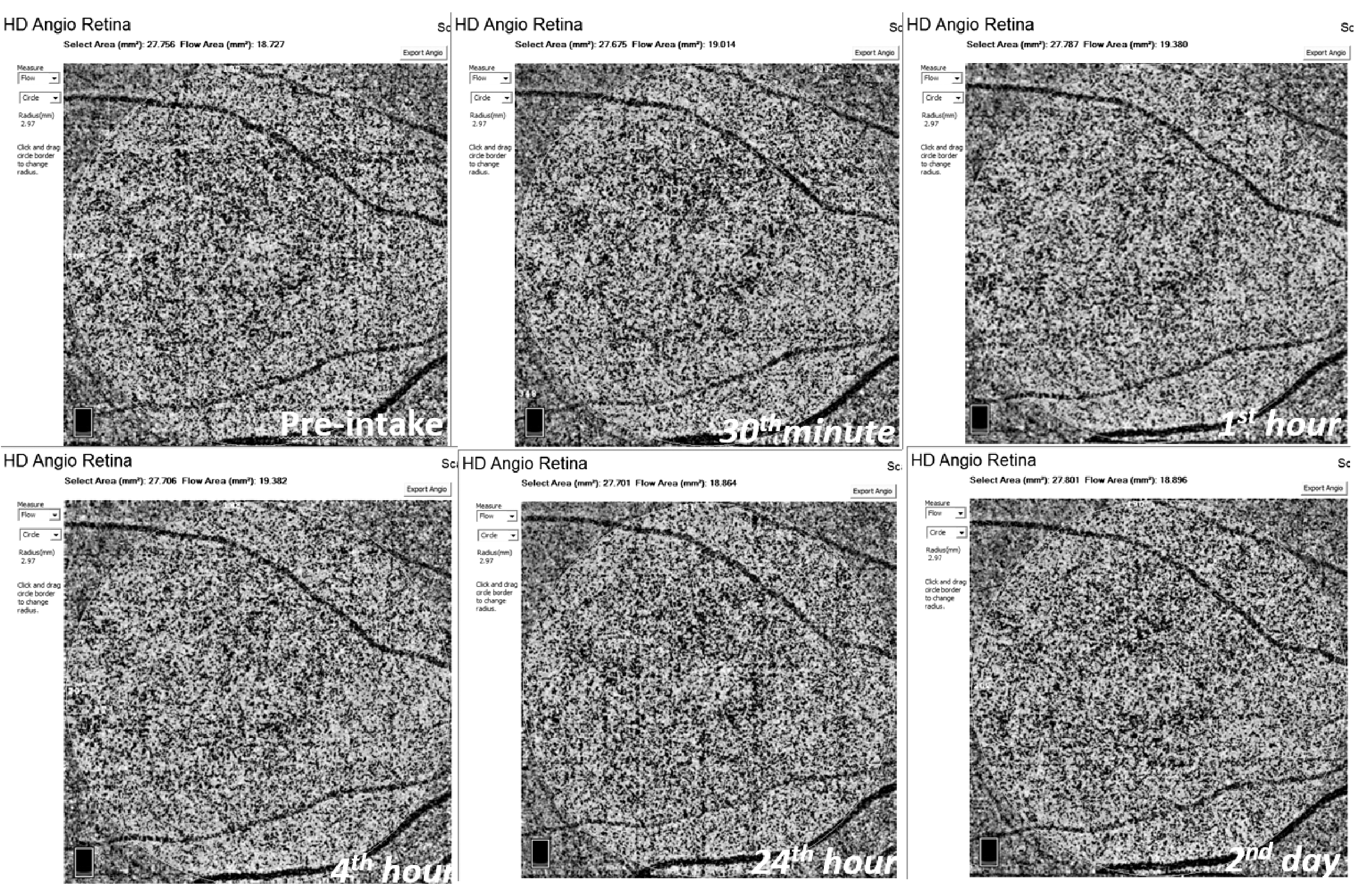
Displaying consecutive measurements of the choriocapillaris flow area (in 2.97 mm radius macular circle) using OCTA (RTVue XR Avanti with the AngioVue, Optovue, Fremont, CA). Note the increased choroidal flow area at the 30th minute, the first and fourth hour compared with preintake, and it returns to normal after 24 h. OCT, optical coherence tomography; OCTA, OCT angiography.
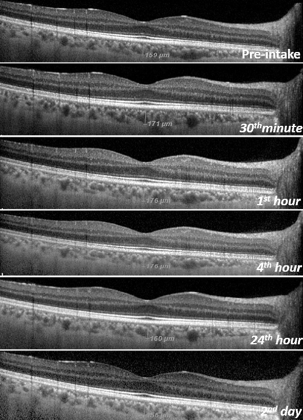
Images of semiautomatic subfoveal choroidal thickness' consecutive measurements using SD-OCT with EDI mode (SD–OCT; Heidelberg Engineering, Inc., Heidelberg, Germany). Note the increased choroidal thickness at the 30th minute, the first and fourth hour compared with preintake, and it returns to normal after 24 h. EDI, enhanced-depth imaging; SD-OCT, spectral domain OCT.
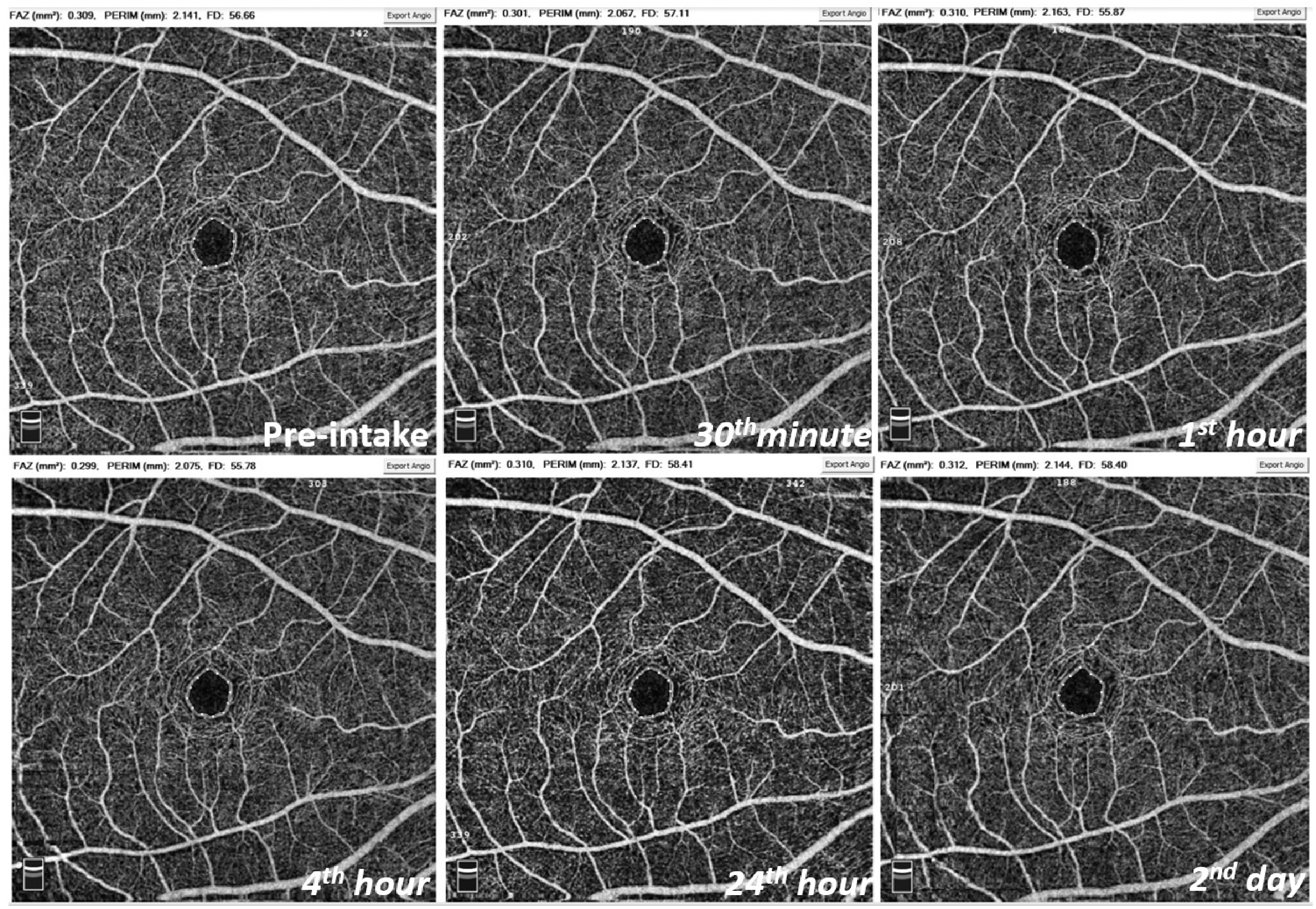
Display of the consecutive measurements for features of the foveal avascular zone: Area, perimeter (PERIM) and FD (Vessel density in a 300 μm wide region) using OCTA (RTVue XR Avanti with the AngioVue, Optovue). Note no change in foveal avascular zone (FAZ) properties.
Results and Comparisons of Consecutive Measurements After a Single Oral Dose of 5-mg Tadalafil
The mean and standard deviation of the parameters are shown.
Friedman test results to compare the difference between all consecutive measurements. Wilcoxon signed-rank test binary comparison according to preintake are shown: p1: 30th minute and preintake, p2: 1st hour and preintake, p3: 4th hour and preintake, p4: 24th hour and preintake, p5: 2nd day and preintake, p6: 3rd day and preintake, p7: 4th day and preintake, and p8: 7th day and preintake. P < 0.05 was considered statistically different and stated bold.
FAZ, foveal avascular zone; PERIM, foveal avascular zone perimeter in mm; FD-300, vessel density 300 μm from the fovea and CT, subfoveal choroidal thickness.
Although no serious side effects were observed, 3 participants had a mild headache, 2 had nonsevere nasal congestion, and 2 had mild flushing on the face (Table 3).
Observed Adverse Effect Rates in the Study Group
Serious adverse effect: heart attack, stroke, irregular heartbeat, and death.
A statistically significant positive correlation was found between the change in CT and choriocapillaris flow area (P < 0.001, r = 0.726). There was no correlation between the change in CT and AL (P = 0.660, r = 0.99), IOP (P = 0.239, r = −0.262); between the change in choriocapillaris flow area and AL (P = 0.241, r = 0.261), IOP (P = 0.544, r = −0.137).
Discussion
In the present study, we found that a single dose of oral 5-mg tadalafil causes an increase in choriocapillaris flow and CT in healthy volunteers. These effects began to appear half an hour after the tadalafil intake and were detected even after 4 h. At the end of the 24th hour, it was observed that these effects completely returned to normal. The detection of these effects in this study group indicates that tadalafil affects choroidal vascular circulation. The reason for these temporary effects has been attributed to vasodilation due to tadalafil-dependent PDE5 inhibition.
Ocular side effects of oral tadalafil are thought to be secondary to PDE5 and PDE6 inhibition.7,8 The potential adverse effects on the retinal and choroidal microvascular morphology should be evaluated. A comprehensive understanding of the mechanisms associated with these situations can help us improve our current understanding of the relationship between vascular flow and other chorioretinal diseases, such as CSCR, as well as the prevention of adverse effects observed in tadalafil.10,15
Aslan et al. 16 showed that choroidal and choriocapillaris thickness increased with the use of daily routine 5-mg tadalafil. Kim et al. 17 and Vance et al. 18 found similar results with 50 and 100 mg of sildenafil. Sildenafil is a potent and selective PDE5 inhibitor like tadalafil.18,19 Similarly, in this study, an increase in choroidal thickness, which lasted at least 4 h and returned to normal at 24 h, and did not increase again, was observed after the use of oral 5 mg of tadalafil. Kurtulan et al. 19 showed that mean cavernous artery peak systolic flow velocity increased significantly after 100 mg of sildenafil with color Doppler ultrasonography (CDU). However, they showed no effect on central retinal arterial circulation based on CDU. 19 They argued that ocular side effects seem to be the result of sildenafil that inhibits PDE6 molecules at the retinal photoreceptor level. 19 However, the vascular supply of the choroid is not limited to only the central retinal artery, 20 which limits the study to evaluate all effects of sildenafil on the retina and choroid. It is known that vascular dilatations detected by CDU are at a more macroscopic level according to OCTA measurements. More minor and sensitive differences can be detected using OCTA. Berrones et al. 10 showed that a single dose of oral 50 mg of sildenafil increases choroidal thickness and outer retinal blood flow, which they thought were probably due to sildenafil-induced vasodilation, using OCTA. Similarly, in our study, following the use of oral 5 mg of tadalafil, there was an increase in choroidal thickness. However, differently, we also found an increase in the choriocapillaris flow area and not observed any outer retinal blood flow differences. Although the same OCTA device (RTVue XR Avanti with the AngioVue, Optovue, Fremont, CA) was used in our study and the study of Berrones et al., 10 we made choriocapillaris and outer retinal flow area measurements within the 2.97 mm radius macular circle, whereas their study did not contain information about how and where they made the measurement flow area. These findings were attributed to tadalafil making a potent PDE inhibition, even at low doses. Although there were choroidal effects, retinal vascular densities were found to be unaffected in this study. This event has been attributed to the intact retinal barrier in healthy individuals. Retinal vascular density changes can be expected in diseases such as uncontrolled diabetes and hypertension that can disrupt the blood/retinal barrier.
On the other hand, there are many reported cases of CSCR associated with tadalafil in the literature.21–23 A dramatic spontaneous recovery after discontinuation of tadalafil has been reported in almost all case reports. Although there is no complete consensus, the hypothesis put forward is that, due to the increased hydrostatic pressure within the choroid, it influences the capability of the overlying retina pigment epithelium to pump fluid from the retina to the choroid. 21 In our study, the increase in choriocapillaris flow and CT supports this situation. However, CSCR was not observed in our study.
Additionally, the number of side effects (n:7) (Table 3) associated with tadalafil in this study may be related to the fact that this study was conducted with young volunteers without any systemic disease. The frequency of adverse effects may vary in chronic use, in the elderly and those with systemic disease.
The main strengths of this study are that it included a cohort of healthy volunteers with many repeated measurements (9 in total), and apart from macular vascular density measurements, the fact is that choriocapillaris, outer retinal flow, and CT were also measured. To the best of our knowledge, this article is the first prospective OCTA study which showed that tadalafil affects the choriocapillaris flow using OCTA.
This study has several limitations. The study is conducted only on a small sample size of healthy and young Caucasian male volunteers. Results may vary in the elderly, different races, and those with systemic disease. Also, due to the small sample size, a type 2 error probability is high. Variability in biometric values can affect the volume of distribution of the drug among users. Also, more potent effects may have been unobserved with a single low dose of 5 mg tadalafil. Thus, appropriate further studies are needed in a large sample size with a higher dose of tadalafil or in the elderly population with or without systemic diseases. Differently, erectile dysfunction may have organic causes such as diabetes, as well as retinal microvascular change in patients with organic erectile dysfunction. 11 Therefore, further studies are needed in these patient groups.
In conclusion, this study showed that a single dose of oral 5-mg tadalafil causes an increase in choriocapillaris flow and CT. It should be kept in mind when evaluating patients with a recent history of tadalafil use. Inhibition of PDE5 and 6 is probably responsible for this temporary effect lasting at least 4 h. To evaluate the effects of tadalafil on the retina and choroid, an OCTA assessment may be helpful.
Informed Consent
All participants were informed of the study design, and written consent was obtained from each.
Footnotes
Authors' Contributions
All authors attest that they meet the current ICMJE criteria for authorship. All authors made substantial contributions to conception and design, acquisition of data, or analysis and interpretation of data, took part in drafting the article or revising it critically for important intellectual content, gave final approval of the version to be published, and agree to be accountable for all aspects of the work.
Acknowledgments
The authors specially thank Vildan Altinoz, Fatma Telek, and Yasar Alpay for their help in acquiring figures. The article has been read and approved by all the authors.
Author Disclosure Statement
The authors report no conflict of interest for this research. The authors alone are responsible for the content and writing of this article. All authors certify that they have no affiliations with or involvement in any organization with any financial interest or nonfinancial interest in the subject matter or materials discussed in this article.
Funding Information
No funding was received for this article.