




Abstract
Objective:
In the treatment of severe cases of bacterial keratitis, conventional eye drops containing antibiotics should be applied daily and very frequently. The aim of this study is to develop low-dose high-effect formulations with the prepared nanostructured lipid carrier (NLC) formulations to reduce antibiotic resistance and increase patient compliance.
Methods:
NLC formulations were loaded with besifloxacin HCl (BHL) and the besifloxacin HCl: sulfobutyl ether beta-cyclodextrin (SBE-CD) complex. Positive charge was gained with chitosan, and corneal permeation and resolubility were increased with SBE-CD. In vitro characterization studies, permeability studies, and cytotoxicity and ex vivo transport studies were carried out.
Results:
In this study, it was found that SBE-CD increased BHL's solubility by 8-fold based on phase solubility studies. The optimized NLCs were small in size (13.63–16.09 nm) with a low polydispersity index (0.107–0.181) and adequate BHL drug loading efficiency. In vitro release studies showed that formulations were released approximately for 8 h and at levels over the minimum inhibitory concentration of Pseudomonas aeruginosa and Staphylococcus aureus. NLC formulations had a better corneal permeation rate than the marketed product during 6 h of ex vivo studies.
Conclusions:
According to in vitro and ex vivo data, it was determined that the most favorable NLC formulation was the formulation containing BHL/SBE-CD that was covered with chitosan. It has the highest drug loading capacity and one of the highest ex vivo corneal passage levels, along with desired drug release. The formulation containing BHL/SBE-CD and chitosan can be a potential alternative for the treatment of bacterial keratitis.
Introduction
The eye, generally divided into anterior and posterior chambers, is a complex structure with each of its parts vulnerable to various diseases. It is safeguarded from foreign elements by its anatomically and physiologically unique barriers, namely the tear film, cornea, conjunctiva, and blood–aqueous and blood–retina barriers, causing ophthalmic drug delivery to be a major challenge.1,2 Poor ocular bioavailability and short retention period of drugs on the eye can result in drugs not achieving their intended effects without increasing doses to levels that are high enough to potentially enter the systemic circulation and cause side effects. 3 Thus, novel drug delivery methods have been developed to increase ocular bioavailability.4–6
Acute infections of the cornea caused by bacteria, fungi, viruses, or amoeba are called keratitis, the most common being bacterial infection, causing 90% of all cases. 7 Unless the infection source is eliminated, various eye pathologies and ocular morbidities such as corneal scarring and vision damage might happen. While the ocular surface is often exposed to external pathogens, keratitis is a rare condition. Physical barriers are formed by the eyelids, corneal epithelium, and tear film.8,9 Pseudomonas aeruginosa (PA) and Staphylococcus aureus (SA) are commonly seen pathogen sources of keratitis. 10
Fluoroquinolones (FQs) are the antibiotic group of choice in treatment of bacterial keratitis, which are also affected from the spreading antibiotic resistance. Therefore, the newest addition to FQs, besifloxacin, the first topical chlorofluoroquinolone, was chosen for ocular disease treatment as its unique molecular structure offered enhanced antibacterial activity. Besifloxacin proved potent in vitro against a wide array of bacteria that are resistant to both other fluoroquinolones and antibiotics. Potent against Gram-positive and negative bacteria alike, 0.6% besifloxacin suspension was approved by the FDA in 2009 for treatment of bacterial conjunctivitis.11–13
Nanostructured lipid carriers (NLCs) are formed by solid lipid matrices containing liquid lipid and are a new breed of solid lipid particles. Conventional solid lipid particles have poor drug loading, are unstable, and leak loaded drugs as the lipid structure changes overtime, but NLCs do not share these drawbacks. NLCs are highly biocompatible as they are made of biodegradable low-toxicity lipids and can be produced at scale in the industry. Furthermore, their solid outer surface prevents coalescence (merging droplets), as per observations in emulsions.14,15
Literature review has laid out that NLC formulations increase precorneal retention time and therefore drug permeation through the cornea. 16 It has also been pointed out that permeation enhancers added to the NLC structure increase the corneal passage. 17 In addition, electrostatic interactions of cationic polymers add to the mucoadhesive effects, leading to enhanced drug transmission through the cornea with NLC preparations containing cationic polymers. 18
In the light of the information given above, this study was planned to take a step further than our previous study, in which besifloxacin HCl-loaded inserts were developed and promising results were obtained. 19 It was also desired to take advantage of the NLCs. 20 In this study, 4 different formulations were prepared and examined in vitro and ex vivo to analyze the results of increasing the solubility of besifloxacin by forming a complex with cyclodextrin and coating the NLC surface with chitosan, a positively charged mucoadhesive polymer.
By increasing the solubility, it is aimed to increase corneal permeation. Another goal is to increase the residence time on the eye surface by coating NLCs with chitosan and to show that chitosan-coated NLCs also have suitable properties. The current study is significant in paving the way toward an alternative to conventional eye drops.
Methods
Materials
Besifloxacin HCl, sulfobutyl ether beta-cyclodextrin, Tween 80, chitosan, Dulbecco's modified Eagle's medium/nutrient mixture F-12 (DMEM/F-12), fetal bovine serum, penicillin–streptomycin solution, sodium dodecyl sulfate (SDS) and tryptic soy broth and thioglycollate broth were purchased from Sigma-Aldrich. Compritol ATO 888 and Gelucire 44/14 were kindly offered by Gattefossé.
All solutions used in the study were of high-performance liquid chromatography (HPLC) grade and supplied by Sigma.
Design of besifloxacin HCl-carrying NLC formulations
The following NLC formulations were prepared (Fig. 1) and characterized (Table 1):
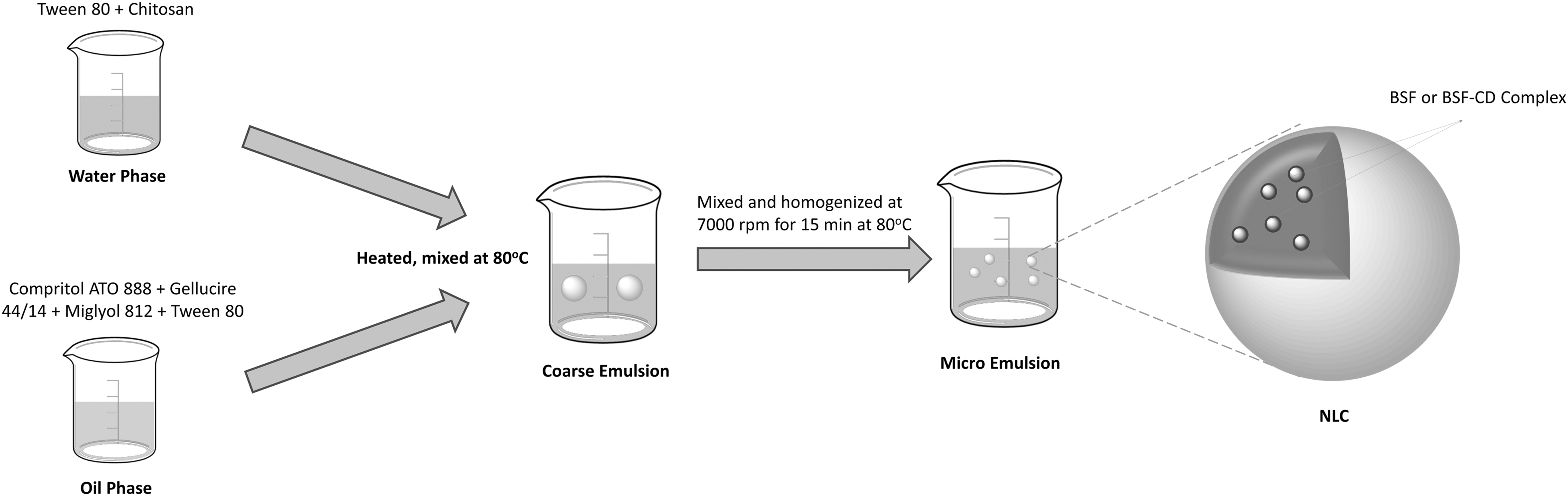
Preparation of NLCs. NLC, nanostructured lipid carriers.
Besifloxacin HCl-Containing Nanostructured Lipid Carrier Formulations
NLC, nanostructured lipid carrier.
Besifloxacin-only formulation (BSF)
Besifloxacin HCl-SBE cyclodextrin complex formulation (BSF-SBE)
Besifloxacin HCl and chitosan formulation (K-BSF)
Besifloxacin HCl-SBE cyclodextrin complex and chitosan formulation (K-BSF-SBE)
NLC formulations were prepared using a combination of emulsification–ultrasonication. 21 For preparation of NLC formulations containing BHL or the BHL/SBE-CD complex, the water phase was obtained by adding Tween 80 (for all formulations) and chitosan (for K-BSF and K-BSF-SBE) into water and then mixing at 80°C. Although chitosan is normally insoluble in water, it was made soluble by adding 0.1% acetic acid. 22
For the oil phase, Compritol ATO 888, Gelucire 44/14, Tween 80, and Miglyol 812 (for all formulations) were mixed at 80°C in a beaker (BHL was added to the oil phase, and the BHL/SBE-CD complex was added to the water phase). Then, when the oil phase was mixed at 7,000 rpm in a mechanical mixer, the water phase was added dropwise onto the oil phase and mixed for 15 min.
Formation of cyclodextrin–drug complex
Phase solubility studies
Loftson and Brewster's methods were utilized to carry out phase solubility studies. 23 Increasing concentrations (0–10 mM) of the SBE-CD solution were added into a fixed amount of BHL.
Mixtures were then passed through a 0.22-μm filter membrane, and HPLC was utilized to determine the amount of BH in the supernatant. A C8 column (250 × 4.6 mm, 5 μm) with a mobile phase of methanol:acetonitrile:buffer (pH 3) (25:25:50 v/v/v%) at a flow rate of 1 mL/min was used to carry out the HPLC (Agilent 1200) analysis. The BH detection wavelength was at 297 nm. Columns were kept at 25°C while 20 μL of the sample was injected. 24 Experiments were conducted in triplicate (n = 3). BHL concentration as part of the SBE-CD concentration was illustrated to create the phase solubility diagram. 25
Procedures for validating whether the analysis is accurate, specific, and reproducible under specified conditions are collectively called analytical method validation. In our testing of the HPLC method and its results, linearity, precision, sensitivity, accuracy, and repeatability parameters were of significance. 24
The diagram created was assessed based on the Higuchi and Connors
26
classification that consists of AP, AL, AN, BS, and BI diagram models. Complexation efficacy and the complex stability constant were calculated, referencing equations defined in an earlier study.
23
where S indicates intrinsic BHL solubility, which is 0.17 mM, and Slope indicates the linear regression's slope of the phase solubility diagram.
Complexation efficacy was calculated in accordance with Eq. (2).
23
Preparation of the cyclodextrin–drug complex
The freeze–drying method was used to prepare cyclodextrin–drug complexes. 27 Each of the SBE-CD and BHL substances (equal molar ratio of 1:1) was dissolved either in water or methanol, respectively. It took 24 h for the SBE-CD and BHL solutions to be mixed before methanol was evaporated, then the sample was lyophilized. Fourier-transform infrared spectroscopy and differential scanning calorimetry were used to check whether development of inclusion complexes succeeded.
Characterization studies
Particle size and zeta potential
Particle size and distribution have a great influence on the stability, solubility, release rate, and biological behavior of the final product. Particle size, polydispersity index, and surface charge (zeta potential) of the prepared NLCs were determined by dynamic light scattering at 25°C (Malvern Zeta Sizer Nano Series Nano ZS instrument).
Nanoparticles obtained before and after the lyophilization process were suspended in Milli-Q water and then measured. The measurements were performed in triplicate (n = 3).
Drug loading efficiency
Drug loading efficiency of NLC formulations was assessed after being lyophilized. Ten milligrams of each lyophilized formulation (n = 3) was weighed out and then transferred into 2.5-mL Eppendorf tubes. One milliliter of ethanol was then added and the mixture was vortexed for 5 min, followed by addition of 1 mL of 0.1% acetic acid. Ultrasonication at 10% power for 1 min was done, following partial dispersion. The end mixture was diluted with buffer at a 1–4 ratio, followed by filtration through a 22-μm pore size filter. Drug loading efficiency was determined using the HPLC method specified in phase solubility studies.
Release study
An in vitro release study on NLC formulations was conducted by utilizing a dialysis membrane (Sigma; D9277) with a molecular cutoff value of 14.000 MW. Roughly 10 milligrams of lyophilized formulation was dispersed in 1 mL of pH 7.4 PBS before being positioned on the dialysis membrane. It was then put in a 15-mL centrifuge tube with 10 mL of PBS 7.4. Drug release was determined using the HPLC method specified in phase solubility studies.
NLC stability
At the end of the 30th day, particle size and zeta potential of the prepared NLCs were measured using the Malvern Zeta Sizer Nano Series Nano ZS instrument. The measurements were performed in triplicate (n = 3).
Ex vivo
Ex vivo corneal transport studies were conducted using sheep corneal tissue locally sourced from butchers. Formulations were tested against commercial preparations to assess passage and involvement of BHL through corneal tissue. Transport studies were conducted with n = 3. Five different formulations were tested, with one of them being a commercially available preparation (Besivance®). The “Ussing Chamber” transport system was used for transport studies. 28
Tissue transport cells were positioned at acceptor and donor sides, with the donor side facing the outer corneal surface and contact area measuring 0.34 cm2. Four milliliters of PBS (pH 7.4) was added as the buffer on the acceptor side, and the donor side had 0.005% BHL containing 4 mL of formulation added to it. The system was kept at a constant 32°C in a light-proofed container without adding any stirring element. Samples were drawn from the recipient cell into vials after 2 h, and the BHL drug loading capacity was assessed with HPLC (Agilent 1200).
Cytotoxicity study
Cell cultures were exposed to prepared formulations to measure cytotoxicity, which were then assessed for cell viability by 3-(4,5-dimethylthiazol-2-yl)-2,5-diphenyltetrazolium bromide (MTT; Sigma, M5655) assay.
MTT cytotoxicity assessments were conducted using human retinal pigment epithelial cells (ARPE-19) (ATCC CRL-2302). The ARPE-19 culture medium was prepared by supplementing DMEM/F-12 with 10% volume per volume of fetal bovine serum and 50 μg/mL streptomycin and 50 U/mL penicillin. Ninety-six-well plates were loaded with ARPE-19 cells, with 5,000 cells per well. Following an overnight incubation to let cells stabilize, media were removed off the plates and diluted formulations were added and incubated for 4- and 24-h periods.
Cytotoxicity studies were conducted with both empty and BHL-containing formulations to see whether the carrier system itself had any effects. Drug-containing samples were prepared to contain 0.1 mg of BHL, while drug-free samples were arranged to include the corresponding amount of nanoparticles in 2 mL of water. One milliliter of it was topped up to 10 mL using cell medium. After the cells were exposed to formulations, 25 μL of MTT (5 mg per mL) solution was added to the plates.
After 4 and 24 h of incubation, 200 μL of DMSO was transferred to the wells for dissolving formazan crystals, after which the plates were incubated for another 4 h. Absorbance was evaluated through the ELISA plate reader at 570 nm to assess cell viability. The absorbance value was assumed to be 100% for the control group. If cell viability was measured as less than 70%, the tested sample was assessed as cytotoxic. 29
Permeability studies
ARPE-19 cells were again utilized for in vitro permeation studies. ARPE-19 cells were kept in the medium previously used for cytotoxicity tests. Cell seeding was done on cell culture inserts, with the density being 15 × 104 cells per insert, after which they were put into 12-well plates. Each well had 1 mL of complete medium, and 0.5 mL of it was present with cells on the inserts for cell growth. Plates were left to incubate under 5% CO2 at 37°C. Every 2 days, the medium and wells were swapped for fresh ones.
Following 15–21 days of seeding, the obtained cell monolayer's integrity was examined for its transepithelial resistance (TEER) value using the Millicell® ERS Voltohmmeter. Permeation studies were conducted between 200 and 250 Ωcm2 of measured resistance; 0.5 mL of produced formulations (containing 70 mM of BHL) dispersed in PBS or marketed formulation was added to the apical surface of cell monolayers, and 1 mL of PBS was added to the basolateral surface.
Wells were put on a shaker set to 30 rpm to incubate for 2 h at 37°C. At certain points, samples were taken from the basolateral side to be assessed with HPLC.
Statistical analysis
Statistical analysis was conducted with GraphPad Prism 5.02 (La Jolla) by either utilizing the unpaired 2-tailed Student's t-test or one-way ANOVA with Tukey's multiple comparison test (illustrated as ***P < 0.001, **P < 0.01, *P < 0.05, and ns = not significant).
Results and Discussions
BHL is a commercially available preparation used in treatment of keratitis and conjunctivitis. Commercial forms are used 3 times a day. BHL-loaded NLC formulations that we have developed in this study enabled usage of lower doses of BHL, which can stay above the minimum inhibitory concentration (MIC) value. These formulations also helped the drug to pass physiological barriers and regulate the drug release. 30
Stability and thermal properties of the cyclodextrin–drug complex
Phase solubility studies are usually the preferred method for determination of the efficacy of CD drug complexation on drug solubility.23,31 The 1:1 drug/CD complex is the most common type of association where a single drug molecule is included in the cavity of one CD molecule, with a stability constant K1:1 for the equilibrium between the free and associated species. When solubility diagrams (Fig. 2) were inspected, it was observed that SBE-CD had a linear increase. In accordance with the defined methodology, the diagram of SBE-CD was classified as “AL” type.

Besifloxacin HCl phase solubility diagram.
From the straight line of SBE-CD (r 2 = 0.9309), the slope was determined as 0.5911. The complexation efficiency (EC) was 1.68 and stability constant (KS) was 9,922 M−1. Upon literature review, it has been determined that the stability constant between 100 and 10,000 M−1 is the ideal value for formation of the drug:CD complex. However, with the AL-type solubility curve determined by examining the diagram, the drug:CD complex ratio was decided to be 1 mM:1 mM.27,32
However, it was determined that solubility of BHL in water increased 8-fold in the presence of SBE-β-CD. When the literature was examined, it was determined that SBE-B-CD significantly increased the solubility of drugs having poor solubility and accordingly increased the ocular efficiency of drugs.33,34
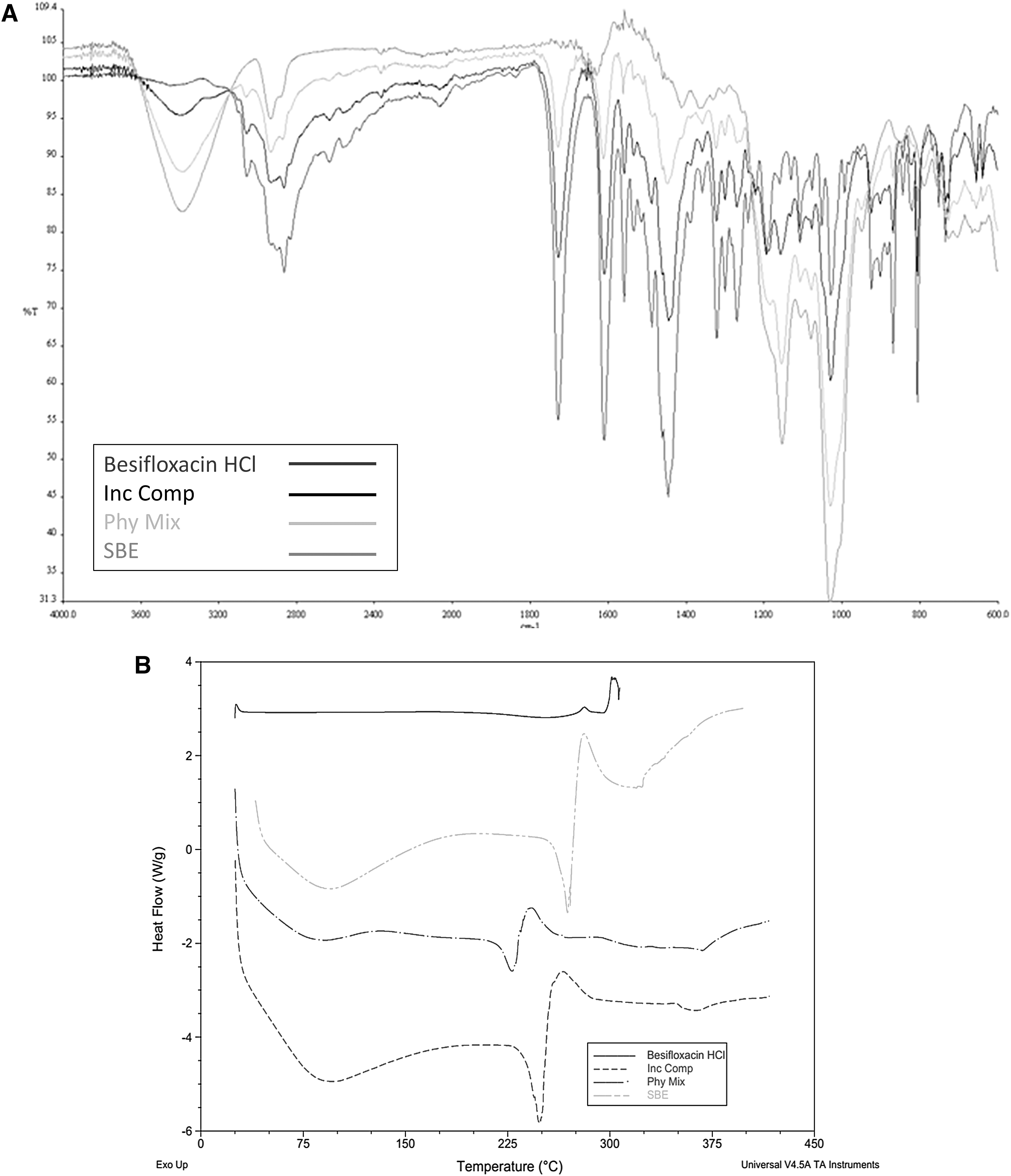
Using the DSC thermograms of BHL, CDs, BHL/SBE-CD, and physical mixture complexes were compared (Fig. 3A); BHL's endothermic peak (melting point) at 321.5°C was missing, pointing to a lack of crystalline BHL. The BHL/SBE-CD complex also lacks the melting endotherms of BHL, validating complex formation. 6 When FTIR outputs were assessed for confirmation of the BHL/SBE-CD complex, 3 peaks (2,864, 1,726, and 1,446 cm) normally present on the base drug were absent in the complex utilizing freeze–drying (Fig. 3B). In addition, SBE-CD peaks were present, further validating BHL/SBE-CD complex formation (Fig. 3B). 27
Particle size and zeta potential
When developing an ocular drug delivery system, particle size proves itself to be a key measure to assess irritation and discomfort. Sizes beyond 10 μm are poorly tolerated by eyes. Larger particles lead to tearing, thus the drug can be washed away from the eye surface, diminishing contact duration of the drug with the conjunctival sac and negatively impacting bioavailability.35,36 As a thumb rule, smaller particle sizes can enhance drug penetration and uptake while lowering irritation.
Four separate NLC formulations were arranged and categorized for PDI, zeta potential, and size. PDI was observed to be less than 0.25 for all tested samples, attesting to low polydispersity of the distribution.
Upon analysis of particle size results, it was observed that NLC particle sizes varied depending on the water phase. In 2 of the formulations, despite using chitosan, no significant change in particle size was detected, and 0.5% fat phase and 3% surface active material containing formulations yielded desirable particle sizes.
Decreasing lipid ratios would enable more surfactant on newly produced surfaces, resulting in formation of smaller sized particles with hot microemulsion. This shows that lipid increase causes decreased emulsification efficacy and aggravates particle agglomeration.37,38 The reduction in particle size minimizes irritation to ocular tissues, increases precorneal residence time, and minimizes dose-related toxicity.
Furthermore, decreasing the particle size increases the corneal permeability of the drug. The ability of particles smaller than 200 nm to escape from phagocytosis causes them to accumulate more in the areas of inflammation, which gives this formulation an advantage. 39 Upon literature review, decrease in particle size was found to reduce ocular irritation and increase corneal passage. 40 Increased lipid concentration also increases molten lipid phase viscosity.
PDI increase was also proportional to lipid content, likely due to decreased homogenization efficiency caused by changes in viscosity. The lack of significant increase in chitosan formulations is due to the concentration of chitosan used. A study in the literature found that particle diameters of NLC formulations vary due to increased chitosan concentration. 18
Zeta potential is a measure of particle charge and electrostatic repulsion, thought to be an indicator of physical stability and mucoadhesive properties. It is the measured potential at shearing surfaces, displayed in millivolts. In formulations, the electrical charge in terms of zeta potential varied between −0.84 ± 0.09 and +33.8 ± 1.95 mV (Table 2). The zeta potential value increased when chitosan was added into the formulation.
Nanostructured Lipid Carrier Classification by Particle Size, PDI, and Zeta Potential
Measurements were performed in triplicate (as mean ± SD for 3 freshly prepared formulations).
The increase observed in zeta potential values of formulations hinted that the polycationic chitosan could be adsorbed to the particle surface. However, since the ocular surface has a negative charge, formulations with positive zeta potential have much higher adhesion to the ocular surface than those with negative zeta potential. 41
Physical stability of NLC formulations is one of the key points of evaluation. The most important problems of NLC formulations are that they are heterogeneous and thermodynamically unstable. For this reason, NLCs tend to lose their physical stability during storage. 42 The NLC formulation's stability study was done by measuring the particle size, PDI, and zeta potential at 4°C. Four NLC formulations that were kept at room temperature yielded similar particle size distribution results to fresh NLCs (Fig. 4). When kept at +4°C, a physical stability review of the formulations produced did not find meaningful signs of instability.
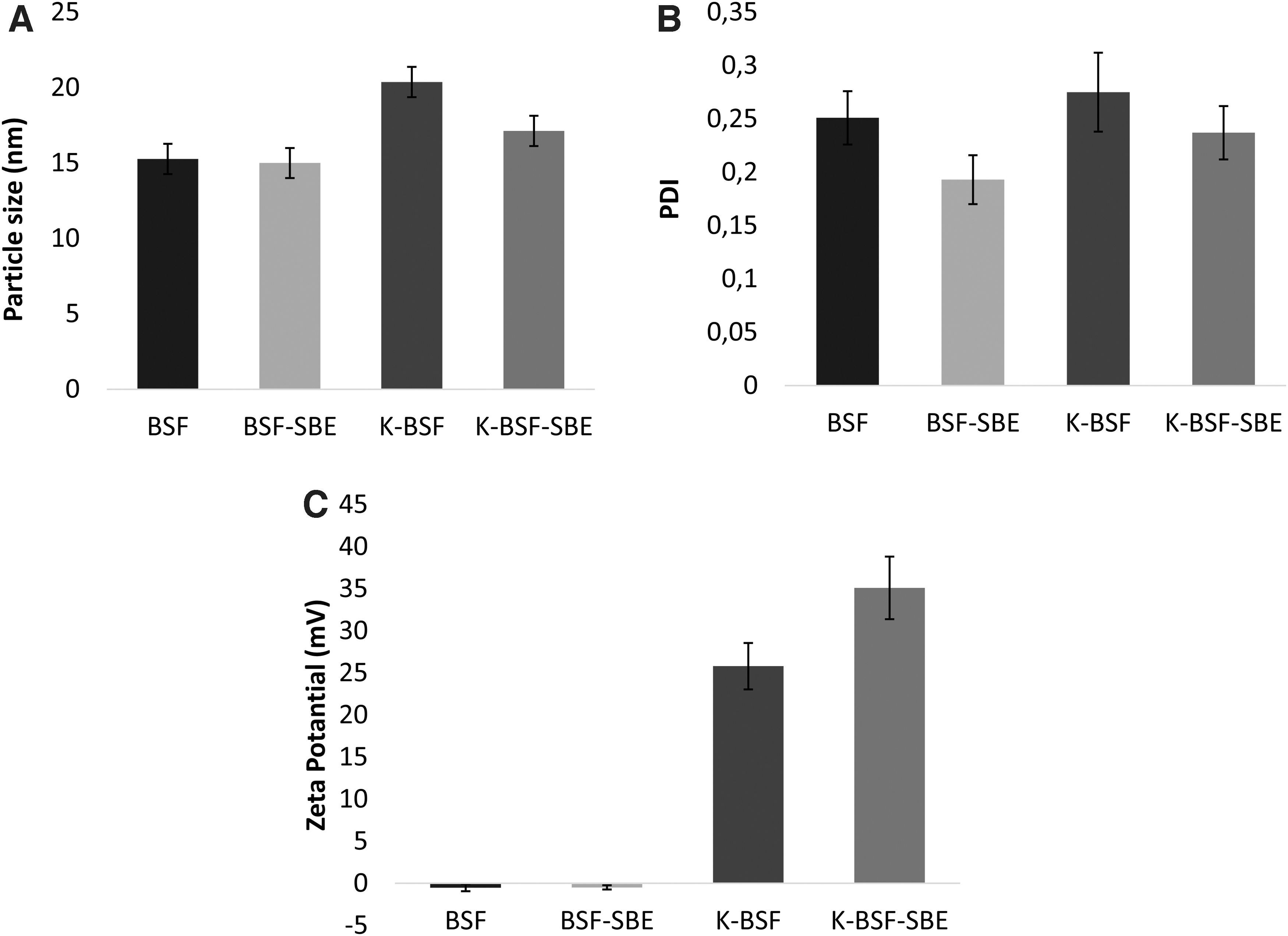
NLC characterization after storage at 4°C for 30 days.
Drug loading efficacy
The HPLC method was used to determine the percentage of drug loaded in NLC formulations, and findings are presented in Fig. 5. Upon inspection, formulations containing cyclodextrin complexes were found to have higher drug loading percentages (P < 0.0001). Literature review suggests that formulations containing cyclodextrin–drug complexes exhibit higher drug loading capacities when compared with drug-only formulations.43,44 When cyclodextrin complex-containing formulations were compared with each other, the chitosan coating was seen to increase drug loading (P < 0.05).
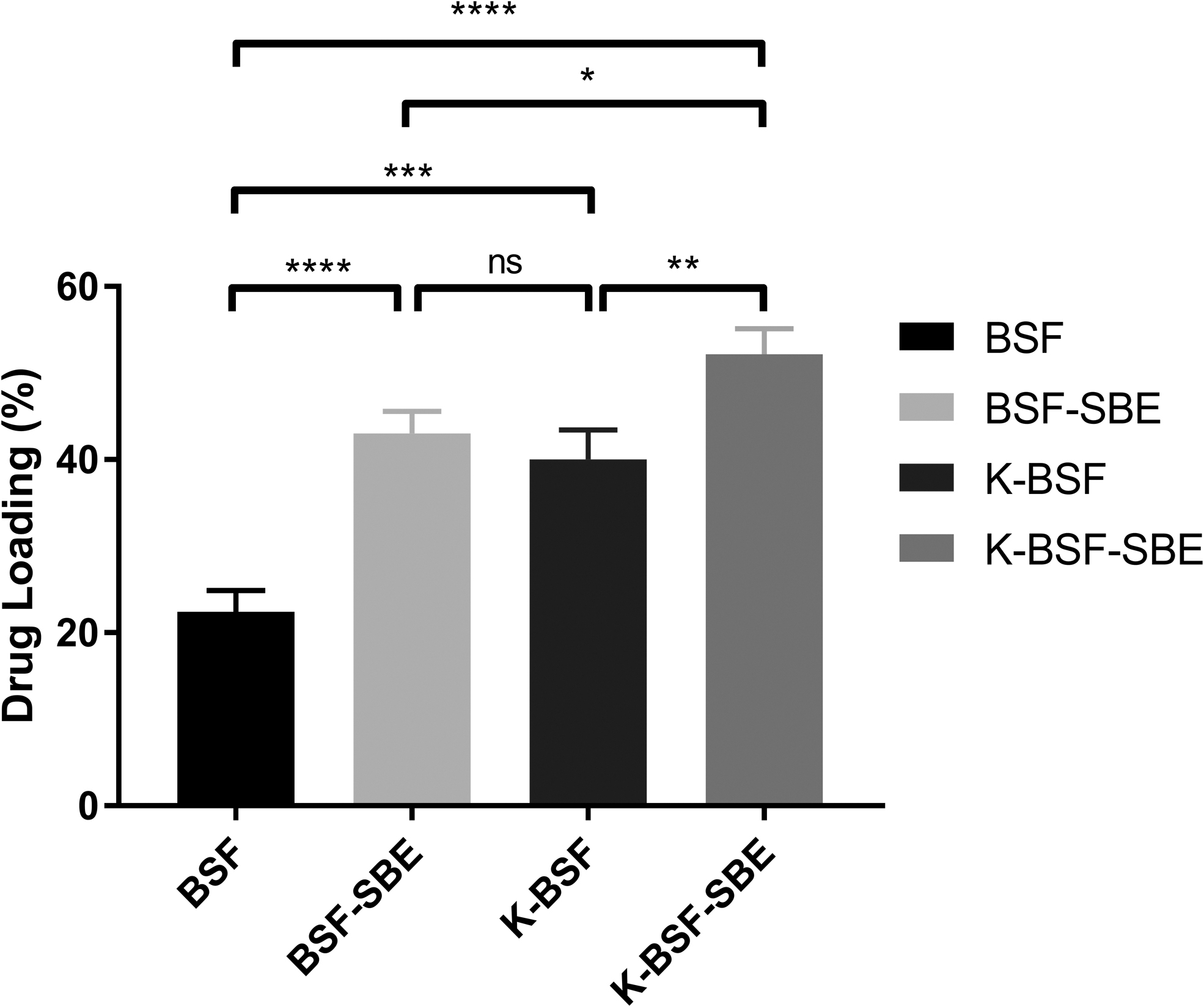
Drug loading given in percentages for besifloxacin HCl. The measurement was performed in triplicate (as mean ± SD for 3 freshly prepared formulations). Statistical significance was obtained with unpaired one-way ANOVA with Tukey's multiple comparison test (illustrated as *P < 0.05, **P < 0.01, ***P < 0.005, ****P < 0.0001, and ns = not significant).
Overall, formulations had between 20% and 50% drug loading. This was thought to be due to intrinsic qualities of NLCs. Drug loading to these systems made of a mixture of solid and liquid lipids is difficult due to structural imperfections of the crystals forming said systems. 45 In addition, it is documented that as the lipid phase decreases, drug loading efficacy also decreases.46,47
Drug release
When particle size results were analyzed, it was seen that NLC particle sizes varied depending on the water phase. In 2 of the formulations, despite using chitosan, no significant change in particle size was detected, and 0.5% fat phase and 3% surface active material containing formulations yielded desirable particle sizes.
The in vitro BHL release from formulations using a dialysis membrane during a period of 8 h is shown in Fig. 6A. All formulations were seen to demonstrate similar release properties, and at the end of the 8-h period, they had all finished releasing BHL, caused by particle sizes of the formulations. Upon literature review, decrease in the particle size correlated with the NLC release period. 48
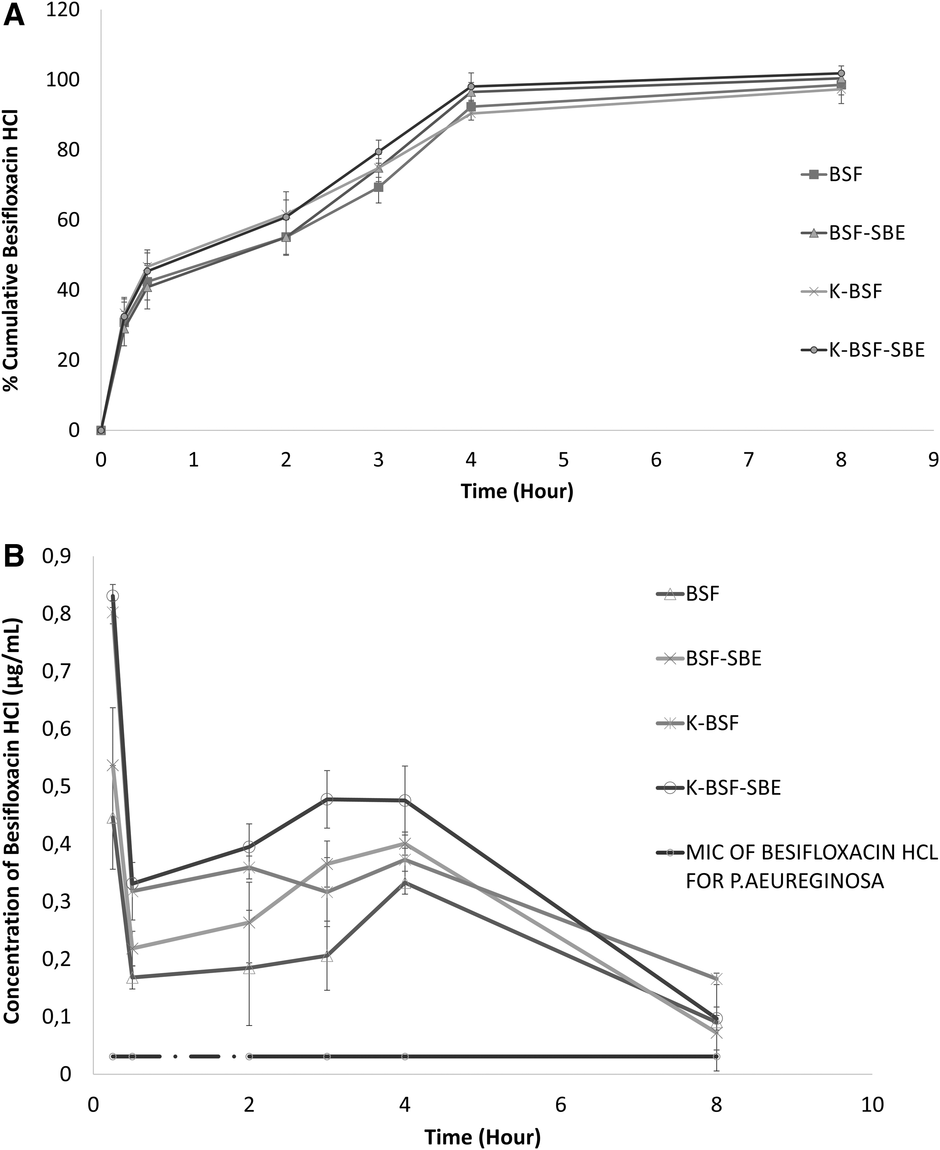
All formulations were found to exhibit controlled release through the 8-h period. Release studies found that when the first half-hour was examined, 42%–46% of the formulations were seen to release the drug. This initial burst effect may be due to drugs loaded into the outer shell of NLCs. The initial burst effect may provide an advantage for treatment of bacteria-induced diseases. Thanks to the burst effect, high doses of drugs that can kill bacteria can be administered.
When the literature is examined, it is seen that ocular eye drops for treatment of diseases such as keratitis are applied in high doses to provide therapeutic efficacy on the first day.19,49 This is followed by slow release of the drug trapped in the core of the NLC. When the literature is examined, it is seen that there is a burst effect first and then a controlled release of the drug from the NLC formulations.50,51 Generally, when release profiles of formulations were compared, chitosan-containing formulations had higher release rates compared with those that did not.
The MIC of BHL for PA was measured at 0.031 μg/mL. 52 As illustrated in Fig. 6B, all examples had BHL concentrations above the MIC, meaning all formulations were effectively delivering the drug for the entirety of the release duration.
Cytotoxicity study
BSF-, K-BSF-, BSF-SBE-, and K-BSF-SBE-containing and empty NLC formulations were used in cytotoxicity studies (Fig. 7). Upon evaluation of cell viability findings, cell death at significant levels (P < 0.001) was seen in formulations with empty NLC, either neutral or containing chitosan. This observed toxicity seems to be dependent on dosage. Increase in dilution brought toxicity levels closer to the control group. Furthermore, from 1/2 to 1/16 dilution levels, cell viability results stayed over 70% when compared with the control.
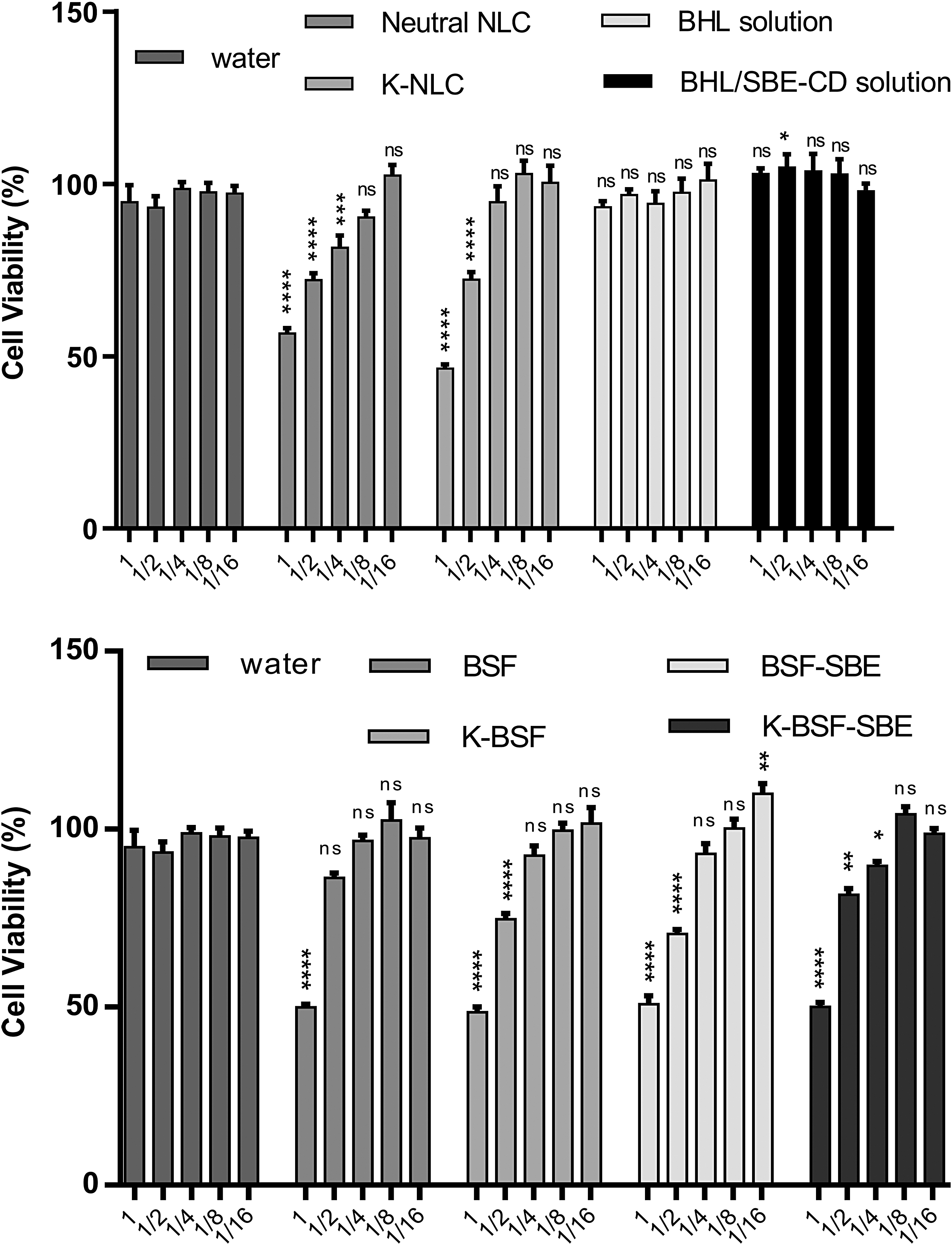
Toxicity studies performed by exposing ARPE-19 cells to BSF, K-BSF, BSF-SBE, K-BSF-SBE, and BSF-free (neutral NLC and K-NLC) formulations, BHL and BHL/SBE-CD solutions, and their following dilutions. Cell viability is stated in percentages. Data are indicated as mean ± SEM for n = 5. Two-way ANOVA analysis with Bonferroni post-test (water as control) yielded a statistical value of note (illustrated as *P < 0.05, **P < 0.01, ***P < 0.005, ****P < 0.0001, and ns = not significant).
It was observed that BHL and BHL/SBE-CD solutions did not show cytotoxicity, and nanoparticles without BHL showed little toxicity at high concentration and this toxicity disappeared with dilution. When nanoparticles without BHL were compared, it was determined that the cytotoxicity of nanoparticles containing chitosan was lower.
It is known that chitosan shows concentration-dependent toxicity in some cell lines. Vllasaliu et al. showed that chitosan nanoparticles have lower cytotoxicity than the chitosan solution on the Calu-3 cell line. 53
Permeability studies
In permeability studies using the ARPE-19 cell line (Fig. 8), it was observed that permeability of BHL decreased in the presence of chitosan. While this decrease was statistically significant in formulations without SBE (P < 0.01), it was not observed for SBE-containing formulations. Permeability decreased slightly with addition of SBE to the formulation, but this decrease was not statistically significant.
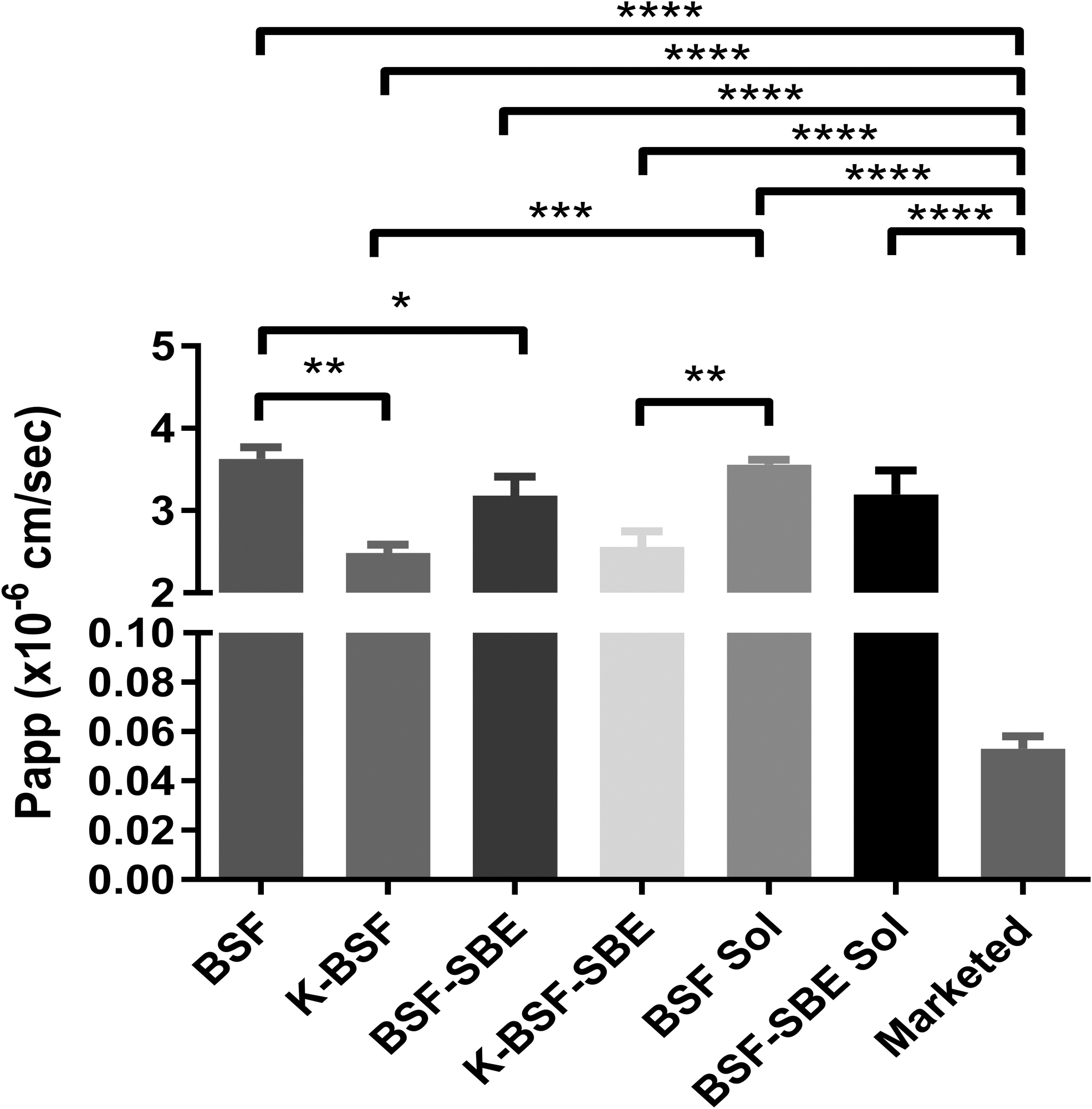
Permeability of BSF, K-BSF, BSF-SBE, and K-BSF-SBE formulations, BHL and BHL/SBE-CD solutions, and marketed formulation. The measurements were performed in triplicate (as mean ± SD for 3 freshly prepared formulations), illustrated as *P < 0.05, **P < 0.01, ***P < 0.005, ****P < 0.0001.
Compared with the BHL solution, K-BSF (P < 0.001) and K-BSF-SBE (P < 0.01) formulations were significantly reduced. The permeability of BHL from the market preparation was statistically significantly (P < 0.001) lower than all other samples. The most important reason for this dramatic difference is thought to be the high density and high concentration of the marketed formulation.
The Papp values may allow rating the permeable compounds as poorly, moderately, and highly permeable (Papp <1 × 10⁻⁶, 1–10 × 10−6, and >10 × 10−6 cm/s, respectively). 54 Considering this, it was determined that the BHL-containing NLC formulations were moderately permeable in ARPE-19 cell lines (2–4 × 10⁻⁶ cm/s).
Ex vivo results
Transport studies on sheep corneal tissue were conducted with NLC formulations along with a commercially available drug (suspension) within a 6-h duration, and the tissue was not impaired. 55 Findings (Fig. 9) and permeability coefficients (Papp, cm/s) (Table 3) demonstrated that NLC formulations provide an increased drug concentration over Besivance. Upon literature review, similar findings were often reported.
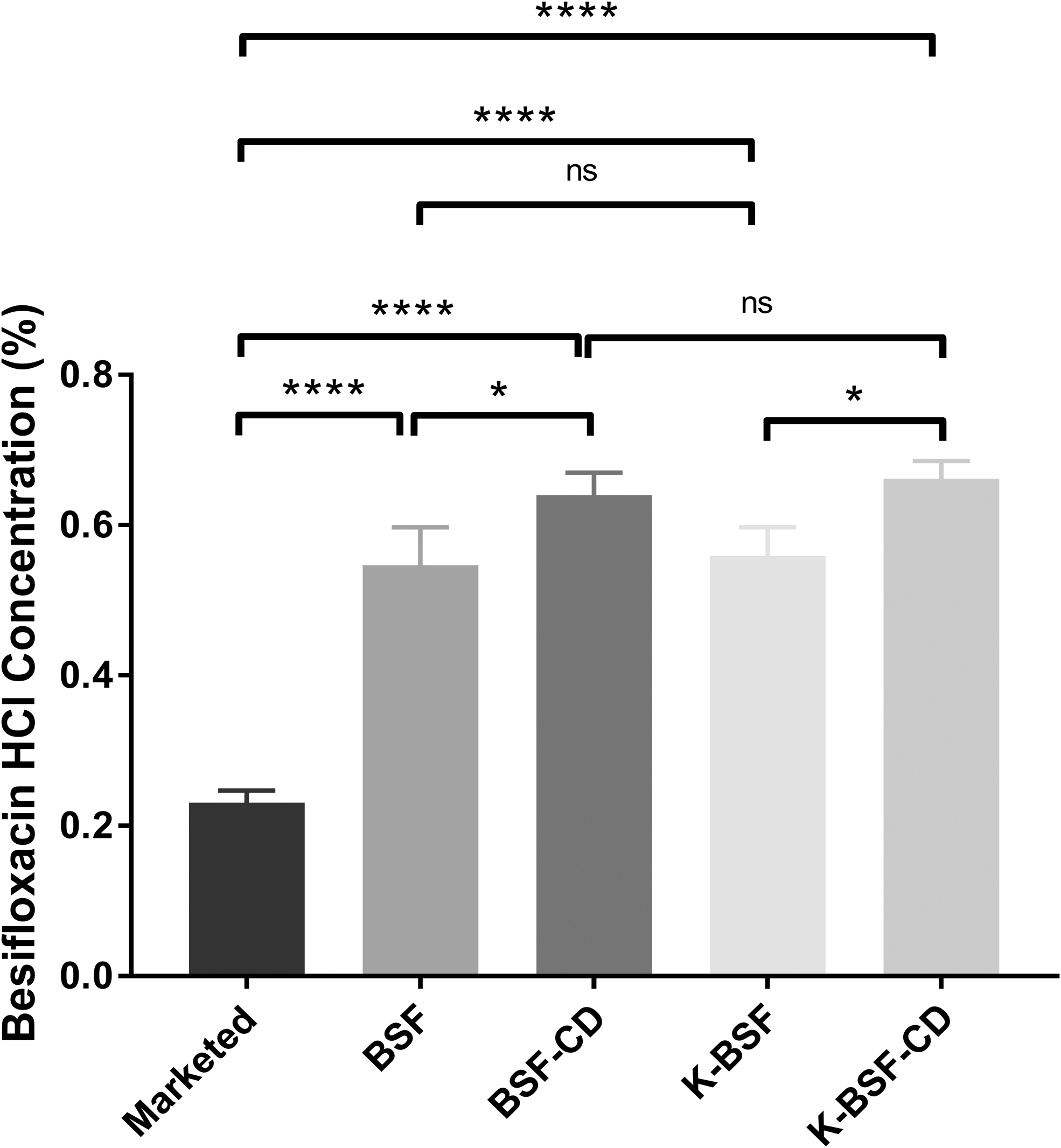
Cumulative corneal transport of formulations and a commercially available drug. Statistical significance was found using unpaired one-way ANOVA with Tukey's multiple comparison test (illustrated as *P < 0.05, ****P < 0.0001, and ns = not significant).
Apparent Permeability Coefficients (Papp, cm/s) for Ex Vivo Transport Studies
According to an ex vivo study conducted by Üstündağ-Okur et al., their clarithromycin-loaded NLC formulations reported higher corneal passage when compared with commercially available alternatives. 47 Another ex vivo study on timolol maleate- and brinzolamide-containing NLC formulations again demonstrated higher corneal passage over suspensions with the same active ingredients. 56
Furthermore, in our study among the developed formulations, BSF-SBE and K-BSF-SBE (containing CD) demonstrated the highest transportation ability, which can be credited to an increase in drug solubility due to CD being a part of the formulation (P < 0.05). In addition, Yavuz et al. 57 found that increase in drug solubility is a factor that positively affects corneal passage. In the ex vivo study conducted by Polat et al., cyclodextrin-containing formulations were found to have higher corneal passage rates compared with those not containing cyclodextrin.
Conclusions
Treatment of ophthalmological diseases remains a difficult task, despite the advances in drug research and technology, due to the innate protective barriers in eyes, causing severe limitations in transporting drugs to their target locations without complications. As a novel drug delivery system, NLC was prepared to circumvent these limitations. In this study, BHL was chosen as the drug to be loaded on NLCs. BHL is an active ingredient used in treatment of various ocular bacterial diseases as an antibacterial agent. This makes BHL an attractive drug for researchers.
This study aimed to ease passage of active ingredients through the epithelium by using a permeation enhancer. Subsequently, a positive charge was added to increase adherence to negatively charged epithelium by introducing chitosan to the formulation, increasing its transition. In conclusion, NLCs proved to be a significant improvement over marketed drugs with respect to corneal permeability. However, further research is needed to investigate the potency of this novel formulation for clinical use.
Footnotes
Author Disclosure Statement
All authors declare that they have no conflicts of interest.
Funding Information
No funding was received for this article.