




Abstract
Purpose:
This study evaluated the ocular distribution and plasma pharmacokinetics (PKs) of 8-oxo-2′-deoxyguanosine (8-oxo-dG) in rabbits and rats, respectively.
Methods:
A test formulation containing radiolabeled [14 C]8-oxo-dG and unlabeled 8-oxo-dG was ocularly administered to rabbits as a single dose of 1 mg per body and intravenously injected to rats as a single dose of 5 mg/kg. The ocular distribution of [14 C]8-oxo-dG was evaluated using autoradiography until 48 h postdose. Plasma radioactivity in rabbits and rats was determined until 72 and 168 h, respectively.
Results:
After ocular instillation, [14 C]8-oxo-dG distributed into ocular tissues, and high radioactivity concentrations were observed in the ciliary body, conjunctiva, and cornea. The maximum plasma concentration (Cmax) and area under the concentration–time curve (AUC0-t) were highest in the ciliary body and conjunctiva, respectively. In the conjunctiva, cornea, and aqueous humor, time to reach Cmax (Tmax) was 0.5 h, and the half-lives were 11.2, 30.2, and 15.1 h, respectively. The radioactivity of [14 C]8-oxo-dG in plasma of rabbits displayed a double-peak phenomenon with the second peak considered as Cmax (37.9 ± 3.1 ng eq./mL) occurring 24 h postdose. After systemic exposure of [14 C]8-oxo-dG in rats, a rapid decline in the initial phase and a terminal half-life of 56.1 ± 31.3 h were observed.
Conclusions:
Rapid ocular distribution and high concentrations in anterior ocular tissues with minimal systemic exposure were observed after the ocular instillation of 8-oxo-dG in rabbits. These PK profiles are favorable for the treatment of ocular surface diseases.
Introduction
RCI001 is a novel therapeutic candidate for treating ocular inflammatory diseases, and 8-oxo-2′-deoxyguanosine (8-oxo-dG), the active component of RCI001, is an oxidized derivative of deoxyguanosine released after a chemical reaction in DNA when the guanine base is damaged.1,2 Exogenous administration of 8-oxo-dG demonstrates excellent anti-inflammatory and antioxidative effects in several experimental inflammatory models.1,3–6 We previously showed that RCI001 dose-dependently restored corneal damage and markedly inhibited ocular surface inflammation in ethanol-induced and alkali burn ocular injury models.7,8
In particular, the anti-inflammatory potency of RCI001 was not inferior to 1% prednisolone solution, the most potent corticosteroid eye drops commercially available. 7 In addition, RCI001 suppressed Rac1 activation and NLRP3 inflammasome/Caspase-1/IL-1β axis in an alkali burn model and improved tear secretion and ocular surface damage from desiccating stress in environmental and inflammation-related dry eye models.2,8 Nonetheless, to date, no pharmacokinetic (PK) profiles from animal models have been reported to assess the intraocular distribution or systemic exposure after single administration of 8-oxo-dG.
Elucidating the absorption, distribution, metabolism, and excretion of RCI001 eye drops is important for interpreting and determining the pharmacological and toxicological properties of this agent. In this study, the ocular distribution of RCI001 in the conjunctiva, cornea, aqueous humor, and other intraocular tissues, and its absorption into systemic circulation were investigated after a single topical instillation of RCI001 eye drops into the eyes of rabbits. In addition, plasma PKs were investigated in rats. [ 14 C]8-oxo-dG was employed for these experiments because 8-oxo-dG is an endogenous bioactive material.
Methods
Test formulation and analytical conditions
[ 14 C]8-oxo-dG, at a specific radioactivity of 1.92 GBq/mmol (6.74 MBq/mg), was synthesized and radiolabeled by Curachem (Cheongju, Korea). The radiochemical purity of [ 14 C]8-Oxo-dG in the test formulation was >98%, which was established by high-performance liquid chromatography (HPLC) equipped with a radioactivity detector (525TR, PerkinElmer, CT, USA), using an Atlantis T3 column (5 μm 4.6 × 250 mm, Waters, MA, USA). The mobile phase consisted of 0.1% trifluoroacetic acid in water and 0.1% trifluoroacetic acid in acetonitrile at a flow rate of 1.0 mL/min. The column temperature was maintained at 25°C.
Animals
Male New Zealand White (NZW) rabbits (Kitayama Labs Co. Ltd., Nagano, Japan) weighing 1.5–1.7 kg at 8 weeks of age were used in PK studies after eye drop instillations. Crl: CD (SD) rats (Charles River Laboratories Japan, Inc., Kanagawa, Japan) weighing 260.5–296.7 g at 7 weeks of age were used in PK studies after intravenous (IV) infusion of the test formulation. The animals were housed in air-conditioned, temperature-controlled (17.9°C–20.8°C for rabbits and 21.7°C–25.2°C for rats), and humidity-controlled (39%–55% for rabbits and 35%–64% for rats) rooms under 12 h light/12 h dark cycles.
Rabbits were fed a 120 g standard diet (LRC4; Oriental Yeast Co. Ltd., Tokyo, Japan) once a day and rats were fed a standard rodent diet (MF; Oriental Yeast Co. Ltd.) ad libitum. All experimental animals were handled according to the Guide for the Care and Use of Laboratory Animals of the National Institutes of Health, and the animal use protocol was reviewed and approved by the institutional animal care and use committee at the testing facility (Sekisui Medical Co., Ltd., Japan). Animals were treated in strict accordance with the Association for Research in Vision and Ophthalmology Statement for the Use of Animals in Ophthalmic and Vision Research.
Administration of [14C]8-oxo-2′-deoxyguanosine
The [ 14 C]8-oxo-dG test formulation was prepared by accurately weighing out 8.921 mg of labeled [ 14 C]8-oxo-dG and 11.094 mg of unlabeled 8-oxo-dG before dissolution in 4 mL of vehicle solution (0.1% ethylenediaminetetraacetic acid, 2.0% glycerol, 0.15% Tween 80 in PBS pH 7.2) to achieve a final solution concentration of 5 mg/mL. To determine ocular distribution and PKs in rabbits, the test formulation was instilled into the conjunctival sac of both eyeballs at a dose volume of 50 μL per eye considering a general volume of 1 drop with a maximum solubility of 0.5%.
Dosing was repeated once more at least 5 min after the initial administration to avoid loss of drug, thus achieving a total administered volume of 100 μL per eye (i.e., 200 μL per body or 1 mg per body). The final dose containing [ 14 C]8-oxo-dG that was ocularly administered was equivalent to 3 MBq per body. For evaluation of PK profiles after IV administration in rats, 5 mg/kg of the test formulation was injected through the tail vein.
Determining radioactivity levels
Approximately 0.5 mL of blood sample was obtained at 0.5, 1, 2, 4, 8, 24, 48, and 72 h after ocular instillation from the auricular ambient vein in 3 male rabbits to determine the concentrations of radioactive material. Approximately 240 μL of blood was collected from the jugular vein in 3 male rats at 5, 15, 30 min, 1, 2, 4, 8, 24, 48, 72, 96, 120, 144, and 168 h after single IV administration. The blood was centrifuged at 8000 g for 5 min at 4°C and the obtained plasma (100 μL) was dissolved with 2 mL of tissue solubilizer, Soluene-350, and mixed with 10 mL of scintillator Hionic-Fluor. Radioactivity was determined using a liquid scintillation counter (LSC; 2700 TR and 3100TR for rabbit, and 2500TR, 2700TR, 3100TR, and 3110TR for rats, PerkinElmer)
Ocular distribution by autoradiography
A single animal was euthanized by IV infusion of thiamylal sodium (100 mg/kg) at each predetermined time point (0.5, 2, 8, 24, and 48 h) postinstillation. After the animal was dead, its eyeballs were washed with saline (∼10 mL per eye) and the hair on the head was removed. The nasal cavity, external ear canal, and anus were filled with 4% carboxymethylcellulose sodium (CMC-Na). The rabbit carcasses were frozen in a dry ice–acetone mixture, and the head region was surgically removed from the frozen animal. The head was embedded in 4% CMC-Na and frozen again in a dry ice–acetone mixture before preparing 30-μm-thick frozen sections.
After drying, the frozen sections were covered with a 4-μm-thick protective membrane and exposed to the imaging plate (BAS SR2040, Fujifilm, Tokyo, Japan) for 24 h. Next, the radioactivity recorded on the imaging plate was analyzed to prepare autoradiograms using a bioimaging analyzer (Fujix BAS 2500; Fujifilm). The analytical conditions were as follows: reading resolution 50 μm, gradation 256, sensitivity 10,000, and latitude 4. For the quantification of radioactivity concentration, quality control (QC) samples containing the known 3 radioactivity levels were exposed simultaneously with animal sections. The accuracy of QC samples was within ±15% of the theoretical value.
PK analyses
The primary PK parameters for radioactivity concentrations of [ 14 C]8-oxo-dG were estimated using a noncompartmental method of Phoenix® WinNonlin® 8.0 (Certara, L.P., Princeton, NJ, USA). The maximum plasma concentration (Cmax) and time to reach Cmax (Tmax) were accepted from the observed data. The area under the plasma concentration–time curve from time 0 to the last detectable time point (AUC0–t) was calculated using the linear trapezoidal method for rabbit and linear-up/log-down trapezoidal method for rats.
Results
Radioactivity concentrations of [ 14 C]8-oxo-2′-deoxyguanosine after ophthalmic administration in rabbits
After single ocular instillation, radioactivity level of [ 14 C]8-oxo-dG in plasma displayed a double peak. The initial peak was observed at 19.8 ± 2.8 ng eq./mL at 2 h after administration, and the second peak, which was higher than the first peak, was attained at 24 h. The initial peak was ∼52.4% of Cmax. The Cmax of radioactivity concentration was 37.9 ± 3.1 ng eq./mL and the AUC0–t was 1362.7 ± 230.1 ng eq.·h/mL with coefficients of variation (CV) of 8.2% and 18.7%, respectively. The CV values were greater for AUC0–t. The lowest plasma concentration of the radioactivity during the observational period of this study was 6.2 ± 0.8 ng eq./mL, which was ∼16.2% of Cmax.
To investigate the distribution pattern of [ 14 C]8-oxo-dG, the radioactivity was measured in various ocular tissues after a single ocular instillation (Fig. 1). The PK parameters are given in Table 1. [ 14 C]8-oxo-dG penetrated into ocular tissue and relatively higher radioactivity was observed in the ciliary body, conjunctiva, and cornea. Radioactivity was barely detected in the lens and vitreous body when visualized on an autoradiogram. The observed Cmax was highest in the ciliary body followed by the conjunctiva and cornea. The rank order of AUC0–t in ocular tissues was as follows: conjunctiva > ciliary body > iris > cornea > aqueous humor.
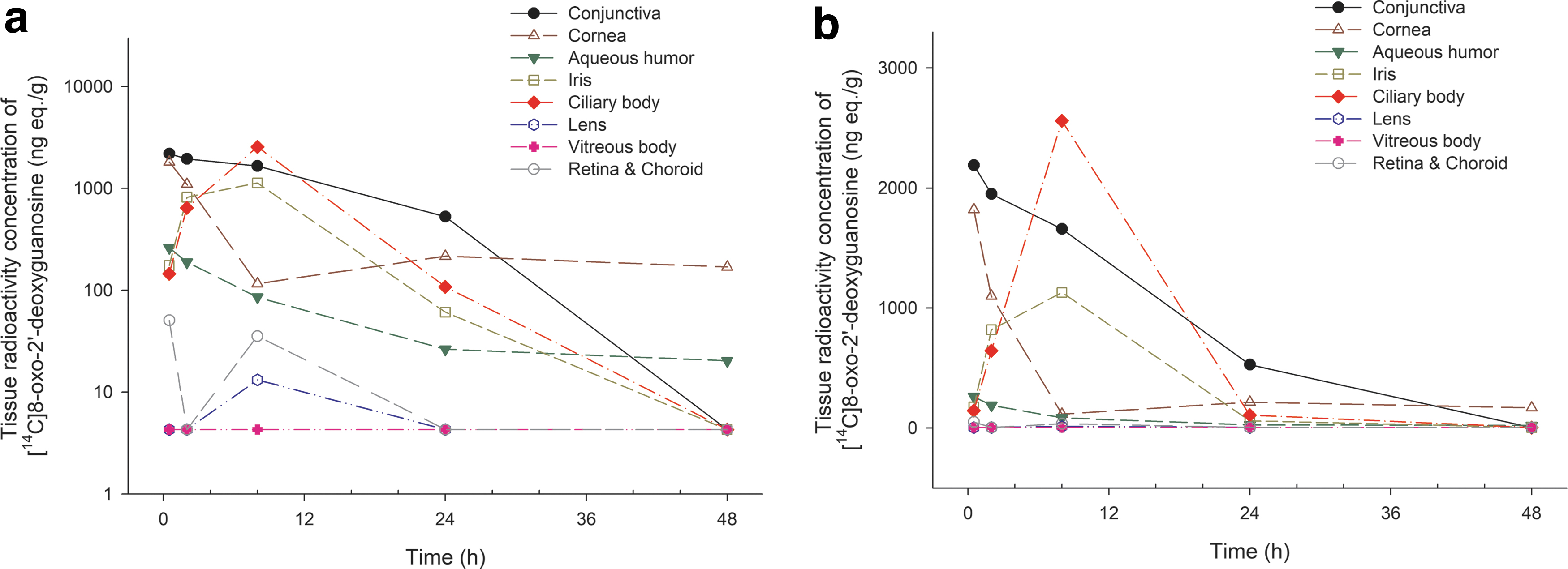
Radioactivity concentration–time profile in ocular tissues after single ocular instillation of [
14
C] 8-oxo-2′-deoxyguanosine in male rabbits.
Summaries of Pharmacokinetic Parameters of Radioactivity in Ocular Tissues After Single Ocular Administration of [ 14 C] 8-Oxo-2′-Deoxyguanosine at a Dose of 1 mg per Body in Male Rabbits
Data were obtained in autoradiography (n = 1 at each time point, total n = 5).
Data in plasma are presented as mean ± SD (%, CV) for Cmax (ng eq./mL) and AUC0–t (ng eq.·h/mL), and median [min, max] for Tmax. (n = 3).
AUC0–t, area under the concentration–time curve; NC, not calculated; SD, standard deviation.
The half-lives were estimated only in the conjunctiva, cornea, and aqueous humor, and were 11.2, 30.2, and 15.1 h, respectively. In the conjunctiva and cornea, concentrations of [ 14 C]8-oxo-dG rapidly reached Cmax after 0.5 h. However, slower Tmax was observed at 8 h in the ciliary body and iris. The radioactivity of [ 14 C]8-Oxo-dG in ocular tissues, except the cornea and aqueous humor, was lower than the limit of quantification at 48 h.
Plasma radioactivity concentration of [ 14 C]8-OxodG in rats
Plasma radioactivity of [ 14 C]8-oxo-dG in rats was plotted as a function of time after IV bolus injections (Fig. 2). Systemic exposure of [ 14 C]8-oxo-dG resulted in a double-peak phenomenon being observed on log-scaled radioactive concentration–time profiles. There was a rapid decline in radioactivity after attaining Cmax (4766.7 ± 226.8 ng eq./mL at 0.08 h). The first lowest radioactivity concentration was observed at 4 h with a mean value of 30.0 ± 7.4 ng eq./mL. A secondary peak was observed at 111.5 ± 49.7 ng eq./mL, which was much lower than the Cmax and appeared 8 h after dosing. The AUC0–t was 6503.0 ± 534.6 ng eq./h/mL and terminal half-life was estimated to be 56.1 ± 31.3 h (Table 2).
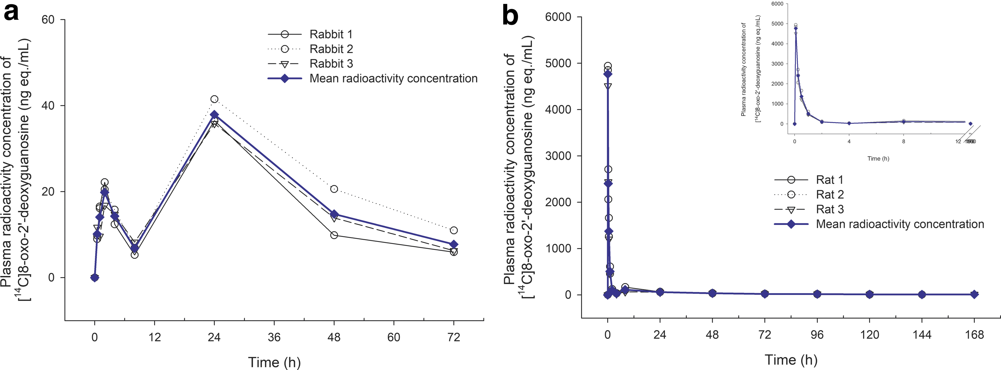
Radioactivity concentration–time profiles in plasma:
Summaries of Pharmacokinetic Parameters of Radioactivity in Plasma After Intravenous Infusion of [ 14 C] 8-Oxo-2′-Deoxyguanosine in Male rats
Data are presented as mean ± SD (%, CV).
Tmax values are presented as median [min–max].
Discussion
8-oxo-dG is an endogenous oxidized derivative of deoxyguanosine released from the damaged guanine base in DNA and is a useful biomarker of oxidative stress and carcinogenesis.9,10 In previous studies, it was suggested that topical application of 8-oxo-dG shows anti-inflammatory effects comparable with corticosteroids in a murine ocular alkali burn model, through Rac1 and the NLRP3 inflammasome/IL-1β axis suppression.2,7 To date, no PK profiles from animal models have been reported to assess the intraocular distribution or systemic exposure after single administration of 8-oxo-dG. It is essential to understand preclinical PK profiles of new drug candidates to further investigate their clinical applications as therapeutic agents.
We investigated the preclinical ocular and plasma PKs of 14 C-labeled 8-oxo-dG in rabbits and rats, respectively, for the first time. After ocular instillation in rabbits, the concentrations of radioactivity determined in ocular tissue including the conjunctiva, cornea, aqueous humor, iris, and ciliary body were relatively higher than in other tissues. Considering the intraocular pathway through topical administration, rapid ocular distribution with a very short Tmax was observed in anterior ocular tissues such as the conjunctiva, cornea, and aqueous humor, with subsequent delivery to the iris, ciliary body, and lens presenting Tmax at 8 h.
The extent of absorption of an ophthalmic drug is affected by physiological factors including permeability of membrane barriers, blood flow, and physicochemical drug properties.11,12 In general, conjunctival uptake is greater than corneal uptake because of the relative leakiness of the membrane, rich blood flow, and large surface area of conjunctiva, and an impermeable corneal barrier. 11 In our study, consistent with this phenomenon, the targeted anterior ocular tissues, especially the conjunctiva, showed that high concentrations of radioactivity of [ 14 C]8-oxo-dG were maintained. This implies that 8-oxo-dG mainly distributes to key ocular tissues and have favorable therapeutic outcomes in the treatment of ocular surface diseases including dry eye disease. 13
After ocular administration, the radioactivity of [ 14 C]8-oxo-dG was detected in plasma. Although PK parameters were not estimated in the same subjects, very low radioactivity of [ 14 C]8-oxo-dG was detected in the systemic circulation; Cmax and AUC0–48 in plasma were 1.7% and 3.4% in the conjunctiva, respectively. In systemic circulation, Tmax was observed at 24 h, which was the second peak in the plasma concentration–time profile. The multiple peaks are partly due to the various absorption pathways of topically administered ocular drugs. 14 In early phase of absorption, high levels of [ 14 C]8-oxo-dG were observed in the conjunctiva and cornea. However, absorption and distribution patterns can differ based on factors such as the nature of the vasculature and the transport process. 14
In general, topical ocular drug delivery involves the corneal and noncorneal routes. 14 The corneal route that represents the main absorption path involves passive diffusion for most drugs. 15 The noncorneal route, which spans across the conjunctiva and sclera, transports administered drugs into systemic circulation through capillary beds. 14 This route is important for hydrophilic and large molecules due to their poor corneal permeability. 16 In a previous study, we determined the partition coefficient of 8-oxo-dG to be 0.06, showing hydrophilic properties. In this preclinical PK study, the initial peak in plasma mainly reflected the noncorneal pathway and the latter higher peak was likely to be associated with the corneal pathway.
However, after a single ocular instillation of [ 14 C]8-oxo-dG, Cmax and AUC0–t in anterior target ocular tissue were much higher than those in plasma, suggesting that systemic adverse effects would not be significant and therapeutic effects can be anticipated at the application site. The PK profiles after IV infusion in rats exhibited a double-peak phenomenon, with a rapid initial decrease and slow terminal decrease (56.1 h of terminal half-life). The first peak represented the Cmax and the second peak occurred at 8 h. In blood concentration–time curves, multiple peaks are generally associated with factors such as the chemical properties of drugs, formulation-related factors including the excipients, gastric emptying, and site-specific absorption. 16
In this study, the secondary peak may be partially explained by enterohepatic recycling based on the IV infusion process. 16 However, in the previous study, 14.0% and 68% of single dose of 8-oxo-dG were excreted in bile and urine, respectively, within 48 h after IV infusion (data not shown), and it was known to be excreted in urine, the second peak by reabsorption appeared to be insignificant. 17 Nevertheless, this study has a few limitations.
The results were obtained in a small number of animals, especially male animals, and in discrete animal groups after a single administration of the test formulation. Moreover, this was an explorative PK study, and the pharmacodynamics or efficacy of the study drug cannot be concluded. Therefore, additional large-scale clinical assessments based on multiple dosing are required in human subjects to allow for the clinical translation of these findings.
Conclusions
In this study, we demonstrated the preclinical PKs of [ 14 C]8-oxo-dG after ocular administration in rabbits and IV infusion in rats. After ocular instillation, [ 14 C]8-oxo-dG was rapidly distributed and yielded high concentrations in anterior ocular tissue, with minimal detection in systemic circulation. IV infusion in rats showed a rapid decline of [ 14 C]8-oxo-dG after administration. These PK profiles suggest favorable effects in the treatment of ocular surface inflammation or dry eye disease. Nevertheless, this study has a few limitations. The results were obtained in a small number of animals, and in discrete animal groups after a single administration of the test formulation. Therefore, additional large-scale clinical assessments based on multiple dosing are required in human subjects to allow for the clinical translation of these findings.
Footnotes
Authors' Contributions
H.C. contributed to conceptualization (equal), writing the original draft, and formal analysis (lead). Y.H. was involved in conceptualization (equal), reviewing (equal), methodology, and data curation (lead). Y.H.K. carried out conceptualization (equal), data curation (equal), and reviewing (equal). D.H.K. was in charge of conceptualization (lead), data curation (equal), reviewing, and editing (lead). D.S. was involved in writing the original draft and formal analysis (lead), reviewing, and editing (lead).
Compliance with Ethics Guidelines
Ethical review and approval were waived for this study, due to anonymized names of the patients.
Author Disclosure Statement
D.H.K. is the inventor of the patent for the usage of 8-oxo-dG to treat various ocular inflammatory diseases. RudaCure company transferred the aforementioned patent of 8-oxo-dG. Y.H. and Y.H.K. are an employee and CEO of Rudacure, respectively. The rest of the authors have no proprietary interest in this article.
Funding Information
This research was supported by the Starting growth Technological R&D Program (S3030809) funded by the Small and Medium Business Administration (SMBA, Korea) and by the Korea Government (MSIT) (Grant No. NRF-2020R1F1A1072586).