




Abstract
Purpose:
Brimonidine is a highly alpha-2 adrenergic agonist, which provides a potential myopia control effect. This study aimed to examine the pharmacokinetics and concentration of brimonidine in the posterior segment tissue of eyes in guinea pigs.
Methods:
A liquid chromatography-tandem mass spectrometry (LC-MS/MS) method was successfully used for brimonidine pharmacokinetics and tissue distribution research in guinea pigs following intravitreal administration (20 μg/eye).
Results:
Brimonidine concentrations in the retina and sclera were maintained at a high level (>60 ng/g) at 96 h postdosing. Brimonidine concentration peaked in the retina (377.86 ng/g) at 2.41 h and sclera (306.18 ng/g) at 6.98 h. The area under curve (AUC0–∞) was 27,179.99 ng h/g in the retina and 39,529.03 ng h/g in the sclera. The elimination half-life (T1/2e) was 62.43 h in the retina and 67.94 h in the sclera.
Conclusions:
The results indicated that brimonidine was rapidly absorbed and diffused to the retina and sclera. Meanwhile, it maintained higher posterior tissue concentrations, which can effectively activate the alpha-2 adrenergic receptor. This may provide pharmacokinetic evidence for the inhibition of myopia progression by brimonidine in animal experiments.
Introduction
In recent years, the prevalence of myopia has been increasing, and the age of onset is gradually becoming younger. Uncorrected refractive error and complications caused by high myopia (such as cataract, retinal detachment, macular hole, choroidal neovascularization, and glaucoma) have brought a huge global economic burden.1,2 So it is important to find effective methods to prevent myopia onset and control myopia progression. 3
Numerous drugs have been tested in animal models, among which atropine, pirenzepine, and 7-methylxanthine have been evaluated in many clinical trials.4,5 Brimonidine is a highly alpha-2 adrenergic agonist approved by the United States Food and Drug Administration (FDA) for the treatment of ocular hypertension and glaucoma. 6 Besides lowering intraocular pressure (IOP), previous studies suggest that brimonidine can protect the retina/optic nerve and visual field damage.7–9 Evidence of animal experiments also provides a potential myopia control effect of brimonidine. Brimonidine has been reported to inhibit axial elongation in lens-induced myopia in guinea pigs 10 and form deprivation (FD) in chicks. 11 Our previous study demonstrated that intravitreal brimonidine at 4 μg/μL significantly reduced the progression of FD myopia in guinea pigs. 12
A drug should be delivered to intraocular tissues in adequate concentration to exert lasting effects. Numerous studies have proposed that there is a “signal cascade theory” in the pathogenesis of myopia. Visual input changes are transmitted from the retina to the retinal pigment epithelium (RPE) and then to the choroid, finally causing scleral remodeling and eye growth.13,14 It can be said that the signal cascade is triggered in the retina and act on the sclera, which play an important role in myopia progression. The direct target tissue of brimonidine on intravitreal injection is the retina.
As a highly hydrophilic and small-molecule compound, brimonidine can penetrate Bruch's-choroid complex and eventually be delivered to the sclera. 15 However, brimonidine has a short half-life after intravitreal injection and will be cleared quickly. 16 Therefore, frequent injections are required to achieve effective and sustained concentration. In our previous experiment, intravitreal injections of brimonidine were performed every 96 h. Intravitreal injection allows efficient drug binding to the target receptors in the posterior segment of the eyes. Drugs combine with the target receptor to release signals to produce a cascade effect, inhibiting changes in visual input and subsequent signal pathways caused by the environment during the onset and progression of myopia. 13 Thus, it is necessary to evaluate its tissue distribution and pharmacokinetics in vivo to provide solid proof for future research.
In contrast to traditional high-performance liquid chromatography (HPLC), liquid chromatography-tandem mass spectrometry (LC-MS/MS) is more efficient, more sensitive, and has higher accuracy, 17 which is currently the most commonly used quantitative assay in biological drugs. This study in guinea pigs was performed by LC-MS/MS to characterize the ocular pharmacokinetics of single-dose (20 μg/eye) brimonidine following intravitreal administration (Fig. 1). The purposes of this study were (1) to investigate the posterior tissue distribution of brimonidine after intravitreal injection with an effective concentration on myopia progression in guinea pigs and (2) to evaluate the ocular pharmacokinetics of brimonidine in the retina and sclera.
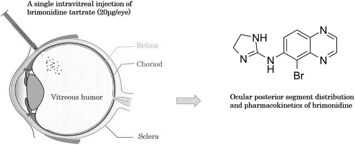
Schematic representation of the study. Both eyes of each guinea pig were injected. Brimonidine pharmacokinetic and tissue distribution were investigated in guinea pigs following intravitreal administration (20 μg/eye).
Methods
Reagents and chemicals
Brimonidine tartrate (purity >99%) was purchased from MedChemExpress (Shanghai, China). Brimonidine standard (purity >98%) was purchased from Shanghai Yuanye Bio-technology Co., Ltd. (Shanghai, China). Acetonitrile (HPLC grade) was purchased from TEDIA. Methanol (HPLC grade) was purchased from Sigma-Aldrich (Shanghai, China). Milli-Q purified water was prepared for this study (Millipore, MA).
Animals
Male tricolor guinea pigs (n = 48) weighing ∼120–150 g were obtained from Changsha Tianqin Biotechnology Corporation (Changsha, China). All guinea pigs were raised in a temperature-controlled environment (25°C) and a 12-h light/12-h dark cycle. Food, water, and fresh vegetables were provided ad libitum. All experiments were conducted in accordance with the Association for Research in Vision and Ophthalmology Statement for the Use of Animals in Ophthalmic and Vision Research.
Sample preparation
Brimonidine tartrate solution was prepared in sterile phosphate-buffered saline (Procell Life Science&Technology Co., Ltd.), and the concentration of intravitreal injection was 4 μg/μL. Guinea pigs were anesthetized by the intraperitoneal injection of 1.5% sodium pentobarbital (37.5 mg/kg). The conjunctiva was disinfected with 5% povidone-iodine, rinsed with normal saline, and pupils were dilated with compound tropicamide eye drops. Proparacaine hydrochloride was used to anesthetize the ocular surface to prevent eye movement. The solution was injected using a 33 gauge needle and a 5 μL Hamilton microsyringe.
The injection was performed ∼2 mm from the superior temporal rim, through the sclera into the vitreous body, and 5 μL of the solution was injected into each eye. Both eyes of each guinea pig were injected. Guinea pigs were euthanized by an overdose of sodium pentobarbital at 0.5, 1, 2, 4, 8, 12, 24, or 96 h after administration. After sacrifice, the eyeballs were enucleated and were quick-frozen in dry ice, then anterior segment tissues and vitreous body were removed, the retina was bluntly separated from RPE-choroid complex, and the choroid attached to sclera was gently scraped with a blade; finally, retina (n = 6 eyes/time point) and sclera (n = 6 eyes/time point) were collected. All tissue samples were stored at below −80°C until analysis.
LC-MS/MS conditions
All samples were analyzed for brimonidine concentration using a Waters UPLC Quattro Premier triple quadrupole ultra-performance liquid chromatography-mass spectrometry system (Quattro Premier XE series) with an ACQUITY UPLC series for liquid chromatography. The chromatographic column is a Waters column ODS C18 UPLC HSS T3 series (1.8 μm, 100 × 2.1 mm) and the guard column is a Waters XBridge C18 (1.8 μm, 20 × 2.1 mm). The mobile phase was A: B = 0.1% formic acid aqueous solution: acetonitrile, with gradient elution according to Supplementary Table S1. The flow rate was 0.2 mL/min, the column temperature was 25°C, the running time was 5 min, and the injection volume was 5 μL.
Multiple reaction monitoring scan mode was selected for mass spectrometric detection. The MS conditions were as follows: the spray voltage was set at 3.5 kV, cone voltage at 30 V, collision voltage at 20 V, ion source temperature at 150°C, desolventizing temperature at 300°C, desolventizing gas flow rate at 700 L/h, collision gas flow rate was 0.15 mL/min, and the injection volume was 5 μL.
Method validation
The method validation of this study follows the FDA guidelines for bioanalytical method, 18 including specificity, linearity, accuracy, precision, recovery, matrix effect, and stability.
Pharmacokinetic analyses
The mean and standard deviation of the mass concentration of brimonidine at each time point were calculated, respectively. Drug and Statistics version 3.0 pharmacokinetic statistical analysis software (Anhui Provincial Center for Drug Clinical Evaluation) was used for atrial chamber fitting and calculation of pharmacokinetic parameters.19–21 Moreover, the elimination rate constant (Ke), elimination half-life (T1/2e), absorption rate constant (Ka), absorption half-life (T1/2a), the time to Cmax (Tmax), the maximum drug concentration (Cmax), and the total area under the curve (AUC) from time zero until infinity (AUC0-∞) were calculated. The results of tissue distribution data and pharmacokinetic parameters are presented as mean ± SD.
Results
Method validation
In this study, the corresponding drug standards were well separated from the impurities. The linear relationship of brimonidine was good in the concentration range of 1.0–200.0 ng/mL (Fig. 2). The slope, intercept, and correlation coefficient were calculated using the linear regression method, and the standard curve equation and correlation coefficient were as follows: y = 17184x–44.724 and r = 0.9999. Under the given extraction method and assay conditions, the accuracies of brimonidine in tissue homogenates reached more than 95%, and the intraday and interday relative standard deviations (RSD) were less than 10% (the results are presented in Table 1).

The standard line of mass concentration of brimonidine.
Accuracy and Precision of Brimonidine in Tissue Homogenates (n = 5)
RSD, relative standard deviations.
The recoveries and matrix effects are presented in Table 2. The recoveries of brimonidine were 91.2% ± 7.13%, 92.77% ± 5.25%, and 96.08% ± 3.11% at the low, medium, and high QC concentrations, respectively. The corresponding RSDs were 7.82%, 5.66%, and 3.24%, respectively. The mean matrix effect for QC samples was 99.85% ± 6.12%, 98.77% ± 3.24%, and 99.63% ± 5.37%, respectively, and RSDs were less than 6.13%.
Recovery and Matrix Effect of Brimonidine in Tissue Homogenates (n = 5)
The results of stability are shown in Table 3. It indicated that the accuracies for stability after 24 h at room temperature, 3 freeze-thaw cycles, and 7 days at −20°C at 3 concentrations were with the ranges 95.36%–98.61%, 98.38%–99.31%, and 98.67%–99.37%, respectively. The corresponding RSDs were 3.66%–5.53%, 6.15%–7.18%, and 5.13%–6.22%, respectively.
Stability of Brimonidine in Tissue Homogenates (n = 5)
Pharmacokinetic and tissue distribution studies
The concentration-time profiles of the retina and sclera after a single intravitreal administration of brimonidine (20 μg/eye) are shown in Figure 3. Within 96 h after intravitreal injection, the effective drug concentrations were in the range of 46.95 to 407.71 ng/g in the retina and 88.83 to 396.31 ng/g in the sclera.
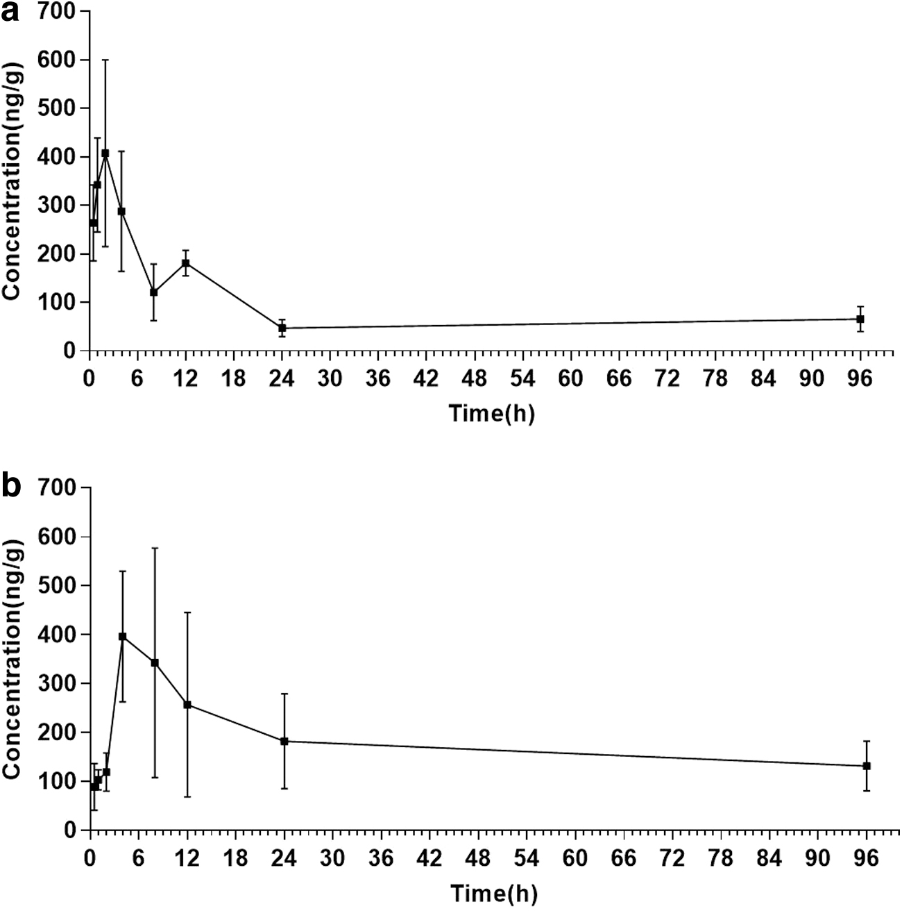
Mean(±SD) concentration of brimonidine in the retina
The primary pharmacokinetic parameters of brimonidine are presented in Table 4. Brimonidine concentration peaked in the retina (377.86 ng/g) at 2.41 h and sclera (306.18 ng/g) at 6.98 h. The area under curve (AUC0–∞) was 27,179.99 ng h/g in the retina and 39,529.03 ng h/g in the sclera. The elimination half-life (T1/2e) was 62.43 h in the retina and 67.94 h in the sclera.
Pharmacokinetic Parameters for Brimonidine Following a Single Intravitreal Injection (20 μg/eye) in Guinea Pigs
Discussion
Brimonidine has shown the potential to inhibit myopia progression in guinea pigs10,12 and chicks. 11 This study evaluated brimonidine's posterior segment tissue distribution and pharmacokinetics after a single bilateral intravitreal administration in guinea pigs.
The results suggested that matrix effects and recoveries do not significantly affect the analysis of samples. Under this assay condition, brimonidine possessed a high accuracy, while RSDs were less than 10% (including intraday and interday), indicating that the instrumentation and precision met the assay criteria. The study results also suggested that brimonidine has good chemical stability under different storage conditions. It indicated that LC-MS/MS is accurate and precise for determining the mass concentration of brimonidine in the retina and sclera.
The retina and sclera are important sites for drug action on myopia progression. Visual signals are transmitted from the retina to the sclera. After myopia is induced, retinal gene expression is severely affected, then affecting scleral remodeling through the retinal-scleral pathway.4,22 Thus in this study, we gave priority to conducting the pharmacokinetics in these 2 target tissues. Intravitreal injection allows the drug to be delivered directly to the posterior segment, allowing it to remain in sufficient concentration in the posterior segment tissue to exert its therapeutic effect. Our data showed that the Cmax of the retina reached 377.86 ng/g at 2.41 h and the sclera reached 306.18 at 6.98 h; it indicated that brimonidine was rapidly absorbed and diffused to the retina and sclera after a single intravitreal administration of 20 μg/eye. Hence, brimonidine has a high affinity for the retina and sclera, which is consistent with the previous studies.23,24
Furthermore, our results showed that the Tmax of brimonidine was earlier in the retina than in the sclera, and the Cmax was also higher than the sclera. Drug absorption in the eyes involves transmembrane transport, of which passive transport is one of the most common and important modes of drug transport. The driving force of passive transport depends mainly on the concentration difference between the 2 sides of the membrane, and it is maintained at a dynamic and stable level when the concentration between the 2 sides of the membrane reaches equilibrium. After intravitreal administration, brimonidine gradually diffused to the retina and sclera in the direction of the concentration gradient. Besides, the retina of guinea pigs is avascular, only having an outer retinal barrier composed of the tight junction between RPE cells, and the retinal nutrition supply provided by the choroidal circulation. 25
Due to the hindrance by RPE and choroid, brimonidine penetrating from the vitreous cavity to the sclera is slow. Brimonidine has also been shown to reversibly bind to ocular melanin for storage and to preferentially distribute to pigmented tissues.23,24 Drugs combined with melanin or protein in the tissues will affect the bioavailability of ophthalmic administration. Likewise, the high levels of exposure of brimonidine in posterior ocular tissue might be due to brimonidine bound to melanin and slowly released to the retina and sclera in vivo. 16 In that case, the drug level measurement of pharmacological targets or intraocular metabolism may be overestimated. 26
There are 2 main ways to eliminate drugs following intravitreal administration: on the one hand, drugs can penetrate through the retinal barrier and eventually be eliminated by choroidal blood flow in the posterior segment. On the other hand, intravitreal drugs can also be eliminated through aqueous humor circulation and uveal blood flow in the anterior segment of the eyes. 27 Generally speaking, lipophilic and small molecular drugs have higher permeability in the retinal barrier, and most of them are eliminated in the posterior segment of the eye. 28 While brimonidine is highly hydrophilic, it seems that brimonidine was most likely to be eliminated by the anterior route, which leads to a slower elimination procedure.
The exact mechanism by which brimonidine inhibits myopia progression is not clear. In this study, brimonidine concentrations in the retina and sclera were still maintained at a high level (>60 ng/g) at 96 h postdosing, which is 100-fold higher than the necessary concentration to activate the alpha-2 adrenergic receptor (0.6 ng/mL or 2 nM). 29 As a highly selective alpha-2 adrenergic agonist, brimonidine is effective in lowering IOP. 30 The relationship between IOP and myopia progression has been controversial. Liu et al. attributed one of the reasons for the inhibition of myopia progression in guinea pigs by brimonidine to the lowering of IOP. 10 Carr et al. also suggested that brimonidine can counteract myopia progression by lowering scleral compliance through lowering IOP. 11
Some growth factors are involved in experimental myopia progression. Brimonidine can regulate the expression of basic fibroblast growth factor 31 and transforming growth factor beta. 32 These 2 growth factors may be involved in scleral remodeling in myopic eyes by regulating matrix metalloproteinase-2, which influences the progression of experimental myopia.33–35 In addition, studies in animal models have indicated that retinal dopaminergic function is crucial for the development of myopia.36,37 There is also evidence that retinal alpha-2 adrenergic receptors may regulate the activity of tyrosine hydroxylase, the rate-limiting enzyme for dopamine synthesis, 38 which may be a potential mechanism of brimonidine inhibiting myopia progression. Obviously, further research is needed in the future.
There are still some limitations in this study. First, we believed that the target of brimonidine in retarding myopia progression is the retinal-scleral pathway. However, we did not conduct choroid in this study, considering the presence of melanin and proteins, which will affect drug delivery and distribution. Thus, further studies need to focus on the drugs melanin bound and distribution in other eye tissues. Second, as a classic species used for myopia models, guinea pigs have an avascular retina, which may have an effect on the absorption and elimination of the retina. There exist species differences between animals and humans; it is impossible for any animal model to completely simulate the role of drugs in human eyes. It is suggested to use a combination of multiple models to verify the efficacy of brimonidine in the future.
Conclusion
In summary, a specific and sensitive LC-MS/MS method for brimonidine in the posterior segment ocular tissue of guinea pigs was established and validated. The method was successfully used for pharmacokinetic and tissue distribution research in the eyes of guinea pigs. In this study, brimonidine was rapidly absorbed and diffused to the retina and sclera. Meanwhile, it maintained higher posterior tissue concentrations even 96 h following a single intravitreal administration at 4 μg/μL of brimonidine. It may provide pharmacokinetic evidence for the inhibition of myopia progression by brimonidine in animal experiments. Further investigation is required to elucidate the mechanism of brimonidine, slowing down myopia progression.
Footnotes
Ethics Approval and Consent to Participate
The whole experimental procedure was approved by the Medical Ethics Review Committee of Hainan Eye Hospital.
Acknowledgment
The authors gratefully acknowledge Hangzhou Hebei Technology Co., Ltd. (Hangzhou, China) for technical assistance.
Authors' Contributions
A.X. and H.H. conceived and designed the study. A.X., J.Y., and Y.L. performed the experiment. H.Y. and A.L. prepared the figures and tables. A.X. wrote the main article. X.Z. supervised the study and revised the article. All authors read and approved the article.
Author Disclosure Statement
The authors declare that they have no competing interests.
Funding Information
This work was supported by grants from the National Natural Science Foundation of China (82271116), Hainan Province Clinical Medical Center, and Science and Technology Planning Project of Hainan Province (ZDYF2022SHFZ326).

References
Supplementary Material
Please find the following supplemental material available below.
For Open Access articles published under a Creative Commons License, all supplemental material carries the same license as the article it is associated with.
For non-Open Access articles published, all supplemental material carries a non-exclusive license, and permission requests for re-use of supplemental material or any part of supplemental material shall be sent directly to the copyright owner as specified in the copyright notice associated with the article.
