




Abstract
Purpose:
To evaluate the effects of bevacizumab in 3 different application methods, subconjunctival injection (SCI), hyaluronic acid retardant (HAR), and eye drop (ED), on attenuating scar formation in the filtering bleb.
Methods:
Trabeculectomy (TRAB) was performed on New Zealand rabbits. TRAB rabbits were intervened with bevacizumab SCI, HAR, ED, or mitomycin C, respectively. Intraocular pressure (IOP) of 1, 7, 14, and 28 days after TRAB was recorded, and the bleb survival rate was analyzed. Bleb height, area, and vascularization were evaluated using anterior segment optical coherence tomography (OCT) and optical coherence tomography angiography (OCTA) at 7, 14 and 28 days after surgery. A histopathology examination of the bleb tissue was performed. The expression levels of vascular endothelial growth factor (VEGF)-A, interleukin (IL)-1α, tumor necrosis factor-alpha (TNF-α), transforming growth factor-β1 (TGF-β1), and α-smooth muscle actin (α-SMA) were measured by Western blot.
Results:
Bevacizumab significantly reduced postoperative IOP and increased the survival of the filtering bleb, especially in the ED group. Less vascularization was shown in the SCI, HAR, and ED groups. Histopathological results showed the fewest levels of scarring and fibrosis in the ED group. The local VEGF-A, IL-1α, and TNF-α expression levels after bevacizumab ED were decreased, combined with suppression of TGF-β1 and α-SMA.
Conclusions:
Postoperative use of bevacizumab EDs was an effective application method for improving surgical outcomes after TRAB in rabbits. It might be effective in preventing scarring of the filtering bleb by antivascularization and anti-inflammation.
Introduction
Glaucoma, is the world's first irreversible blindness. According to epidemiological data, the number of people with glaucoma is expected to reach 111.5 million worldwide by 2040. 1 Trabeculectomy (TRAB) has been the gold standard treatment option for more than 50 years. 2 At present, it is still one of the most effective ways for lowering intraocular pressure (IOP). 3 The maintenance and assessment of the outcome of glaucoma surgery depends on the function of the filtering bleb. The failure rate of TRAB is ∼30%. 4 The main reason for surgery failure is scar formation, 5 which subsequently increases IOP. Antimetabolites, mitomycin C (MMC), and 5-fluorouracil were introduced to significantly suppress scar formation. 6 However, side effects of MMC and 5-fluorouracil should not be overlooked, such as persistent postoperative hypotony, corneal toxicity, filtering bleb leakage, and endophthalmitis. 7
Angiogenesis and the accompanying scar formation play a decisive role in wound healing. 8 Previous studies have shown that the vascular response to trauma is a key factor in scar formation. 9 Our previous findings showed that the degree of change in the vascular area of the filter blebs area 1 month after TRAB was positively correlated with IOP at 6 months postoperatively, indicating that the vascularization of the filtering blebs influences, to some extent, the outcome of glaucoma filtration surgery (GFS). 10 In other words, with postoperative angiogenesis inhibition, the survival time of postoperative filtering blebs can be increased. Vascular endothelial growth factor (VEGF) is a potent endothelial cell-specific cytokine that enhances microvascular permeability and vascular endothelial cell proliferation and plays a key role in angiogenesis. 11 After surgery, external stimuli and inflammatory responses cause wound tissue to secrete large amounts of VEGF, which promote angiogenesis.
Previous studies have shown that VEGF levels in aqueous humor are maintained at high levels for 30 days after surgery. 12 Therefore, we can intervene earlier on the postoperative VEGF levels to reduce its effect on angiogenesis, reduce scar formation, and subsequently increase the survival rate of the filtering bleb.
Bevacizumab, a full-length humanized monoclonal antibody targeting all isoforms of VEGF-A, has been approved by the Food and Drug Administration (FDA) for the treatment of metastatic colorectal cancer. 13 Although most previous studies performed intravitreal injections of the agent, the usage method of bevacizumab has been a source of debate. 14 Clinical cases have reported that subconjunctival injections (SCIs) of bevacizumab are more effective than antimetabolites in the treatment of recalcitrant scarring of the bleb. 15 However, it has also been reported that subconjunctival bevacizumab and MMC have similar effects on IOP control. It has been reported that the use of anti-VEGF alone to control IOP may not be as effective as MMC. 16 The 2 delivery methods, SCI and eye drops (EDs), have also been shown to be effective in patients with corneal neovascularization and experimental rabbit models.17,18
With the continuous development of modern polymeric nanomaterials, many studies have been conducted based on drug slow-release delivery systems. 19 In clinical practice, the most common form of drug administration to the eye is in the form of EDs, which is more convenient. The regulation of filtering blebs by anti-VEGF drugs after glaucoma surgery, including its efficacy and mode of administration, still need to be further studied.
To avoid invasive injection or reduce the number of injections, we prepared bevacizumab via controlled-release and ED, and compared their effects on the structure and function of the postoperative filtering bleb with SCIs. It is meaningful to provide new options to improve the postoperative success rate in a more convenient and effective way.
Methods
Study design
Twelve-week-old female New Zealand white rabbits (2.0–2.5 kg) were used in this study. The rabbits were placed in the Soochow University Experimental Centre and acclimatized under controlled light conditions (12 h of light; 12 h of darkness) for 7 days. Subsequently, 54 rabbits were randomly divided into 6 groups: group TRAB: TRAB group (n = 9); group HA: TRAB with subconjunctival implantation of hyaluronic acid retardant (HAR) group (n = 9); group B+HA: TRAB with SCI of bevacizumab+HAR group (n = 9); group SCI: TRAB with SCI of bevacizumab group (n = 9); group ED: TRAB with postoperative topical administration of bevacizumab (n = 9); and group MMC: TRAB with intraoperative MMC (n = 9). All procedures were performed by one surgeon on the right eye of each rabbit.
All animal experiments were approved by the Animal Ethics Committee of Soochow University and were performed in accordance with the Association for Research in Vision and Ophthalmology (ARVO) Statement on the Use of Animals in Ophthalmic and Vision Research. IRB No. SUDA20221123A01.
Preparation of bevacizumab sustained-release agent and EDs
A mixture of bevacizumab+HAR was made by dissolving 8 mg of sodium hyaluronate solid in 250 μL of phosphate-buffered saline. Next, to 20 μL of the above mixture, we added 10 μL of horseradish peroxidase and 100 μL of bevacizumab stock solution (25 mg/mL), followed by 0.4 μL of dopamine to adjust the hardness of the retardant. We then added a solution to 10 μL of hydrogen peroxide (H2O2) for rapid gel formation within a few seconds to 1 min. The no-load HAR replaced the bevacizumab stock solution with normal saline. For the bevacizumab EDs, we took 0.1 mL of bevacizumab stock solution (25 mg/mL) and added 0.4 mL of 0.9% saline (NaCl) to prepare EDs (5 mg/mL). 20 We then stored them at 4°C away from light, to be used as needed. All the above operations were performed by the same operator in a sterile laboratory environment.
Surgical procedure
Rabbits were anesthetized with intravenous sodium pentobarbital (30 mg/kg) at the ear margins preoperatively with topical anesthesia with proparacaine hydrochloride EDs (5 mg/mL). TRAB was then performed in all rabbits' right eyes by an experienced glaucoma specialist using a previously studied method. 21 In brief, all 54 rabbits were carefully dissected for the domed conjunctival flap, and a 3 × 3 mm partial thickness scleral flap was isolated. After excision of 1 × 2 mm of trabecular tissue, a peripheral iridectomy was performed, and the scleral flap was closed with 10-0 sutures and the conjunctiva with 8-0 nylon sutures. For group TRAB, only the above surgical operation was performed. For group HA, a postoperative injection of 0.1 mL of HAR, as described previously, was administered, using a 1 mL syringe under the conjunctiva away from the periphery of the scleral flap.
For group B+HA, postoperatively, 0.1 mL of prepared bevacizumab+HAR gel was injected into the subconjunctiva around the perimeter of the filter bulb using a 1 mL syringe, away from the peripheral area of the scleral flap. For group SCI, after completion of the filtration procedure mentioned previously, 0.1 mL of bevacizumab stock solution (25 mg/mL) was injected under the conjunctiva away from the periphery of the scleral flap using a 1 mL syringe. For group ED, postoperative drops of the prepared bevacizumab solution (5 mg/mL) were administered to the operative eye twice a day for 28 days. For group MMC, intraoperative application of MMC by surgical sponges immersed in 0.2 mg/mL solution was performed.
The sponges were placed in the conjunctival/tenon's flap for 3 min and then removed and carefully flushed with 10–20 mL of saline before suturing the scleral flap and continuing with subsequent surgical steps. All operated eyes received a combination of topical antibiotic (ciprofloxacin 3.5 mg/g) and corticosteroid (dexamethasone 1.0 mg/g) drops topically 4 times daily for 4 weeks. Three rabbits from each group were killed on postoperative days 7, 14, and 28, and the eyes were removed for analysis.
Clinical examination
Photographs of the rabbit's conjunctiva and filtering bleb were taken with a digital camera, and hand-held slit-lamp examinations were performed before and weekly after surgery to assess the height and extent of the filtering bleb. Anterior segment optical coherence tomography (AS-OCT) was used to scan the filtering bleb and to obtain data on the height and extent of the filtering bleb. The rabbit eyes were anesthetized locally with proparacaine hydrochloride drops (5 mg/mL) and after confirming adequate anesthesia, IOP was measured preoperatively and daily postoperatively for 4 weeks using an I-CARE PRO IOP meter (Finland). IOP was measured 6 times at each examination, and the mean value was taken for analysis. All measurements were performed by the same observer at the same time of day (10:00 am–12:00 pm), and the tonometer was calibrated before use.
Image acquisition and processing
Two masked physicians (J.C. and Y.X.) used optical coherence tomography angiography (OCTA) (Optovue, Fremont, CA) to detect the vascular area in the area of the filtering bleb on postoperative days 7, 14, and 28 in each group of rabbits. The OCTA scan mode was 3.0 × 3.0 mm HD Angio Retina with a probing depth of 2.0 mm. Vascular data from the scanning laser fundoscopy mode OCTA images were compiled by a physician (J.C.) using ImageJ (National Institutes of Health, Bethesda, MD). Vascular density data were extracted in 3 separate sessions, and the mean of the 3 measurements was calculated. The vascular area of the surgical area before TRAB was used as the baseline. The scanned area was focused according to the initial detection, and the bleb morphology was determined by OCT of the anterior segment of the eye (AS-OCT) using the corneal cross-line scheme.
Western blot and histological analysis
On postoperative days 7, 14, and 28, 3 rabbits from each group were killed by intravenous administration of an overdose of sodium pentobarbital. Tissue from the filtering bleb area of each group was collected, and total protein was extracted using RIPA lysate (Beyotime, Shanghai, China) containing a 1% protease inhibitor. Protein concentration was determined using a BCA protein concentration assay kit (Beyotime, Shanghai, China). Equal amounts of protein samples were subjected to gel electrophoresis, transmuted using polyvinylidene difluoride (PVDF) membranes, closed with 5% skimmed milk powder for 1 h at room temperature, and incubated overnight at 4°C with primary antibody. PVDF membranes were washed 3 times with TBST for 10 min/time and incubated with the corresponding secondary antibodies for 1 h at room temperature. Protein bands were developed using an enhanced ECL chemiluminescence detection kit (Merck, Darmstadt, Germany).
The bands were quantified in gray using ImageJ software (National Institutes of Health). β-Actin was used as an internal reference. The eyes of rabbits in each group on postoperative days 7, 14, and 28 were removed and fixed in 4% paraformaldehyde solution and then dissected at the equator and embedded in paraffin. Using the TRAB suture point as a reference, serial sections of a 4-μm thickness were cut from the center of the surgical site and stained with H&E and Masson for histological examination of the filtering bleb area.
Statistical analysis
All data were expressed as mean ± standard deviation. Statistical analysis was performed using SPSS 26.0 statistical software (IBM, Armonk, NY). Differences between groups were analyzed using unpaired t-tests or 1-way analysis of variance. Pearson correlation analysis and 1-way and multiway linear regression analyses were used to compare vascular area data and parameters in rabbits. Kaplan–Meier and Mantel–Cox 2-comparison tests were used for the analysis of filtering bleb survival, and P < 0.05 was considered statistically significant.
Results
Postoperative IOP changes
There was no significant difference in initial IOP among all experimental groups before surgery, with all being within the normal range of 10–15 mmHg. The IOP in TRAB, SCI, and HA groups were within the normal range by postoperative day 14, which was significantly higher than the IOP levels in the other 3 groups. The control of postoperative IOP was better in groups B+HA, ED, and MMC, with all 3 groups in the lower range up to 14 days postoperatively. On postoperative day 21, the IOP of group B+HA first began to rise among these 3 groups and was within the normal range by postoperative day 28. Group MMC had the same upward trend as group B+HA, but the IOP was slightly lower overall than in group B+HA.
The IOP in group ED was below the normal range throughout the experiment but began to rise slowly after postoperative day 14 and approached 10 mmHg on postoperative day 28. In the final stage of the experiment, the IOP levels in the ED group were significantly different from those in the other groups (Fig. 1A).
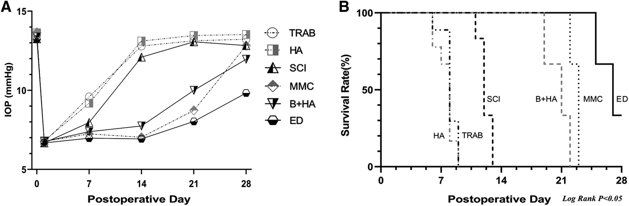
Bevacizumab prolongs the survival time of filtering blebs after GFS
We evaluated the effect of bevacizumab on the survival of the filtering bleb after GFS. Our analysis of filtering bleb survival rates showed significant differences between the ED group and the other groups in terms of time to filtering bleb survival (P < 0.05, Fig. 1B). Rabbits in the TRAB and HA groups exhibited rapid disappearance of the filtering bleb after TRAB, and few filtering blebs were observed on postoperative day 7 (Fig. 1B) and largely disappeared on postoperative day 14 (Fig. 2).

Morphological changes at 1, 7, 14, and 28 days postoperatively in each group with filtering blebs.
The survival time of the filtering bleb was significantly longer in groups B+HA, SCI, ED, and MMC compared with groups TRAB and HA. The filtering bleb in group SCI disappeared on postoperative day 14. In contrast, group B+HA, group ED, and group MMC had significant postoperative effects with longer survival of the filtering bleb, with the longest time being around 21 days in group MMC and group B+HA. Group ED still had 30% of the filtering bleb alive at the end of the experimental phase, although the extent and height of the filtering bleb approached very low levels (Figs. 1B and 2). In conclusion, bevacizumab had the same effect as MMC, which resulted in significantly higher rabbit bleb survival compared with the TRAB and HA groups, with a statistically significant difference between the bleb survival rates of the groups (P < 0.05, Fig. 1B).
Postoperative filtering blebs morphology
Slit-lamp examination and OCT images showed no serious inflammatory reaction in the anterior chamber postoperatively in all experimental rabbits, and no other postoperative complications, such as filtering bleb leakage, leukomalacia, or endophthalmitis, were observed. The height and extent of the postoperative filtering bleb were significantly lower in the TRAB and HA groups than in the other groups throughout the experiment (P < 0.05, Fig. 3B, C). Groups B+HA, SCI, ED, and MMC had intact filtering bleb with normal height and extent on postoperative day 7, with no significant differences among the groups. On postoperative days 14 and 28, the height and extent of the filtering bleb in group SCI decreased significantly compared with groups B+HA, ED, and MMC. There was a significant increase in the height and extent of the filtering bleb in the ED group on postoperative day 14 compared with postoperative day 7 (Fig. 3B, C).
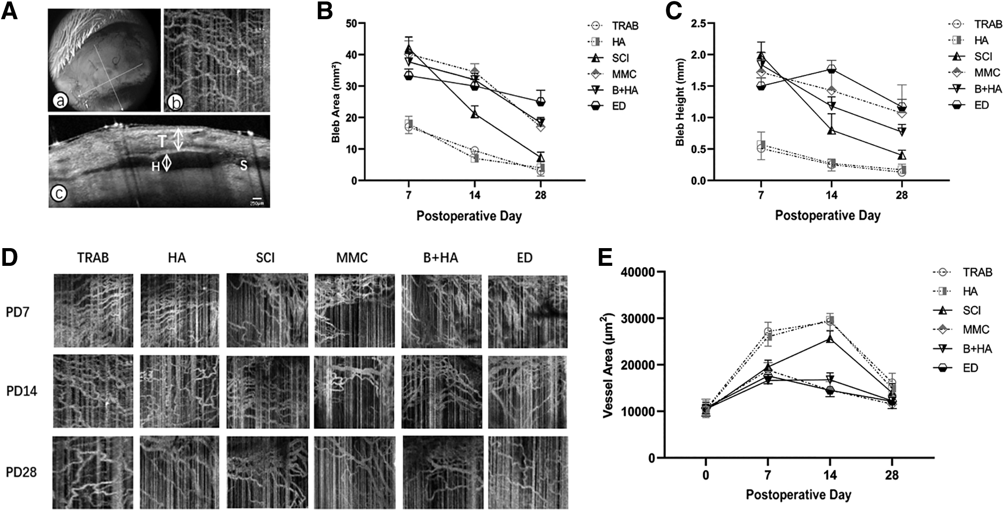
Postoperative level of vascularization in filtering blebs
Visually, it was observed that groups B+HA, SCI, and ED with bevacizumab and group MMC had a lower degree of vascularization in the filtering bleb area than groups TRAB and HA throughout the postoperative experimental period (Fig. 2). The preoperative baseline vascular area was 10,421.3 ± 1,850.2 μm2 and increased on postoperative day 7 in all groups, with a significant increase in vascular area in groups TRAB and HA to 27,183.0 ± 1,968.3 μm, 2 which was significantly different from the other groups; the highest level of vascularization was reached on postoperative day 14 in both groups and then decreased. Group SCI showed a consistent trend of increasing vascular area in the filtering bleb area at 14 days postoperatively, but to a lesser extent compared with the TRAB and HA groups.
The degree of vascularization of the filtration blebs area in groups B+HA, ED, and MMC reached its highest level on postoperative day 7 and then tended to decrease over time in all 3 groups, with no significant differences among the 3 groups. During the final time period of the experiment, the vascularization level in all groups decreased to a state close to the preoperative state, with no significant difference from the preoperative state (Fig. 3D, E).
Effect of bevacizumab on histopathological changes in filtering blebs
Based on the above experimental procedure and the degree of vascular scarring progression in the filtering blebs after TRAB, we selected groups B+HA, SCI, and ED with bevacizumab and the TRAB control group, and performed histopathological examinations of the filtering blebs in the above 4 groups. We did this at 7, 14, and 28 days postoperatively to observe the degree of tissue sparing and fibrosis in the respective filtering blebs to investigate the optimal mode of administration of bevacizumab. The results showed that on postoperative day 7, the tissue in the filtering bleb area of groups B+HA, SCI, and ED was sparse, and the blister-like structure could be seen under the conjunctiva. In group TRAB, the tissue in the filtering bleb becomes dense, with increased fibroblast density, accompanied by deposition of collagenous tissue, increased inflammatory infiltration, and cellular response.
On postoperative day 14, the tissue in the filtering bleb area of groups B+HA and ED were loose, with a reduced density of fibroblasts in the tissue, less collagenous tissue deposition, and a looser subconjunctival stroma, along with a less pronounced cellular response and a lower number of inflammatory cell infiltrates. In group SCI, compared with 1 week after operation, the filtering bleb tissue began to become dense. On the 28th postoperative day, the TRAB and SCI groups had dense tissue in the area of the filtering bleb, with collagen fiber deposition and disorganized arrangement. However, the B+HA and ED groups had relatively loose tissue in the filtering bleb area, and lower levels of collagen fiber deposition with loose subconjunctival stroma, most notably in the ED group (Fig. 4).

Histological characteristics of the filtration site stained with H&E
Effect of bevacizumab on various cytokines in filtering blebs
To investigate the effect of bevacizumab on the postoperative filtering bleb in the animal model of glaucoma filtration from the protein level, we selected groups B+HA, SCI, and ED and the control group TRAB with bevacizumab. We then dynamically observed the levels of VEGF-A, transforming growth factor-β (TGF-β), α-smooth muscle actin (α-SMA), tumor necrosis factor-alpha (TNF-α), and interleukin (IL)-1 in the postoperative bleb area of each group at the postoperative time points of days 7, 14, and 28. Groups B+HA, SCI, ED, and TRAB were analyzed separately for statistical comparison (Fig. 5). VEGF-A levels in groups B+HA and ED were significantly lower than those in group TRAB at all 3 postoperative time points (P < 0.05). Group B+HA had the lowest VEGF levels on postoperative day 14, with a tendency to rebound on postoperative day 28. Group ED showed a decreasing trend in VEGF-A levels throughout the experimental phase.

Expression levels of VEGF-A, IL-1α, TNF-α, TGF-β1, and α-SMA
Group SCI was significantly lower than group TRAB on postoperative days 7 and 14 (P < 0.01), and VEGF-A levels reached their highest on postoperative day 28 but were not significantly different from those of group TRAB. IL-1α and TNF-α, as cytokines in the inflammatory response, play important roles in the proliferation and differentiation of cells in the postoperative filtering bleb zone of glaucoma. On postoperative day 7, the levels of IL-1α and TNF-α molecules in the filtering bleb zone were significantly lower in all 3 experimental groups using bevacizumab than in group TRAB. On postoperative days 14 and 28, the levels of IL-1 and TNF-α molecules were significantly lower in groups B+HA and ED than in group TRAB (P < 0.05). For the use of the SCI of bevacizumab, IL-1α and TNF-α molecule levels were at their lowest on postoperative day 7.
In contrast, the use of EDs kept IL-1α and TNF-α molecule levels at a lower level throughout the experimental phase. TGF-β1, a cytokine closely related to the functions of cell proliferation, migration, differentiation, and apoptosis, can reflect the level of scarring in the filter bleb area after GFS. On postoperative day 7, TGF-β1 levels were significantly higher in group TRAB than in group B+HA and SCI (P < 0.05). At postoperative days 14 and 28, TGF-β1 levels were significantly lower in both group ED and group B+HA than in group TRAB (P < 0.05), with no significant difference between group SCI and group TRAB. As can be seen from the graph, the use of bevacizumab drops was effective in suppressing TGF-β1 levels, which were highest in group ED on postoperative day 7 and then on a decreasing trend.
It is important to note that α-SMA is one of the indicators of postoperative wound fibroblast proliferation and the extent of collagen deposition. Immunoblot analysis showed that α-SMA levels were elevated in all groups postoperatively, with only group TRAB showing significantly higher levels than the other 3 groups on day 7 (P < 0.05). On postoperative day 14, α-SMA levels in group SCI, which received subconjunctival bevacizumab, began to rise, reaching levels similar to those in group TRAB on postoperative day 28.
Discussion
GFS can reduce IOP rapidly in the short term, minimizing damage to ocular structures and functioning under increased IOP. The major causes of surgical failure are rapid postoperative wound healing and vascular scarring. 22 Angiogenesis is an important target for regulating scar formation, and it also affects wound-healing outcomes, mainly during the proliferative and remodeling phases of wound healing. Angiogenesis is at its peak during the proliferative phase and decreases during the remodeling phase. 23 It has been suggested that many functionally immature capillary beds in the early stages of wound healing are detrimental to wound healing and may even contribute to scar formation. 24 It has also been shown that scar formation does not stabilize until at least 6 months after surgery. 25
Therefore, the suppression of angiogenesis with a longer duration can be effective in inhibiting scar formation. In other words, by reducing postoperative angiogenesis in glaucoma, the survival time of the bleb can be prolonged, and the success rate of the surgery can be increased.
Successful filtering of the bleb function is critical for good outcomes after TRAB. The degree of filtering bleb vascularization is an important indicator of the bleb function. In previous studies, we found a correlation between filtering bleb vascularity and filtering bleb function. 10 Studies of surgical outcomes following TRAB with anti-VEGF were inconsistent. This could possibly be related to the route and duration of administration. Previous studies have reported vascularization-suppression with SCIs of anti-VEGF agents. 26 In addition, they also reported that SCIs of bevacizumab can rescue an unsuccessful filtering bleb.27,28 However, in a short period, SCIs should be repeated several times to achieve a sustained effective drug concentration. 29 This greatly increases the risk of postoperative infection complications. Some randomized controlled experimental studies also pointed out that SCI of bevacizumab did not achieve the expected effect. 30 In this study, SCI of bevacizumab was not as effective as the MMC, ED, and B+HA groups.
Previous studies have reported that bevacizumab in the form of EDs is effective in reducing the survival of neovascularization in the anterior segment of the eye.31–33 Thus, we conducted an anti-VEGF strategy using the ED method. We found that EDs are the most effective treatment. They maintain an effective drug concentration and duration of action time, which in turn effectively inhibits angiogenesis and improves the survival rate of the filtering bleb. The EDs are noninvasive and more portable than SCI and HAR. These findings are consistent with the effectiveness of bevacizumab drops in the treatment of corneal neovascularization.20,34–36 The OCTA results showed a continuous antivascularization effect with bevacizumab EDs until 28 days, which was more prolonged than the slow-release and SCIs of bevacizumab. The results of H&E and Masson also indicated that the effect of the ED was not significant in the initial phase of the experiment. Meanwhile, in the middle and late stages of the experiment, the subconjunctival tissue structure of the filtration vesicle area became looser with bevacizumab EDs.
In our study, we found that a reduction in the degree of angiogenesis was followed by a decrease in the levels of proteins associated with inflammation, especially in the ED group. VEGA-A, IL-1α, and TNF-α were all at their lowest levels at 28 days postoperatively following the use of bevacizumab EDs. In addition to the inhibition of inflammatory cytokines, the factors associated with cell proliferation were also inhibited. Levels of TGF-β1 and α-SMA were significantly reduced in the ED group on both postoperative days 14 and 28. The inflammatory response and angiogenesis are 2 interrelated and interacting pathological processes, and the inhibition of angiogenesis can effectively reduce inflammation. Angiogenesis is regulated by a variety of proangiogenic factors, including VEGF, fibroblast growth factor, platelet-derived growth factor, and TGF-β1.37–39 We observed the most significant inhibition of scar formation in the continuous use of bevacizumab in terms of EDs.
In summary, bevacizumab locally administered as an ED could prolong the life of the filtering bleb and maintain the function of the filtering bleb after TRAB (Table 1). Keeping EDs up for 28 days was an ideal protocol for antivascularization, compared with the extended release and SCI. The reason that extended-release agents do not show better results is that they are administered only once and at a fixed dose. As for SCIs, several repeated doses and no surgical complications are required to achieve the desired effect.40,41 Previous studies have found that intravitreal injection of bevacizumab is the most effective route of intraocular drug delivery. 15 Here, we did not adopt intravitreal injection of the drug, considering that the blebs were on the relative anterior and surface of the eye, which may lead to additional risks such as increased postoperative infection, retinal detachment, and endophthalmitis. 42
A Summary of the Comparison of Each Parameter Between the Main Groups and the Control Group Obtained from Figures 1 to 5
“−” represents the control group; “+++” represents the best postoperative outcome relative to the control group.
α-SMA, α-smooth muscle actin; ED, eye drop; IL-1α, interleukin-1α; IOP, intraocular pressure; SCI, subconjunctival injection; TGF-β1, transforming growth factor-β1; TNF-α, tumor necrosis factor-alpha; TRAB, trabeculectomy; VEGF, vascular endothelial growth factor.
With modern polymeric nanomaterials and science and technology, drug delivery systems for slow-release delivery are beginning to be used in clinic. 19 It would be an important breakthrough if we could solve the problem of carrying the highest sustained dose with the smallest sustained release volume and the longest sustained duration. 43 EDs have become the most used drug delivery option in ophthalmology clinics because of their safety, sustainability, efficacy, and portability. At present, bevacizumab EDs are mostly used in animal models, and much research is needed to support their use in the clinic. For patients, EDs are an optimal choice.
In conclusion, bevacizumab can improve the success of GFS in anti-inflammatory and anti-scarring ways as an antiangiogenic drug. In our study, we found that the anti-VEGF effect varied depending on the mode of administration of the drug, and that the use of EDs resulted in better postoperative outcomes, providing a new approach to improve the success of GFS.
Footnotes
Authors' Contributions
Conceived and designed the experiments: P.L., X.Y. Performed the experiments: C.J., X.Y., J.C., G,L. Analyzed the data: C.J., X.Y., P.L. Wrote the article: C.J., P.L.
Author Disclosure Statement
No competing financial interests exist.
Funding Information
This work was supported by the National Natural Science Foundation in China (Grant Nos. 81671641, 82271113), Jiangsu Provincial Medical Innovation Team (Grant No. CXTDA2017039), Suzhou Municipal Health Commission (Grant No. SS202058).