




Abstract
Purpose:
To evaluate the therapeutic efficacy of topical application of a neurokinin-1 receptor (NK1R) antagonist in a rabbit model of nonallergic ocular redness.
Methods:
Nonallergic ocular redness was induced in rabbits by a single, topical application of dapiparzole hydrochloride eye drops (0.5%, 1%, 2%, or 5%). The NK1R antagonist L-703,606 was topically applied to the eye at the same time of induction or 20 min after induction, and phosphate buffered saline (PBS) treatment served as the control. Superior bulbar conjunctival images were taken every 30 s for the first 2 min, followed by every 4 min for 8 min, and then every 10 min until 1 h. The severity of ocular redness was evaluated on the images using ImageJ-based ocular redness index (ORI) calculations.
Results:
The ORI scores were significantly increased after the application of 0.5%, 1%, 2%, or 5% dapiparzole at each time point evaluated, with the most severe redness induced by the 5% dapiprazole that led to a maximal mean increase in ORI score of 14 at 20 min post-induction and thus used for subsequent evaluation of therapeutic efficacy of NK1R antagonism. Topical L-703,606, when applied at the same time as dapiprazole induction, significantly suppressed the increase of ORI scores at all time points (∼40% decrease). Furthermore, when applied at 20 min after dapiprazole induction, L-703,606 rapidly and effectively suppressed the increase of ORI scores at 30, 40, 50, and 60 min (∼30% decrease).
Conclusions:
Topical blockade of NK1R effectively prevents and alleviates nonallergic ocular redness in a novel animal model.
Introduction
Ocular redness is commonly caused by dilation of the conjunctival microvessels in response to a multitude of local stimuli, including allergy, infection, dryness, trauma, foreign body, and contact lens wear.1,2 Therefore, therapeutic management is focused primarily on the underlying relevant etiology.3,4 In cases of conjunctival hyperemia where the etiology is not discernible, ocular decongestants are still the mainstay of current treatment. 5 However, these eye drops do not offer long-term benefits because of their short duration of action. In addition, the adverse effects including tachyphylaxis and rebound redness from continuous use of ocular decongestants present a challenge in treating patients with chronic or recurrent disease.6,7 Therefore, more targeted therapeutic modalities with fewer side effects are needed for the effective management of ocular redness.
Previous studies have demonstrated that vasodilation at the ocular surface can be mediated by the densely distributed corneal and conjunctival nerves innervated by the trigeminal ganglions.8,9 Substance P (SP) is a major nerve-derived neuropeptide present in the central and peripheral nervous systems, exerting proinflammatory and proangiogenic functions via binding and activating its high-affinity neurokinin-1 receptor (NK1R), 10 and thus, NK1R antagonists have been tested in a variety of inflammatory ocular diseases, including dry eye disease.11,12 In addition, SP/NK1R signaling is known to promote vasodilation either through direct action on the microvasculature13,14 or through enhanced mast cell degranulation and release of inflammatory cytokines, 15 suggesting that the blockade of the SP/NK1R pathway may represent a promising targeted treatment for reducing both nonallergic and allergic ocular redness. In fact, a clinical trial in allergic rhinitis has provided indirect evidence demonstrating that the blockade of SP can be an effective treatment for allergic conjunctivitis. 16 Our most recent work has demonstrated topical NK1R blockade effectively reduces allergy-related ocular redness with suppression of conjunctival blood vessel dilation, eosinophil and neutrophil infiltration, and inflammatory cytokine and SP expression in a guinea pig model. 17
In the present study, we first established a novel, robust experimental nonallergic red eye model in rabbits, which recapitulated human patients without any underlying inflammatory pathology. Using the validated ocular redness index (ORI) scale system developed by our laboratory,18,19 we further demonstrated that topical administration of the NK1R antagonist L-703,606 was effective in reducing the nonallergic ocular redness in this model.
Methods
Experimental animals
The study was conducted in male New Zealand white rabbits weighing between 2.5 and 3.5 kg (Charles River Laboratories, Wilmington, MA). All rabbits were confirmed to have normal corneas without abnormalities before experiments. All animal procedures were approved by the Institutional Animal Care and Use Committee for animal experimentation at the Schepens Eye Research Institute and in compliance with the guidelines of the Association for Research in Vision and Ophthalmology for animal experiments.
Induction of ocular redness and topical treatment
The animals were anesthetized by an intramuscular injection of ketamine (35 mg/kg body weight) and xylazine v(5 mg/kg body weight) and then placed under a microscope. To maintain an adequate space for observation, the operative eye of each animal was opened by slightly pulling up the upper lid using a Nexcare sensitive skin tape. To induce ocular redness, 40 μL of a series of increasing concentrations of dapiparzole hydrochloride (an adrenergic antagonist; 0.5%, 1%, 2%, and 5%; Millipore Sigma, Burlington, MA) was dropped onto the surface of the eyeball. PBS was applied as a negative control (Fig. 1A). Animals were followed for up to 1 h post-induction. Some animals were reused after a dapiparzole washout period of 3 days, and animal eyes were examined to confirm back to normal before re-induction of ocular redness. For topical treatment with L-703,606 (Millipore Sigma, Burlington, MA, USA), animals were divided into four groups: 40 μL of 1 mg/mL L-703,606 or PBS was topically applied on the ocular surface of the eyeball at the same time of redness induction (intra-induction treatment) (Fig. 1B) or at 20 min after induction (post-induction treatment) (Fig. 1C).
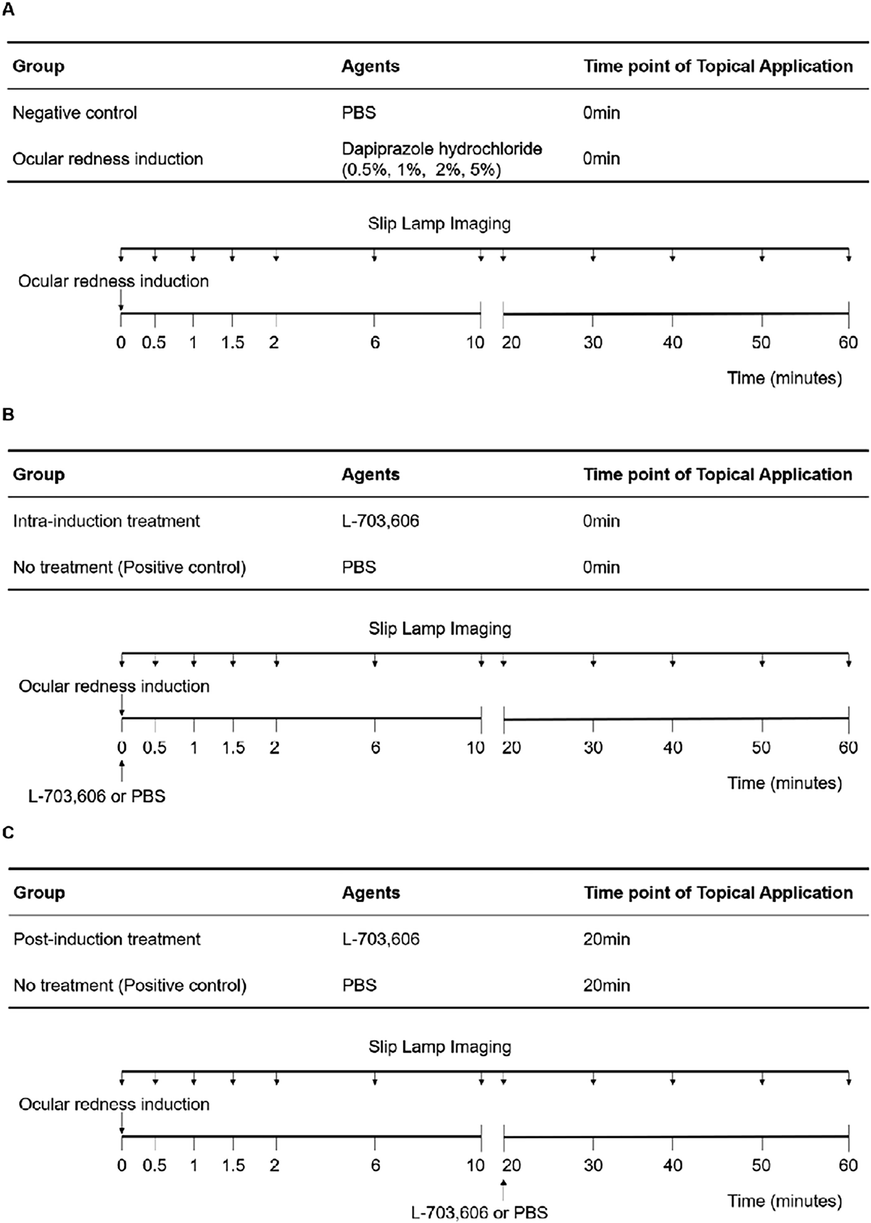
Study design. Schematic diagram of the kinetic evaluation for the development of ocular redness.
Biomicroscopic imaging
A digital camera-equipped biomicroscope was used to acquire the close-up conjunctival images. The animals were anesthetized, and then images were taken at baseline (before redness induction), every 30 s for the first 2 min post-induction, followed by every 4 min for an additional 8 min and then every 10 min until 1 h post-induction.
Image analysis using ORI
ORI was measured on the acquired images in a masked fashion using an ImageJ-based analysis method developed by our lab.18,19 Briefly, the image file was opened on a computer, and a white-balance function was presented to the operator to standardize the color in the image using the white reference mark included in the photograph during its acquisition (Supplementary Fig. S1). The conjunctival area was defined by the observer with a seven-point region of interest selection tool to avoid lids, cornea, and other areas not intended for scoring. The selected area was analyzed by the program, and the ORI was automatically calculated in a continuous numeric centesimal scale, with higher values representing more severe redness.
Statistical analysis
The Mann–Whitney U test was used to evaluate the significance of differences among groups regarding the change in ORI score. SPSS 20.0 software was used for statistical analyses (SPSS Inc, Chicago, IL). P < 0.05 was considered statistically significant.
Results
A single topical application of 5% dapiprazole induces severe and persistent ocular redness
A series of increasing concentrations of dapiprazole (0.5%–5%) in 40 µL volume were tested as a single topical application, and ocular redness was induced in variable degrees in all tested concentrations. During the 1-h observing period post-induction, the changes in ORI score were rapidly increased within 30 s of dapiprazole application in all induction groups, followed by gradual progression to a plateau by 6 min, and then lasted stably until the end of observation. Compared with the vehicle (PBS) group, all dapiprazole induction groups showed significantly higher changes in ORI score at each of the time points evaluated (P < 0.05 for 0.5%, 1%, and 2% dapiprazole compared with PBS, P < 0.01 for 5% dapiprazole compared with PBS, Fig. 2). Moreover, compared with the 0.5% dapiprazole group, the significantly higher change in ORI score was observed in the 1% and 2% dapiprazole groups at the first 0.5 min (P < 0.05), whereas the change of ocular redness in the 2% dapiprazole group increased significantly at 1 min after induction, compared with the 0.5% and 1% dapiprazole groups (P < 0.05). There were no significant differences among the three groups (0.5%, 1%, and 2%) observed until the end of observation (Fig. 2). The maximal mean changes in ORI score in the 0.5%, 1%, and 2% dapiprazole groups were 10, 9, and 12, respectively, all occurring at the end of the observation period (1 h post-induction), whereas the 5% dapiprazole group showed the highest increase in ORI with the maximal mean change of 14 observed at 20 min post-induction (Fig. 2). No infections or other complications were detected during the study period.
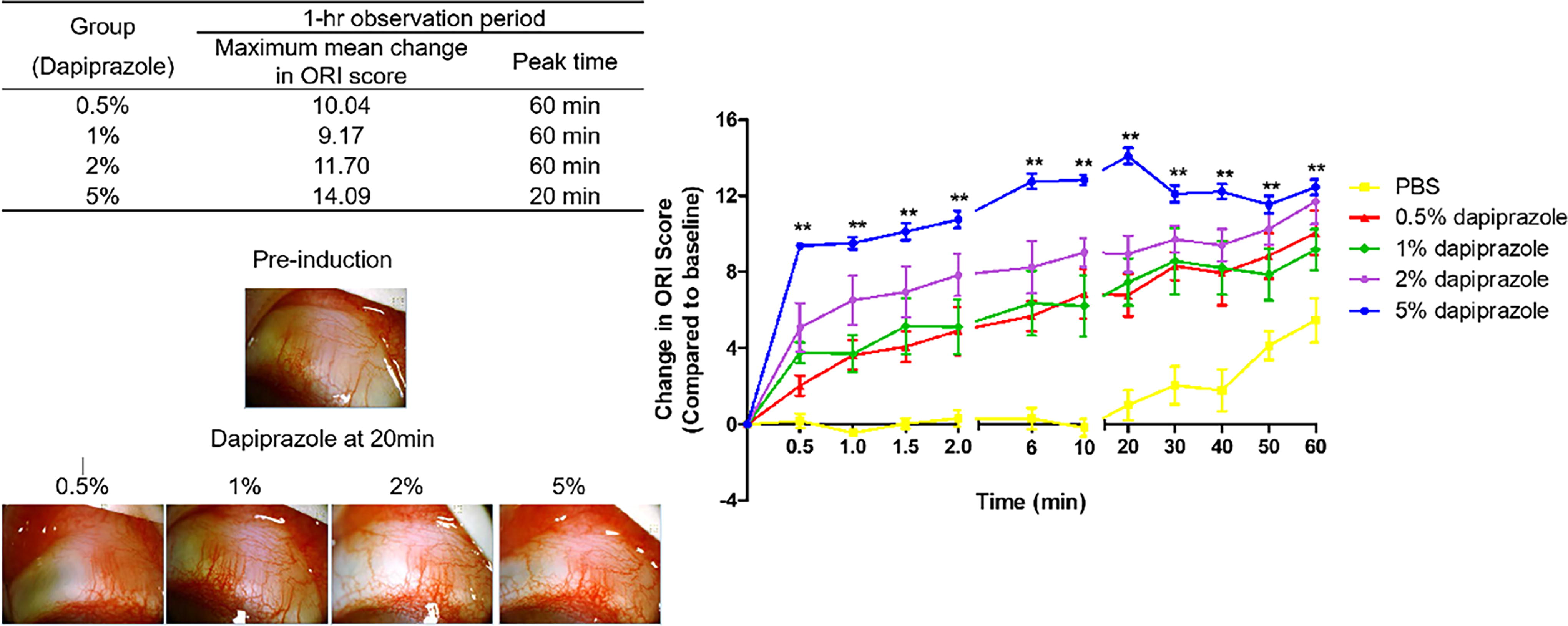
Topical dapiprazole induces ocular redness in rabbits. The table summarizes the maximal mean change in ORI scores post-induction and the time it occurs (peak time) during the 1-h observing period. Representative images show the development of ocular redness at 20 min, and the graph summarizes the kinetics of change in ORI score with data representing mean ± SEM (n = 6–8 per group). **, P < 0.01 for 5% dapiprazole vs. PBS. ORI, ocular redness index.
NK1R antagonism effectively reduces ocular redness
Since the “reddest eye” was induced by 5% dapiprazole in our rabbit model, this concentration was chosen to establish the disease model for testing the therapeutic efficacy of NK1R antagonism. In the intra-induction treatment group, L-703,606 was applied at the same time as dapiprazole instillation. The treatment led to significantly lower changes in ORI score than that untreated group at all time points assessed (around 40% decrease, P < 0.01, Fig. 3), with the maximal mean score change reduced to 8 at 10 min after induction/treatment. Furthermore, in the post-induction treatment group, L-703,606 was applied when the ocular redness reached its peak at 20 min after induction. This single treatment also rapidly and effectively reduced changes in ORI score at 30 min, 40 min, 50 min, and 1 h compared with the untreated group, with >30% reduction at the end of observation (Fig. 4).

Topical NK1R antagonism suppresses the development of ocular redness. Animals were topically treated with 1.0 mg/mL L-703,606 (a highly selective and potent NK1R antagonist) at the same time of redness induction with 5% dapiprazole. The kinetics of change in ORI score with data representing mean ± SEM is summarized, with representative images at 20 min shown (n = 8 per group). **, P < 0.01 as compared with vehicle (PBS)-treated group. NK1R, neurokinin-1 receptor.

Topical NK1R antagonism ameliorates existing ocular redness. Animals were topically treated with 1.0 mg/mL L-703,606 at the peak of the disease (20 min after dapiprazole instillation). The kinetics of change in ORI score with data representing mean ± SEM is summarized, with representative images at 40 min shown (n = 8 per group). *, P < 0.05, **, P < 0.01 as compared with vehicle (PBS)-treated group.
Discussion
Ocular redness is primarily due to conjunctival hyperemia resulting from dilation of conjunctival microvessels because of various local stimuli, and thus, the treatment ideally should, whenever possible, specifically target the underlying causes.3,4 However, some patients suffer from conjunctival hyperemia without a clear underlying ocular surface disorder. Ophthalmical decongestants (vasoconstrictors) and anti-inflammatory ophthalmical medications are the most commonly prescribed drugs for the treatment of this condition, with various adverse effects limiting their long-term use. 5 In the current study, we have successfully established a novel robust rabbit model of nonallergic ocular redness using topical dapiprazole (an adrenergic antagonist leading to vasodilation) and have shown that topical application of an NK1R antagonist L-703,606 effectively treats the nonallergic ocular redness in this model.
The discovery of new therapeutic management for ocular redness requires the use of animal models. Based on the underlying etiology of conjunctival hyperemia, different animal models have been developed over the years.20–22 A majority of the available animal models were established to study mechanisms of specific underlying diseases, such as allergic conjunctivitis, and can hardly provide insights into the conjunctival microvascular dynamics.23,24 To allow studying the precise development of ocular redness in response to nonallergic stimuli, the ideal in vivo model system should have a proper induction time period, provide enough conjunctival captured area, and can be reused to minimize the use of animals. Bimatoprost was reported to induce conjunctival hyperemia during the treatment of glaucoma,25,26 but its induction time was longer than 12 h in both animals and patients, which is too long for us to observe the kinetic development of ocular redness. Clementi and colleagues studied the vasodilatory effects of adrenomedullin in rabbits with intravenous injection, 27 which could induce systemic reactions that prevent the reuse of animals. In the present study, topical dapiprazole hydrochloride was used. As a selective α1-adrenergic antagonist, dapiprazole induces less systemic adverse effects (such as postural hypotension, reflex tachycardia, and adverse gastrointestinal effects) than nonselective α-adrenergic antagonists, such as phentolamine and phenoxybenzamine. 28 Furthermore, topical dapiprazole has been consistently proven to cause conjunctival hyperemia in clinical studies.29–32 Peterson and colleagues demonstrated that the 0.5% dapiprazole hydrochloride successfully induced significant nonallergic ocular redness in 2 min after topical application on human subjects. 33 Given the effectiveness in OR induction, short-acting period, and minimal systemic effects, dapiprazole was chosen as an ideal agent to test its effect in creating a new animal model of nonallergic ocular redness. As expected, our results in rabbits have shown that the topical application of dapiprazole rapidly induces conjunctival hyperemia with a dose-dependent trend, with 5% dapiprazole leading to the reddest eyes peaked at 20 min after induction and maintained stably during the 1-h observation period. Thus, a single topical application of 5% dapiprazole in rabbits represents a robust nonallergic red eye model for further in vivo studies. Notably, we observed that there were almost no significant differences among the three lower groups (0.5%, 1%, and 2%) during the observation time, except for the first 1 min. This finding is consistent with the previous studies, which investigated the different concentrations of dapiprazole eyedrops on reversal of mydriasis tested in patients. Wilcox and colleagues found that the effects of 1 drop of 0.5% dapiprazole were equivalent to 1 + 1 drop and 2 + 2 drop regimens at individual time points on reversal of mydriasis. 32 Hogan’s study also supported this finding. 31
SP is an important neuropeptide that functions as a neurotransmitter and inflammatory mediator, as well as a vascular tone regulator, and thus, it could potentially be involved in the development of red eye.34,35 SP is primarily released from the peripheral nerves and exerts its biological activities via interacting with its high-affinity receptor NK1R. 10 It has been reported that SP levels are increased in the tears of patients with allergic conjunctivitis compared with healthy individuals. 34 A recent study suggested that the ablation of the SP-NK1R signaling pathway in the nasal mucosa can have a therapeutic effect in allergic rhinoconjunctivitis. 36 Our recent work shows topical NK1R antagonism effectively alleviates the red eye in a preclinical model of allergic ocular redness. 17 As a parallel work to this study, we herein demonstrate that topical application of L-703,606 also effectively suppresses the development of nonallergic ocular redness when applied at the same time of disease induction by both lowering the redness severity and shortening the time-to-peak. Furthermore, topical L-703,606 applied at the disease peak effectively treats the existing disease. L-703,606 is a nonpeptidergic antagonist of NK1R. Compared with other NK1R antagonists, such as spantide I and spantide II, L-703,606 has a higher affinity to the NK1R while without pro-histamine-releasing effects.37,38 Moreover, Tao and colleagues demonstrated that L-703,606 could inhibit SP-NK1R-mediated plasma extravasation and suppresses the pathological increase of vascular permeability in the skin tissue. 39 In our case, mechanistically, signals of dapiprazole-induced vasodilation can induce the release of neuropeptides, including SP, because of the dense innervation of the ocular surface, and the increased SP can directly promote the vasodilation of existing blood vessels, resulting in plasma extravasation,13,14 as well as promote an inflammatory response that can further exacerbate ocular redness, and thus, blockade of SP-NK1R signaling pathways could not only inhibit vasodilation of conjunctival tissue but also prevent SP-induced inflammatory response at ocular surface. 40 Our present work serves as a proof-of-concept study that has focused on the therapeutic effect of NK1R blockade on nonallergic ocular redness, and the underlying mechanisms of action (such as dapiprazole-induced vasodilation leading to a burst release of SP) for the treatment warrant further studies. In addition, it should be noted that there are anatomical differences between rabbits and humans. The ORI score system was developed for clinical use with temporal conjunctiva as the ideal captured area.18,19 In our case, since the rabbit was anesthetized during the experiment, we could only capture the superior conjunctival area.
Conclusions
Our data suggest that NK1R antagonism may be an effective therapeutic approach for preventing and reducing nonallergic ocular redness. Further extended and mechanistic studies are required to determine the long-term efficacy/side effects, optimal treatment dosages, and precise vasoconstriction mechanisms.
Footnotes
Authors’ Contributions
R.D. and Y.C. conceived this study. R.D., Y.C., L.L., and J.Y. designed experiments. L.L., S.W., T.B., and S.Z. performed experiments. L.L., S.W., and H.G. analyzed data. L.L., S.W., and Y.C. wrote the original article. R.D. and Y.C. reviewed and revised the article. All authors contributed to the refinement of the study protocols and approved the final article.
Author Disclosure Statement
R.D. and Y.C. are inventors of a patent related to the anti-inflammation of targeting substance P in ocular surface diseases (owned by Massachusetts Eye and Ear).
Funding Information
This work was supported by National Institutes of Health grants EY20889 (R.D.) and P30EY003790 (Massachusetts Eye and Ear).

References
Supplementary Material
Please find the following supplemental material available below.
For Open Access articles published under a Creative Commons License, all supplemental material carries the same license as the article it is associated with.
For non-Open Access articles published, all supplemental material carries a non-exclusive license, and permission requests for re-use of supplemental material or any part of supplemental material shall be sent directly to the copyright owner as specified in the copyright notice associated with the article.
