




Abstract
Purpose:
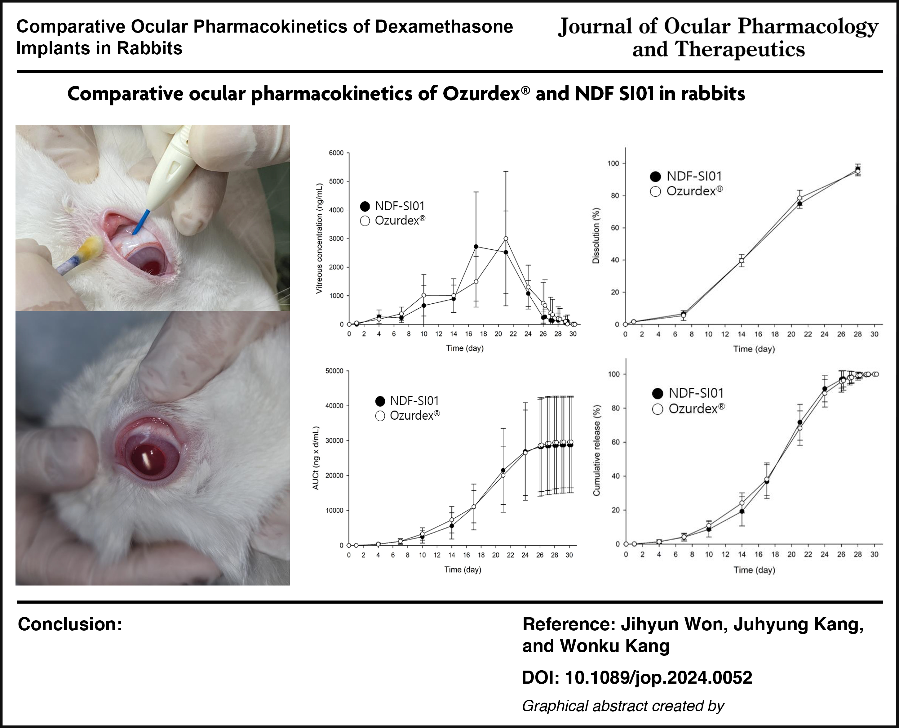
Dexamethasone eye implant has been used to treat macular edema and non-infectious uveitis. To date, its ocular pharmacokinetics are not fully characterized, and the development of generic preparations is in progress, as the patent of the original brand expires soon. Therefore, this work was designed to 1) determine the time course of vitreous dexamethasone concentrations following intravitreal implantation in rabbits and 2) explore the alternative use of NDF-SI01 from a pharmacokinetic point of view compared to Ozurdex®, which is currentlyused in the market.
Methods:
Ozurdex® and NDF-SI01 were implanted into the right and left eyes of the rabbit, respectively. A serial vitreous collection was performed to minimize the sacrifice of animals, and dexamethasone concentrations were measured by HP LC-MS/MS.
Results:
After implantation, dexamethasone concentration reaches the maximum concentration (3.1 μg/mL) in 19.5 days and decreases with a half-life of 40.3 h. AUC and clearance are 683.9 μg·h/mL and 1.29 mL/h, respectively. There is no significant difference in pharmacokinetic parameters between NDF-SI01 and Ozurdex®. The overall patterns of the cumulative release of both implants are similar.
Conclusions:
NDF-SI01 could alternate Ozurdex® in clinics based on the in vivo comparative pharmacokinetic study and in vitro dissolution test.
Introduction
Ozurdex® is an eye implant agent containing dexamethasone as the main ingredient to treat macular edema following retinal vein occlusion, diabetic macular edema, or noninfectious uveitis accompanied by posterior segment inflammation.1,2 The dexamethasone eye implant is manufactured by hot-melt extrusion with two poly(lactic-co-glycolic) acid (PLGA): Resomer® RG 502, which is ester-terminated, and Resomer® RG 502H, which is acid-terminated. 1
Bhagat et al. 3 and Chang-Lin et al. 4 representatively published the ocular pharmacokinetics of dexamethasone in the vitreous humor after implantation of the dexamethasone eye preparation in rabbits. In the former study, the implant was administered after dividing into three pieces and compared with the results with one intact piece. In the latter, the time course of dexamethasone in vitrectomized eyes was compared with that in nonvitrectomized ones. The vitreous body was collected for 28 and 30 days, respectively. However, because the concentration of dexamethasone in the vitreous body was still at the plateau, it was insufficient to obtain information about the appropriate pharmacokinetic parameters, such as half-life and clearance in the vitreous body of the rabbit, even though the comparison of area under the time courses of vitreous concentrations would be enough to assess the vitreous exposure of the drug on two occasions, respectively.
As the patent for Ozurdex expires soon, several companies may prepare to develop generic drugs. However, it should be challenging to carry out a bioequivalence study in healthy human volunteers because of such an invasive administration route. Clinical trials with patients also take time, and recruiting appropriate patients can be complex. In addition, more variables, such as drug–drug interactions, will need to be considered depending on the patient’s situation. Therefore, a proper animal model would require an alternative study design. A review article published by Del Amo and Urtti compared the clearance correlations of various drugs in human and rabbit eyes. 5 They reported that dexamethasone showed a very high correlation. Subsequently, comparing the pharmacokinetics of dexamethasone preparations using rabbits is comparable with clinical trials conducted in volunteers.
This study was designed to precisely investigate the ocular pharmacokinetics of the dexamethasone implant in the rabbit vitreous humor and compare a generic preparation (NDF-SI01) of Ozurdex in vitro and in vivo experiments. A serial vitreous sampling was conducted to minimize the number of animals and interindividual variations.
Methods
Materials
Dexamethasone, diclofenac (as an internal standard), and Resomer RG 502H and 502 were purchased from Sigma-Aldrich Korea. High-performance liquid chromatography (HPLC)-grade methanol and acetonitrile were obtained from Burdick & Jackson (Muskegon, MI, USA). The other chemicals were of the highest analytical grade available.
Preparation of ocular implants
Ozurdex (lot No. E94465) and the test item (NDF-SI01, lot no. SI01-022) were provided by Sangmyung Innovation Co. Ltd. NDF-SI01 was prepared using the double-melt extrusion and shaping process, the same as with Ozurdex. Briefly, the implant was composed of dexamethasone, Resomer RG 502H, and 502 (6:3:1). Extrusion was performed using a Haake miniCTW twin-screw extruder (Thermo Electron GmbH) at 105°C, 100 rpm, fitted with a 0.5 mm round die at the discharge port to target 430–450 µm diameter. The implant was manually cut to 6 mm and inserted into an applicator. After final packing, the preparation was sterilized by electron beam irradiation: The extruded implants were irradiated with 25 kGy at room temperature using a 5 MeV RF linear accelerator on a moving tray (2 m/min). The accelerator was set at a frequency of 25 Hz with 394 mA, and the beam current was scanned to 5.02 mA. Both-side irradiation was performed to achieve an irradiation dose of 25 kGy. All processes were designed to get 700 μg of dexamethasone per implant.
In vitro dissolution test
A dissolution test was conducted to ensure the contents of dexamethasone in NDF-SI01 and Ozurdex for 28 days, as previously reported by Bhagat et al. 3 The implant was taken from the applicator of Ozurdex with the same lot number used in the animal experiment. A Prominence LC-20A series HPLC system (Shimadzu Corp.) with an ultraviolet detector was used to quantify dexamethasone in vitro solution.
Animal study
The animals used in this study were eight 3-month-old New Zealand White rabbits. The animal room was maintained at a temperature of 23 ± 3°C, relative humidity of 50 ± 10% with 10–20 air changes/h, and light intensity of 300 Lux with a 12 h light/dark cycle. The study protocol was approved by the Institutional Animal Care and Use Committee at Chung-Ang University (approval no. 202301020062), and their recommendations for animal care and handling were followed in adherence to the Association for Research in Vision and Ophthalmology statement. After anesthesia with alfaxalone (35 mg/kg) and xylazine (4 mg/kg), Ozurdex and NDF-SI01 were implanted into the right and left eyes of the rabbit, respectively. About 20 μL of vitreous humor was serially taken under anesthesia before and after administration at Days 1, 4, 7, 10, 17, 21, and 24 once a day. All samples for 24 days were stored at −70°C until analysis. From Days 26 to 30, the samples were taken twice in 6 h and immediately prepared for quantification of dexamethasone to ensure the necessity of further sampling.
Quantification of dexamethasone and data analysis
Vitreous samples (10 μL) were mixed with acetonitrile, including an internal standard (100 ng/mL diclofenac, 30 μL), vortex-mixed for 10 s, and centrifuged at 15,000 rpm for 10 min. The supernatant was injected into the API 4000 liquid chromatography with tandem mass spectrometry system (AB Sciex). The analytical method was slightly modified from the previous article published by Gu et al. 6
Calibration standards for dexamethasone ranged from 1 ng/mL to 1,000 ng/mL, and quality control samples were prepared at 1 ng/mL for the lower limit of quantification and 5, 200, and 800 ng/mL for low, intermediate, and high concentrations, respectively. The samples were used to evaluate the intra- and interday precision and accuracy. Extraction recovery, matrix effect, and dilution effect (10-fold) were also examined. The stability of dexamethasone in vitreous humor was examined before the validation process: room temperature for 4 h, 3-cycle freeze–thaw, postextraction for 24 h, and long-term for 4 weeks.
Pharmacokinetic parameters were obtained from the time courses of vitreous dexamethasone concentrations: peak concentration (Cmax) and time to Cmax (Tmax) were directly read from the curve. The elimination rate constant (k) was estimated by linear regression from the log-transformed vitreous concentrations at the terminal phase. The half-life was given by 0.693/k. The area under the vitreous humor concentration-time curve (AUC) was calculated using the trapezoidal rule, and the last vitreous concentration/k was added to get AUCinf. Clearance was calculated by dose/AUCinf.
Statistics
Each pharmacokinetic parameter was shown as mean and relative standard deviation (RSD, %) and statistically compared using a two-tailed unpaired Student’s t-test. Statistical significance was taken as P < 0.05.
Results
Animal study
Figure 1 depicts the time courses of vitreous dexamethasone concentrations following the intravitreous implantation of Ozurdex and NDF-SI01 in rabbits, and the pharmacokinetic parameters of the two preparations are listed in Table 1. The dexamethasone concentration of the test item continued to increase after implantation, reaching the maximum concentration of 3.1 μg/mL (RSD, 57%) in an average of 19.5 days (RSD, 11%). Afterward, it decreased with a half-life of 40.3 h (RSD, 41%). AUC and clearance were 683.9 μg·h/mL (RSD, 48%) and 1.29 mL/h (RSD, 53%), respectively.
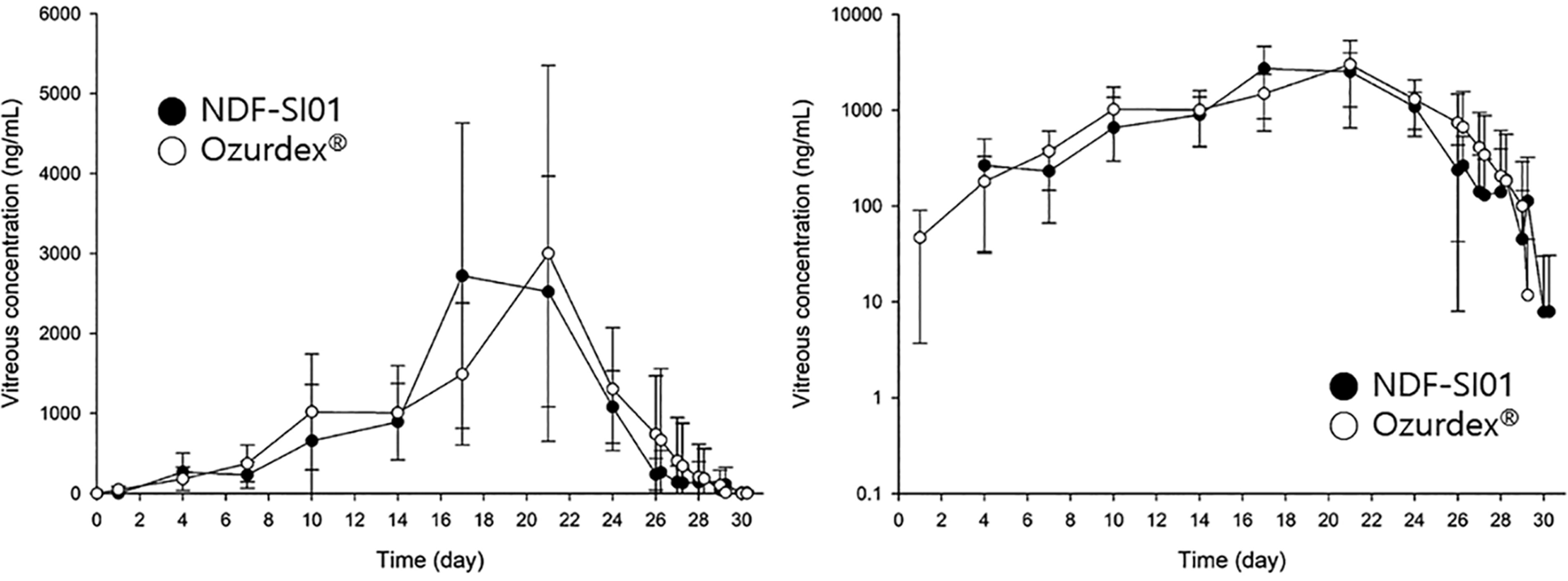
Time courses of vitreous dexamethasone concentration after intravitreous implantation of Ozurdex® (n = 8) and NDF-SI01 (n = 8) in New Zealand White rabbits. The right represents a semilog plot. Error bars indicate standard deviations.
Pharmacokinetic Parameters of Dexamethasone Ocular Implants in Rabbit Vitreous Humor
Data are mean (RSD, %).
AUC, area under the vitreous humor concentration-time curve; RSD, relative standard deviation.
Ozurdex shows a similar pattern to the test preparation and reached 3.3 μg/mL (RSD, 65%) in 20.9 days (RSD, 17%). There is no statistical significance for both parameters. The half-life (35.4 h; RSD, 30%) of Ozurdex is 12% shorter compared with NDF-SI01 without statistical significance. AUC 708.1 μg·h/mL (RSD, 53%) and clearance 1.27 mL/h (RSD, 51%) are also similar.
Figure 2 represents the time course of AUCt. The overall patterns of both preparations are similar, and there is no statistical significance at each time. Figure 2 was reorganized to depict the cumulative release (%) of the implants, that is, AUCt at each time was represented as the percentage of AUCinf by dividing by AUCinf (Fig. 3). Drug release is remarkable between Weeks 2 and 3.
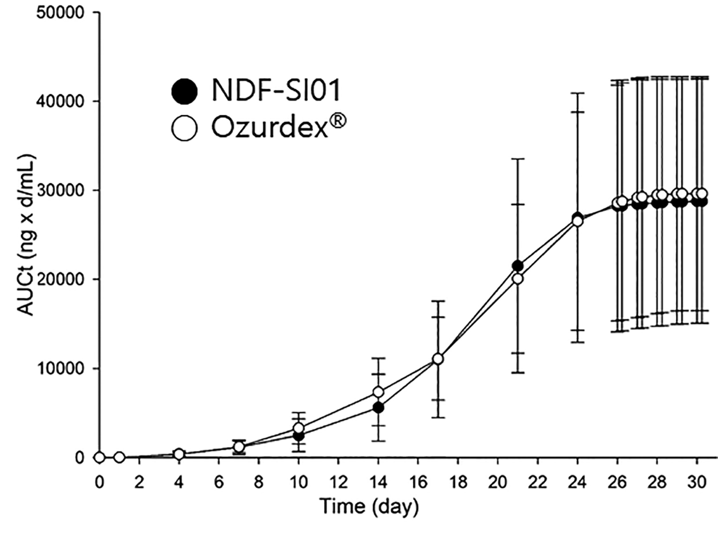
Time courses of cumulative area under the vitreous dexamethasone concentration–time curves after intravitreous implantation of Ozurdex (n = 8) and NDF-SI01 (n = 8) in New Zealand White rabbits. Error bars indicate standard deviations.
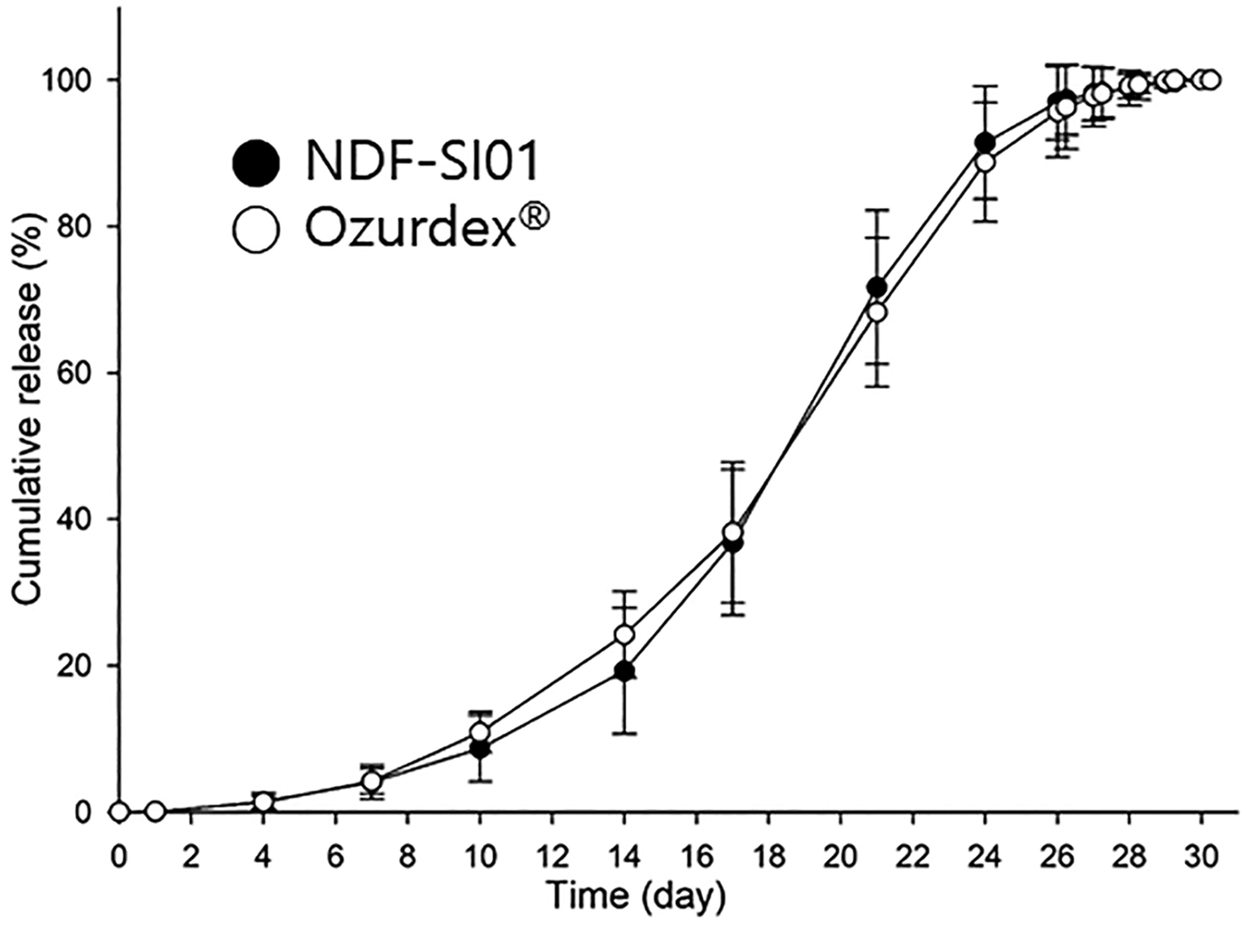
Time courses of fractional AUC (AUCt/AUCinf) after intravitreous implantation of Ozurdex (n = 8) and NDF-SI01 (n = 8) in New Zealand White rabbits. Error bars indicate standard deviations. AUC, area under the vitreous humor concentration–time curve.
Method validation
The calibration curves for determining dexamethasone in rabbit aqueous humor represented good linearity, R2 > 0.999. The detection and quantitation limits were 0.2 and 1 ng/mL at a signal-to-noise ratio of 3 and 5, respectively. The intra- and interday assay precisions were <7.5% and 8.3%, respectively. The intra- and interday assay accuracy ranged from 95.3% to 102.4% and 94.3% to 105.1%, respectively. The mean recovery of dexamethasone was more than 80%, and the matrix effect was more than 92%. Ten-fold dilution with drug-free aqueous humor did not affect the quantitation of the samples over the upper range of the calibration curve. Dexamethasone in vitreous humor was stable for all stability tests.
In vitro dissolution test
Figure 4 represents the in vitro dissolution test for the two preparations. The drug release patterns of both preparations are nearly the same, and no statistical significance was shown at each time. The in vitro results are in accordance with those in Fig. 3.
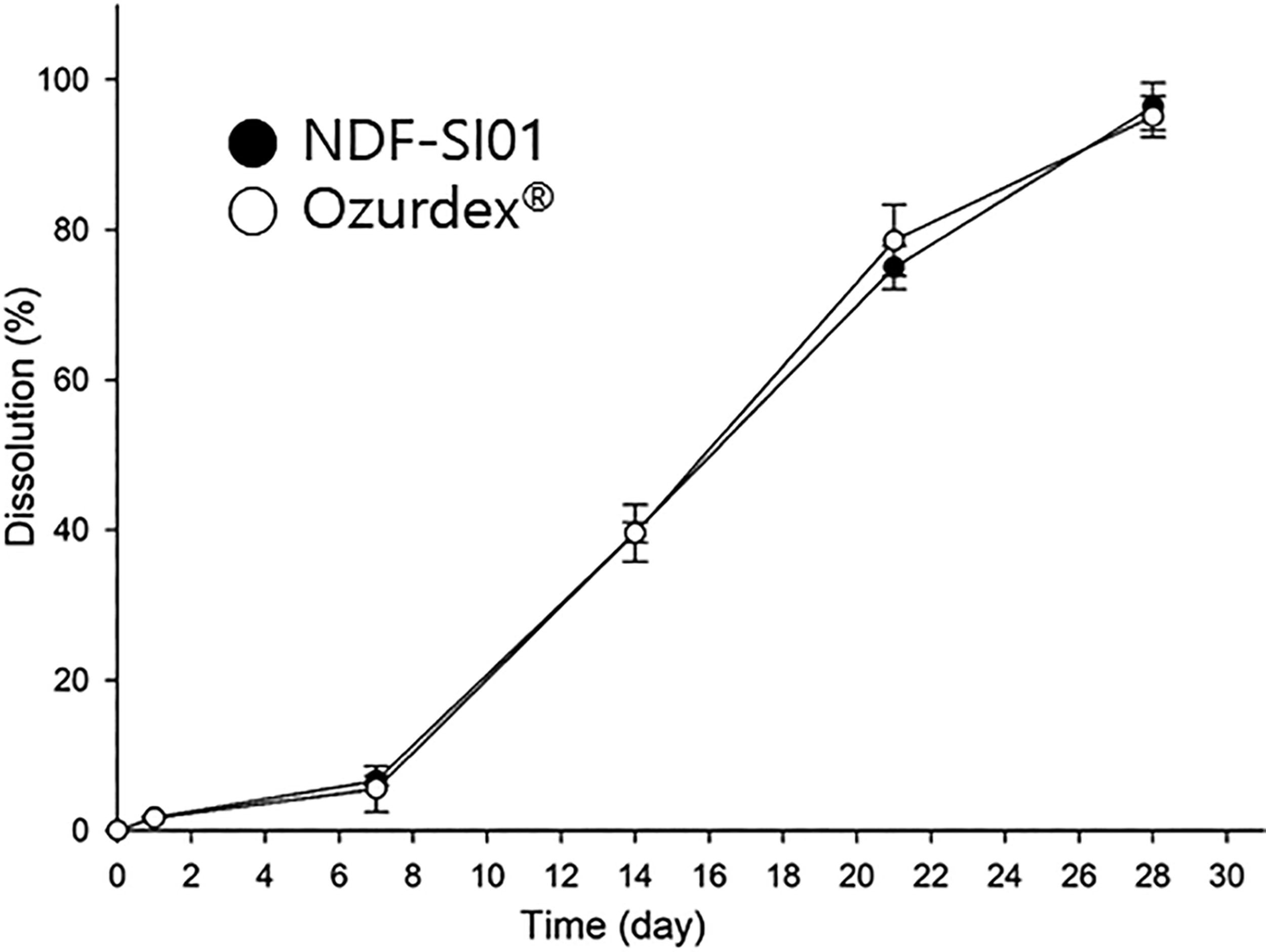
In vitro dissolution test of NDF-SI01 (n = 6) and Ozurdex (n = 6). Error bars indicate standard deviations.
Discussion
Bhagat et al. 3 conducted a study to confirm that fragmentation may occur when administering the implant and that, in this case, dissolution might be accelerated, and exposure of the drug in the vitreous body could be rapidly increased. In contrast to the concerns, there was no difference compared to the results of administering the implant after cutting it into three pieces, inferring that even if the eye implant is divided into segments, the increase in the part in direct contact with the vitreous body is limited to the area (about 0.2 cm2) of the cutting edges. Subsequently, the increase in the dissolution rate should be insignificant. Interestingly, the first sample was collected 3 h after administration, and intravitreal concentrations were measured at about 500 ng/mL. Despite no comments provided by the authors, it was unusual considering the slow release pattern of PLGA preparations. As shown in the present data (Figs. 1 and 3), the eye implant based on PLGA is a sustained-release material that slowly decomposes after contact with moisture, allowing its active ingredient to dissolve. The concentration in the vitreous continued to increase until about 20 days after the administration of both preparations and then decreased and mostly disappeared by Day 28. Chang-Lin et al. 4 also reported comparative ocular pharmacokinetics of dexamethasone after intravitreal implantation of Ozurdex in rabbit eyes in the presence and absence of vitrectomy. There was no significant effect of vitrectomy on the pharmacokinetics of the drug, and the vitreous concentrations of dexamethasone continued to increase until Day 22 and then tended to decrease on Day 31.
At the beginning of this work, we struggled to find reasonable vitreous sampling times, especially after Day 28, to figure out the ocular pharmacokinetics of dexamethasone precisely since information had yet to be shown on the terminal phase of the implant in the eye. The two works cited above only focused on the differences between the two circumstances.
By the way, Graham and Peyman reported a half-life of 3 h. 7 However, that was attributed to an improper calculation in the distribution phase, and if the half-life in the terminal phase is calculated correctly, it should correspond to about 32 h. Unfortunately, the incorrect half-life was recently cited in a review article. 1 In the literature reported by Kwak and D’Amico, 8 the half-life measured after intravitreal dexamethasone injection was also 3.5 h because vitreous humor was not collected long enough, and the concentration was quantified with an instrument representing poor sensitivity. The half-life of dexamethasone in plasma was about 2 h following intravenous administration in rabbits. 9 A slow flow of vitreous humor could be attributable to the long half-life, although no experimental results have been found in rabbits. The vitreous generation and flow in humans (2.5–3 μL/min) could speculate on the slow movement of vitreous humor in the rabbit as well.
Based on the literature, we decided on the sampling times as represented in the section on method and immediately quantified the concentrations of vitreous samples from Day 26 to make sure when the decay could be observed. Consequently, the ocular pharmacokinetics of dexamethasone implants were properly characterized in rabbits for the first time, to the best of our knowledge.
One may be concerned about the impact of such frequent samplings of the vitreous humor. Since, however, 20 μL of the vitreous was taken at each schedule, the amount would not cause any physiological and pharmacokinetic consequences considering the total volume (1.2–1.7 mL), 10 flows, and generation of the vitreous humor in rabbits. 11 In addition, we explored the serial vitreous samplings used in this study compared with point one in the conventional design,3,4,10 which was to sacrifice animals at each sampling schedule before starting the present study. As shown in Fig. 5, no significant difference was found between the two methods. One may speculate that many animals should be needed to figure out the terminal phase, which might hinder the previous works from representing the decay of dexamethasone in the vitreous after implantation.3,4 Each preparation of Ozurdex and NDF-SI01 was implanted in each eye in a rabbit, also contributing to minimizing the animals. The systemic exposure of dexamethasone and its major metabolite, 6β-hydroxy dexamethasone, was below the lower limit of quantitation (0.1 ng/mL) when Ozurdex was implanted in the eye. Therefore, one can rule out the increase in vitreous dexamethasone concentration in the counterpart eye.
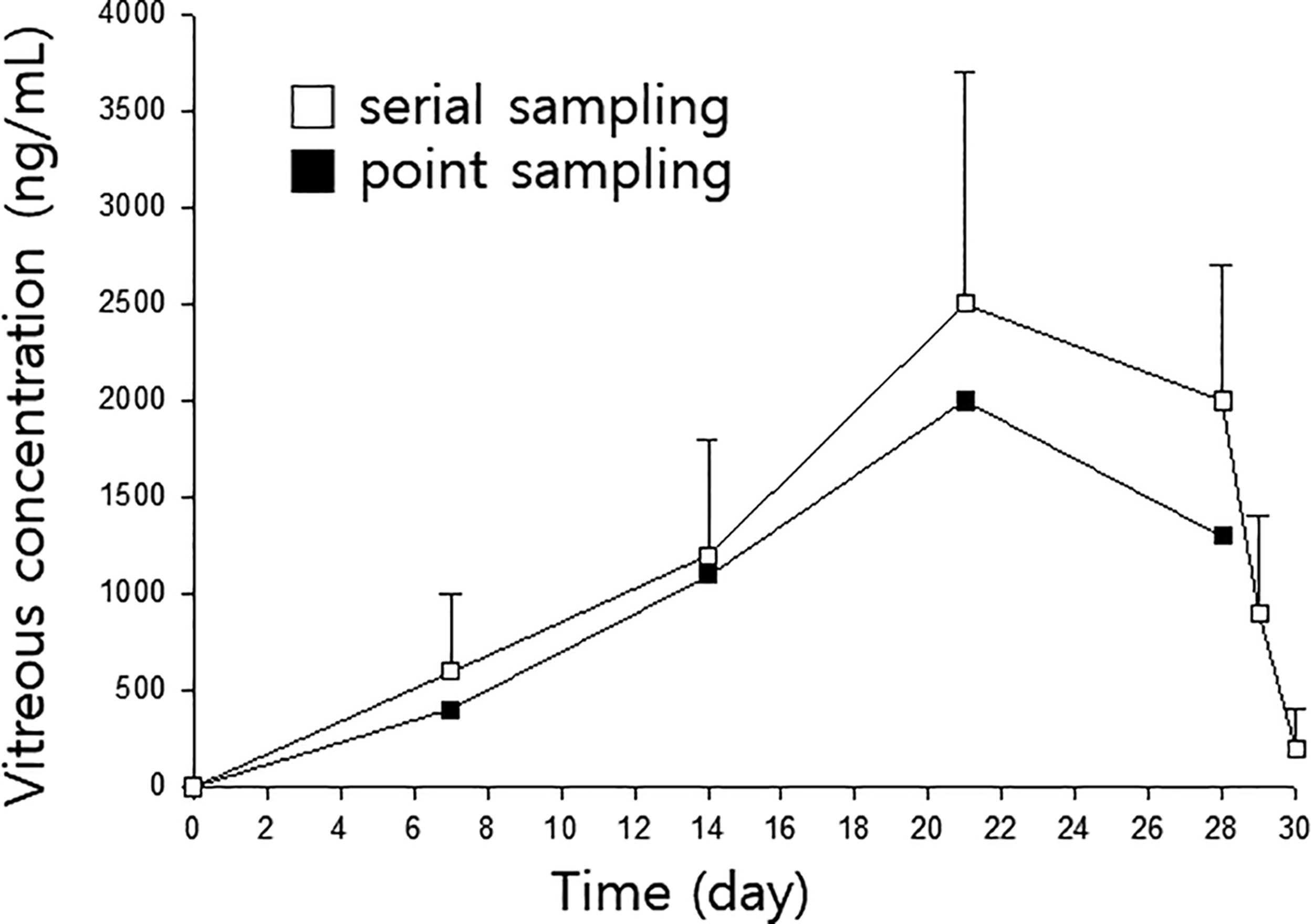
Time courses of the cumulative area under the vitreous dexamethasone concentration–time curves after intravitreous implantation of Ozurdex in New Zealand White rabbits in a pilot study. □, serial sampling in 3 rabbits; ■, point sampling after sacrifice in 4 rabbits. Error bars indicate standard deviations.
The interindividual variability of Ozurdex, representing the RSD, was 54%–133%,3,4 which was relatively significant considering such a small vitreous chamber. We expected the variation to be reduced when serial sampling was conducted on the same subject. However, that was still high, although no such variability was seen in the in vitro dissolution test. Therefore, one may regard the interindividual variation as stemming from the release pattern of the PLGA-based implants in vitreous humor. Further study is needed to dig into the fundamentals.
As a result of the in vitro dissolution test, NDF-SI01 showed no difference from Ozurdex. In addition, even after intraocular application in rabbits, there was no significant difference in the intraocular release, distribution, and disappearance of dexamethasone compared with the reference product. NDF-SI01 could be interchangeable with Ozurdex.
In a review article published by del Amo and Urtti, 5 they compared the clearances of various drugs in human and rabbit eyes. Dexamethasone and ganciclovir represented a very high correlation. Consequently, the results of this study comparing the pharmacokinetics of dexamethasone preparations using rabbits may be comparable to clinical trials conducted on patients.
Summary
In conclusion, a serial vitreous sampling was successfully performed and resulted in the minimal sacrifice of rabbits. The ocular pharmacokinetics of dexamethasone eye implants were fully characterized in rabbits, including their terminal elimination. Based on the in vitro dissolution test and in vivo comparative pharmacokinetic study, NDF-SI01 could be an alternative to Ozurdex in clinics.
Footnotes
Acknowledgment
The authors would like to thank Sangmyung Innovation Co. Ltd. for their support during the study.
Authors’ Contributions
J.W.: Methodology, data curation, and writing. J.K.: Methodology, visualization, and data curation. W.K.: Conceptualization, writing, and review.
Author Disclosure Statement
The authors declare no conflict of interest.
Funding Information
This work was supported by the