
Abstract
Abstract
Certain physical signs can be useful in predicting impending death. We present four terminally ill patients with malignancy who developed petechiae on their bilateral palms and fingers during the week prior to their deaths. Drug-induced eruption, injury, and mechanical stimuli were thought to be unlikely causes of the petechiae. While the phenomenon has not previously been reported, we speculate palmar petechiae may be a clinical cutaneous sign of systemic deterioration in terminally ill patients.
Introduction
Several researchers have stressed the importance of advising caregivers about the common signs that death may be imminent (e.g., deterioration in consciousness, terminal delirium, retained respiratory secretions, audible respirations with mandibular movement, cyanosis of extremities, and no palpable pulse).7,8 Here, we describe four cases in which palmar petechiae appeared to be an indicator of impending death.
Case Reports
Case 1
An 89-year-old male with stomach carcinoma, liver metastasis, and dementia transferred from a nursing home to our palliative care unit for end-of-life care. On admission, he was bed-bound, only able to ingest a small amount of a fluid diet, and suffering from abdominal pain due to liver metastasis (Palliative Performance Scale 9 [PPS]: 30%). Pain was controlled with buprenorphine, and anorexia was refractory to corticosteroids. Four days before his death, palmar petechiae appeared on his bilateral palms and fingers but these were not due to injury or mechanical stimuli (PPS: 30%; Fig. 1). The laboratory findings confirmed a normal platelet count (209 × 103 per microliter) and there were no skin eruptions on any other part of his body. Although he was able to have simple verbal conversation, his health status declined rapidly following the appearance of the palmar petechiae.
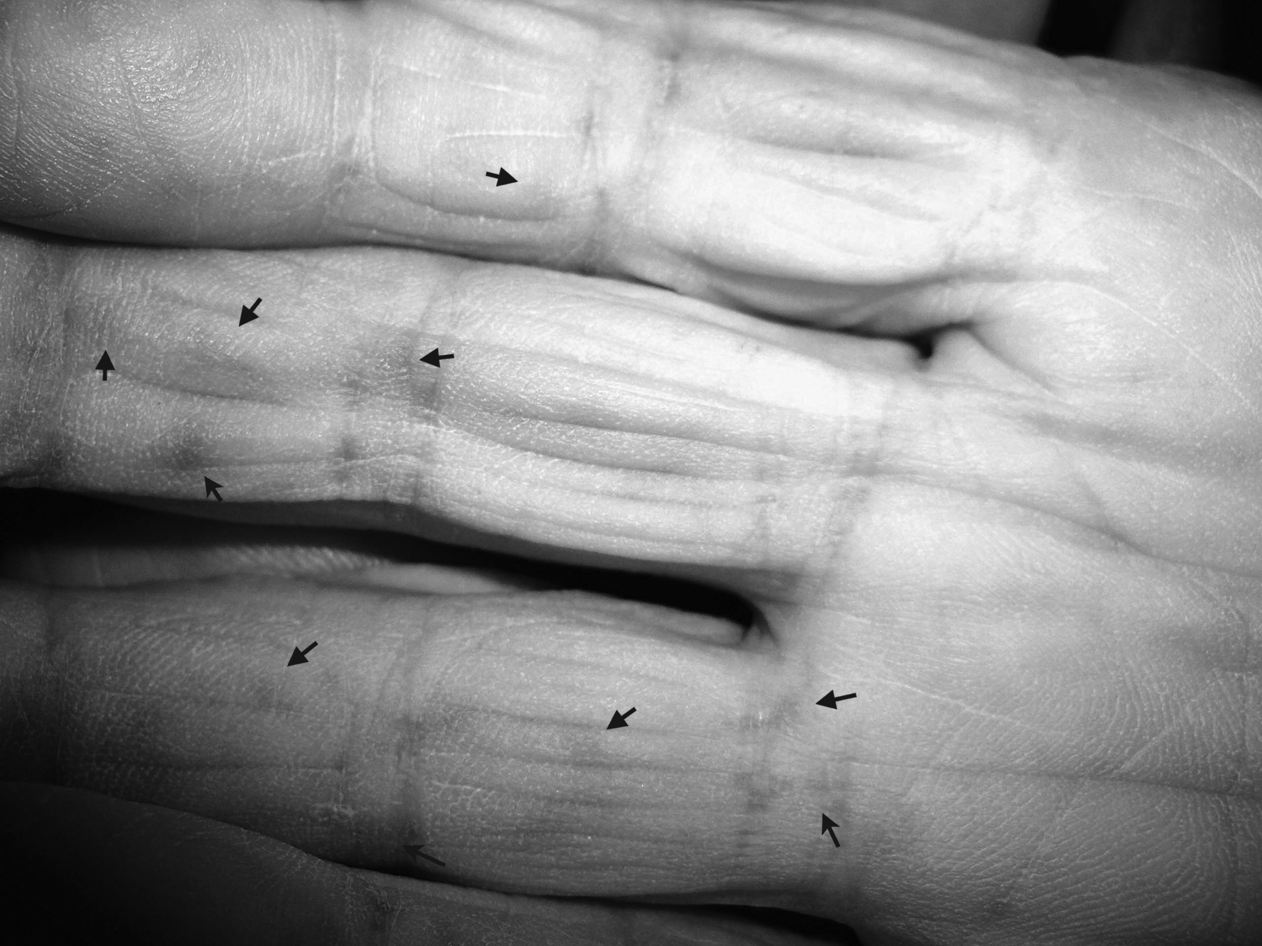
Multiple petechiae on palm and palmar surface of fingers (arrows) in case 1.
Case 2
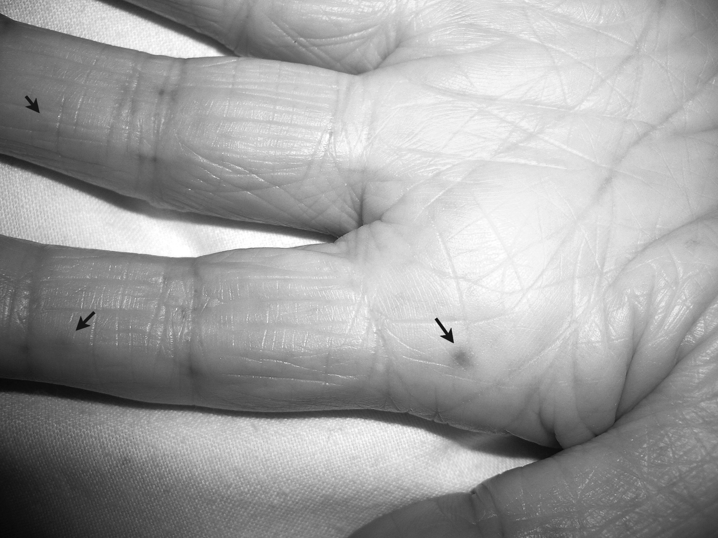
A 67-year-old male with stomach carcinoma, post-total gastrectomy and total pancreatectomy, was admitted to our palliative care unit because of increasing care needs. He continued regular insulin injection despite inadequate oral intake of food. On admission, he was comatose due to hypoglycemia (40 mg/dL). While the laboratory findings revealed thrombopenia (51 × 103 per microliter), no purpura or abnormal skin rash was detected anywhere on his body including his palms and fingers. After administering 50% dextrose, he recovered sufficiently to be able to communicate verbally and eat some food. Five days before his death, palmar petechiae appeared on both his palms and fingers (PPS: 40%; Fig. 2). He was still able to communicate verbally up to 4 hours before his death.

Multiple petechiae on palmar surface of fingers (arrows) in case 2.
Case 3
A 52-year-old male with adenoid cystic carcinoma, multiple bone metastases, and lung metastasis was admitted to our palliative care unit to alleviate dyspnea. On admission (PPS: 50%), he was able to walk around the ward with a portable oxygen tank (2 liters per minute). Providing oxygen and a single thoracentesis kept his dyspnea at a manageable level. The laboratory findings confirmed a normal platelet count (145 × 103 per microliter). Although palmar petechiae appeared on his bilateral palms and fingers a week before his death (PPS: 50%), his appetite and mobility remained satisfactory until 4 days before his death. He was able to converse and say goodbye to his wife 2 days before he died.
Case 4
An 83-year-old female with gallbladder carcinoma, multiple liver metastasis, and peritoneal carcinomatosis was admitted to our palliative care unit because of persistent anorexia. On admission, she was awake, alert and able to express herself despite being bed-bound and having severe anorexia. Hypodermoclysis, 200 mL/d, and dexamethasone, 4 mg/d, were administered to alleviate anorexia. A complete blood panel was not obtained following her admission. She improved sufficiently to be able to ingest a small amount of a fluid diet. However on the second day postadmission (PPS: 20%; Fig. 3), palmar petechiae appeared on her bilateral palms and fingers and she died suddenly 4 days later.
Multiple petechiae on palmar surface of fingers (arrows) in case 3.
The PPS reflects the functional dimensions of ambulation, activity level, self-care, oral intake, and level of consciousness. The PPS is divided into 11 levels, from PPS 0% to PPS 100%, in 10% increments. A patient at PPS 0% is dead, and at 100% is ambulatory and healthy. 9
We confirmed with the relatives of all four patients that the petechiae were not present prior to our detection. The appearance of the petechiae enabled us to inform relatives that the patient's death was likely to occur within a week.
The appearance of palmar petechiae before death occurred in 4 (2.1%) of 193 consecutive admissions to our palliative care unit in a year.
Discussion
All four patients died from their primary disease within 1 week of petechiae appearing bilaterally on their palms. An awareness of the possible associations between internal malignancies and cutaneous manifestations is important in the diagnosis and management of cancer. 10 To our knowledge, there are no previous reports of an association between malignant disease and petechiae. The features of this unique phenomenon are (1) the sudden appearance, (2) the pinhead-like, black, and round shape of the lesions, (3) the limited area and symmetric appearance on palmar surface of the fingers and palm, and (4) the petechiae are not the result of hemorrhage. Following their admission to hospital, all patients were observed carefully by their caregiver and medical staff, and assisted in their daily living. Therefore we believe that this skin reaction did not result from injury, an artificial reaction, or thermal or mechanical stimulation. In general, the petechiae appeared to be discrete, pinhead-like, less than 2 mm in diameter, and did not coalesce but were often clustered in peculiar sites. 11
Cutaneous manifestations may provide a useful indicator of disease activity and prognosis. 10 We speculate that palmar petechiae might be a clinical cutaneous sign that reflects a worsening of a patient's systemic condition. Unfortunately, the lack of an extensive hematological examination and skin biopsy, prevented us from determining the etiology of the palmar petechiae. A drug-induced skin reaction was excluded in all patients because of no temporal relation between drug administration and palmar petechiae appearance. Various forms of cutaneous lesions and their appearance in many different areas of the body characterize drug-induced eruptions. Pigmented purpuric dermatitis occasionally manifests as the characteristic punctate petechiae, but drugs are implicated in a minority of cases. 12 However, the limited area of the petechiae we have reported is not consistent with a drug-induced eruption. In the literature, the main cause of petechiae is thrombocytopenia and not malignancy. If the platelet count improves they may disappear without any trace in 2–3 days. 11
There are a few case reports of trauma as the cause of palmar petechiae. 13 It has been suggested that repetitive lateral shearing forces induced by sudden halting or slamming of the skin rupture the papillary vessels and result in extravasation of blood in the stratum corneum. Because the petechiae are often periductal, the openings of sweat glands may be the vector for transcorneal elimination of the extravasated blood. Other suggested pathogenetic mechanisms for the discoloration may be bacterial pigment formation in the sweat ducts or deposition of rubber particles in the skin. 13
In terms of a differential diagnosis, purpura as a skin reaction occurs in disseminated intravascular coagulation (DIC), thrombotic thrombocytopenic purpura (TTP), idiopathic thrombocytopenic purpura (ITP), mixed cryoglobulinaemia, and cortisone therapy. The rashes tend to appear all over the body as rusty coalescing spots which are larger than petechiae with a tendency to coalescence as result of bleeding and/or embolism.11,14 Acanthosis nigricans, a paraneoplastic cutaneous syndrome, is also clearly distinguishable by its appearance with velvety thickening, hyperpigmentation and larger spots that occur at axillae and flexural sites. 10
In our patients, early recognition of the phenomenon of palmar petechiae was the most accurate predictor of impending death. We were unable to correctly predict the prognosis of Case 2 and Case 3 because of the discrepancy in prognosis resulting from high performance status, as reflected by the patient's PPS. In one study the association between survival estimates with the PPS as measured in 773 palliative patients revealed a correlation between PPS on admission and mean survival times. The mean survival times for a PPS of 30% was 18 (range, 14–22) days, PPS 40% was 36 (range, 29–43) days, and PPS 50% was 51 (range, 40–62) days. 15 It was also found that the average period until death for 129 patients was 1.88 days at 10% PPS upon admission, 2.6 days at 20% PPS, 6.7 days at 30% PPS, 10.3 days at 40% PPS, and 13.9 days at 50% PPS. 9 These findings were not consistent with Case 2 (PPS: 40%) and Case 3 (PPS: 50%) as the patients died within a week of the apprearance of palmar petechiae.
Detecting and recognizing palmar petechiae as soon as they appear may help avoid the need for specialized, painful, and costly examinations and may help patients and families cope with impending death.
In conclusion, the appearance of palmar petechiae might be one indicator of impending death within one week for cancer patients. Further investigation is required to reveal the etiology and more accurately characterize the predictive value of this phenomenon.