




Abstract
Background:
Acne vulgaris is a common skin disease with a large quality of life impact, characterized by comedones, inflammatory lesions, secondary dyspigmentation, and scarring. Although traditionally considered a disease of adolescence, reports suggest it is also a disease of adults, especially adult women. Our objectives were to determine acne prevalence in a large, diverse group of women and to examine acne by subtype and in relation to other skin findings, measurements, and lifestyle factors.
Methods:
We recruited 2895 women aged 10–70 from the general population. Photographs were graded for acne lesions, scars, and dyspigmentation. Measurements were taken of sebum excretion and pore size, and survey data were collected.
Results:
Of the women studied, 55% had some form of acne: 28% had mild acne, and 27% had clinical acne, 14% of which was primarily inflammatory and 13% of which was primarily comedonal. Acne peaked in the teenage years, but 45% of women aged 21–30, 26% aged 31–40, and 12% aged 41–50 had clinical acne. Women with inflammatory acne were younger than those with comedonal acne (p≤0.001), and postmenopausal women had less acne than age-matched peers (p<0.0001). Acne was associated with facial hirsutism (p=0.001), large pores (p=0.001), and sebum excretion (p=0.002). Smokers had more, primarily comedonal, acne than nonsmokers.
Conclusions:
The cross-sectional design precludes conclusions about progression of acne with age. Participation was restricted to women. The photographic nature of the study imposes general limitations. Techniques used in this study were not sufficiently sensitive to identify cases of subclinical acne. More than a quarter of women studied had acne, which peaked in the teens but continued to be prevalent through the fifth decade.
Introduction
Acne vulgaris is a common disorder of the sebaceous follicles thought to develop when the adrenarche-driven increase in sebum production combines with blockage of the follicular orifice by retained keratinocytes, creating the comedone. 1 The inflammation seen in papules and pustules may result from proliferation in the follicle of Propionibacterium acnes or may be the result of another inciting event that causes chemotaxis of neutrophils. 1
The disorder is very common in adolescence, with prevalence estimates ranging from 47% to 90% during the teenage years, 2,3 and has considerable quality of life impact on sufferers. 4 Although most studies report peak prevalence in the midteens to late teens, there is evidence that acne, especially comedonal acne, is seen in premenarchal girls, 5 with one large survey demonstrating acne prevalence of 3% in 7–9-year-olds, 30% in 10–12-year-olds, 78% in 13–15-year-olds, and 93% in 16–18-year-olds. 6 Although acne often resolves in the third decade as adrenal androgens decrease, patients well beyond adolescence seek treatment for acne from dermatologists and primary care physicians.
Prevalence data on adult acne vary based on definitions of both acne and adult and are primarily derived from survey responses. A 1979 survey of patients 18–70 years of age revealed a peak prevalence of 18 years; 5% of women aged 40–49 had mild clinical acne, and 8% of women aged 50–59 had physiologic acne. 7 A 1997 study based on clinical grading showed that 12% of women aged 25–55 had clinical acne, and 48% had physiologic acne. 8 A 1999 survey of Australian women aged >20 established an overall acne prevalence of 13%, with 47% of those aged 20–29 and 30% of those 30–39 having acne. 9 A 2006 survey of Americans reported acne prevalence in women of 66.8% in the teen years, 50.9% in the 20s, 35.2% in the 30s, 26.3% in the 40s, and 15.3% in the 50s or older. 10
This study was designed to determine via clinical grading the relative frequency of acne, its subtypes, and sequelae across different age groups in a sample of racially and ethnically diverse women. We expected, based on published survey data, that acne would be seen in a significant proportion of women through the third, fourth, and even fifth decades. A second aim was to determine if previously implicated factors, such as cigarette smoking, skin sebum production, pore size, hirsutism, or body mass index (BMI), influence acne patterns in any age groups. Additional factors examined included alcohol consumption, caffeine consumption, lifetime sun exposure, education, and income.
Materials and Methods
Participants
The dataset consists of high-resolution digital images of the left side of the faces of 2895 women of five different population groups recruited from the general population in four different cities (Table 1). In Los Angeles, a shopping mall in an ethnically diverse neighborhood was chosen to maximize recruiting of different ethnicities: Caucasian American, African American, Hispanic American, and East Asian American (of Korean, Chinese, or Japanese ancestry). In the other three cities, women were recruited via advertising. London, England, was chosen to sample both northern European Caucasian and Indian Asian women, and Rome, Italy, was chosen to sample southern European Caucasian women. Akita, Japan, was chosen to sample East Asian Japanese women. The study was conducted from November to February to help minimize the influence of seasonal tanning. This study was approved by the Partners Healthcare Human Research Committee and was conducted following the principles of the Declaration of Helsinki. All subjects signed informed consent before face washing, digital photography, noninvasive skin testing, and survey completion.
Total number of subjects from city.
Subject questionnaire
A structured questionnaire was used to collect data on host and environmental factors that might be associated with the severity of acne. Six questionnaire items were submitted to statistical analysis: age, BMI, years of smoking, years menopausal, income, and education.
Imaging
Facial images were collected using a high-resolution digital camera (Fuji DS330) mounted into a standardized illumination rig equipped with head-positioning aids. All subjects in this study had their images collected with the same imaging system. The camera's white balance was set daily. After positioning the subject's face in front of the camera, an image of the entire left side of the face was captured and saved to computer.
Skin measurements
Noninvasive skin testing included skin tone (L*,a*,b*) measurements with a Minolta Chromameter CR300 on the forehead, cheek, and upper inner arm. Forehead and upper inner arm L* value was used as the measure of skin color lightness on sun-exposed vs. sun-protected skin. In addition, a* value was used to measure red to green, and b* value was used to measure yellow to blue. Sebum secretion rate was measured on the forehead with a Sebumeter SM 810 (Courage+Khazaka) 11 30 minutes after cleansing the forehead with a mild detergent followed by a 70% ethanol wipe. Pore size was measured by clinical grading (clinical pore size) and by noninvasive pore count fraction (PCF), which measures the proportion of large pores per skin surface area.
Skin grading
Photographs of the left side of the face were taken with a clinically qualified imaging system using standardized facial platform, lights, temperature, and humidity (Figs. 1 and 2). For each subject, the left side of the face was divided into the forehead, periocular, nose, cheek, and perioral region, and lesion counting was performed for papules/pustules, comedones, scarring, and dyspigmentation. A separate rating was given to comedones on a background of solar elastosis, representing likely Favre-Racouchot's disease. Scoring of upper lip hair was done according to the method of Ferriman and Gallwey 12 : a grade of 0 indicated no hair; 1, a few hairs at the outer margin of the lip; 2, a small mustache at the outer margin of the lip; 3, a mustache extending halfway from the outer margin of the lip; and 4, a mustache extending to the midline of the lip. A single judge graded the images blinded to all patient data other than subject number. Images from the various different cities and ethnicities were randomly mixed before presentation for grading. Within-rater reliability was assessed via repeat ratings of 200 randomly selected images (6.8% of total). Within-rater kappa statistics were 0.97 for papules, 0.87 for comedones, and 0.94 for scarring.
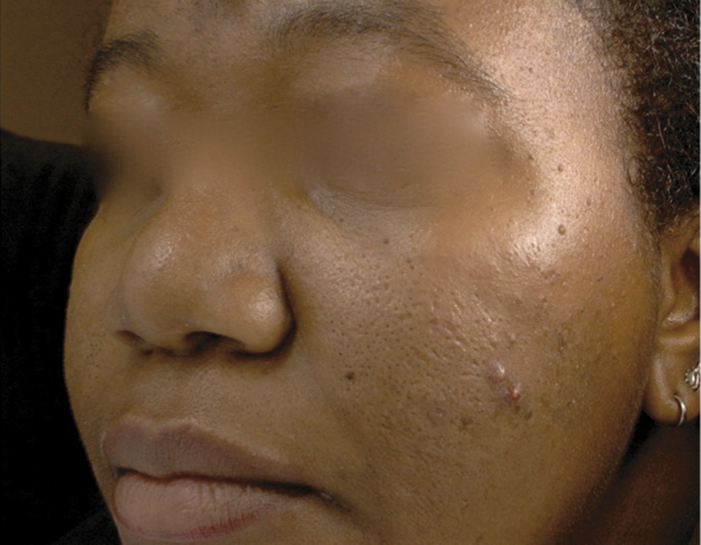
Photograph of African American woman with mild acne.

Photograph of Caucasian woman with mild acne.
Statistical analysis
Many acne grading criteria define acne in terms of both comedones and inflammatory lesions. For example, the popular Leeds criteria define mild acne as mostly comedones with <10 inflammatory lesions, moderate acne as 10–40 inflammatory lesions and 10–40 comedones, and moderate to severe acne as 40–100 inflammatory lesions and 40–100 comedones. 13 Because it was our goal to look at not only acne but also comedonal and inflammatory subtypes of acne, we modified these criteria. Clinical acne was defined as the presence of either inflammatory or comedonal acne. Inflammatory acne was defined as ≥3 papules or pustules on one side of the face and no/mild facial erythema (to distinguish from papular rosacea). Comedonal acne was defined as no more than 2 papules/pustules and ≥5 open or closed comedones on one side of the face (excluding comedones on the nose). Several authors have reported the prevalence of physiologic or mild acne, defined as Leeds score 0.25–0.75, 14 which we converted to 1–2 inflammatory lesions and/or 1–4 comedones on one side of the face.
To assess the differences in acne prevalence between ethnicities and between age groups within ethnicities, the chi-square test was performed. Unpaired t tests were used to examine the relationship between acne and the mean values of continuous variables, such as age, BMI, years of smoking, years of menopause, and sebum production. Analysis of variance (ANOVA) was used to examine the relationship between nominal and continuous variables, such as acne severity or hirsutism and BMI. Logistic regression was used to examine the relationships between continuous variables found to be significantly related to acne on t tests. Kappa statistics were computed to determine within-rater reliability of skin grading. We used an alpha level of 0.05 for all statistical tests.
Results
Demographics
The study population consisted of 2896 women, with the following racial composition: 44% Caucasian (29% from Los Angeles, 37% from London, and 34% from Rome), 19% East Asian (67% from Japan, 33% from Los Angeles), 15% Indian from London, 13% African American from Los Angeles, and 9% Hispanic from Los Angeles. Numbers of subjects and ages by ethnicity and city are reported in Table 1. Nineteen percent of the sample was aged 10–19, 19% was aged 21–30, 18% was aged 31–40, 18% was aged 41–50, 15% was aged 51–60, and 11% was aged 61–70.
Acne prevalence
Seven hundred seventy-three (26.7%) women in the studied population had clinical acne, 370 (12.8%) were classified as comedonal, and 403 (13.9%) were classified as inflammatory subtype of acne. An additional 824 (28.5%) women had mild acne.
Acne and age
In this sample, clinical acne peaks in the teenage years but remains prevalent in older women, with nearly half of women in their 20s, one quarter of women in their 30s, and >10% of women in their 40s affected (Fig. 3A). Additionally, mild acne is seen in a quarter of girls aged 10–15 and maintains a steady moderate prevalence in women of all ages in this sample (Fig 3A). The peak in clinical acne prevalence in the teen years comprises primarily inflammatory acne, whereas the prevalence of the comedonal subtype is steadier across the age groups (Fig. 3B). Within the peak acne prevalence age range, prevalence exceeded 40% by age 12, peaked at age 16, and started to decline after age 18 (Fig. 4). Although the greatest proportion of acne was seen in females aged 10–20 years, more than half the acne seen in this sample was in women >20 years: 26% of acne occurred in women aged 21–30 years, and 18% of the acne was seen in women aged 31–40 (Fig. 5).
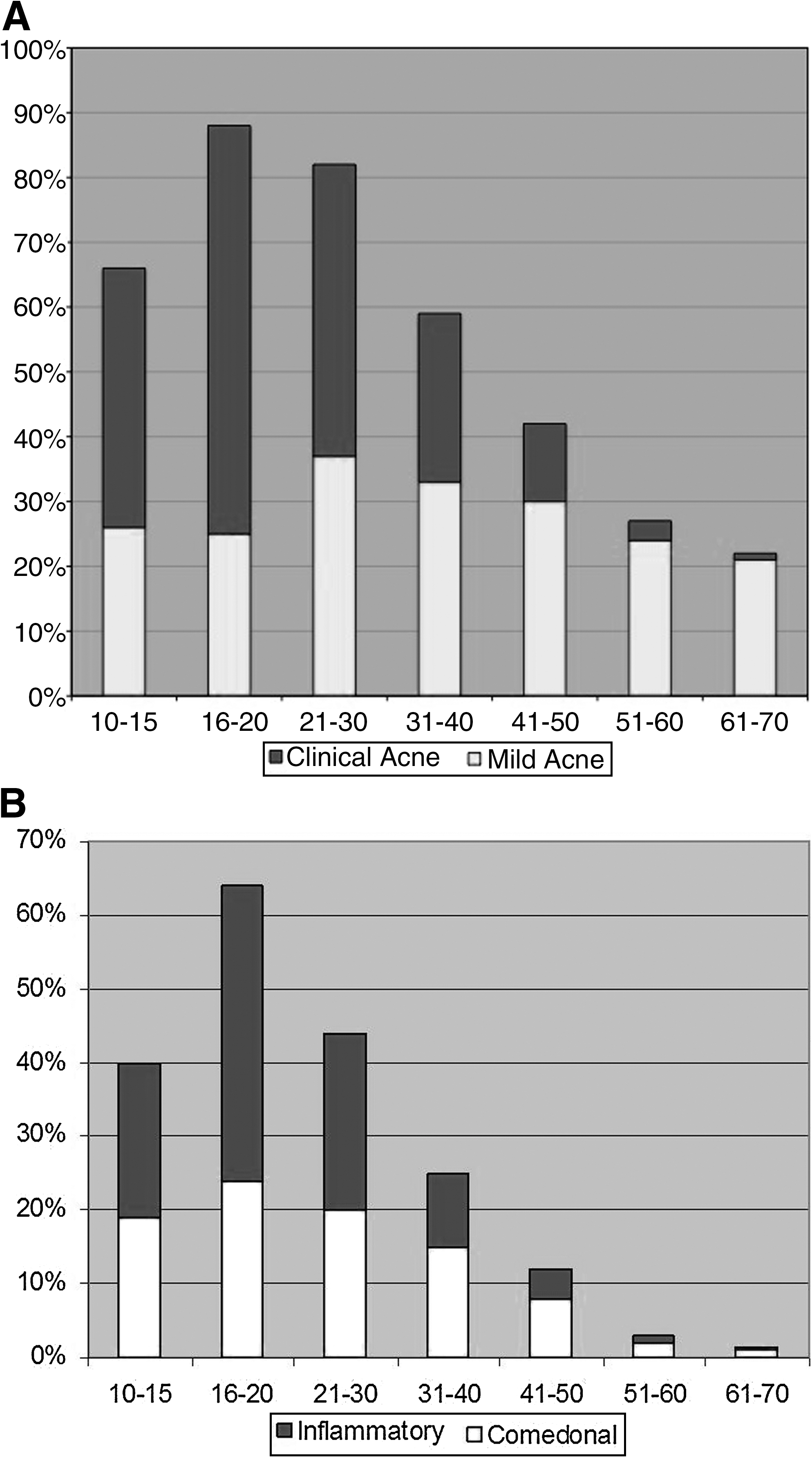
(
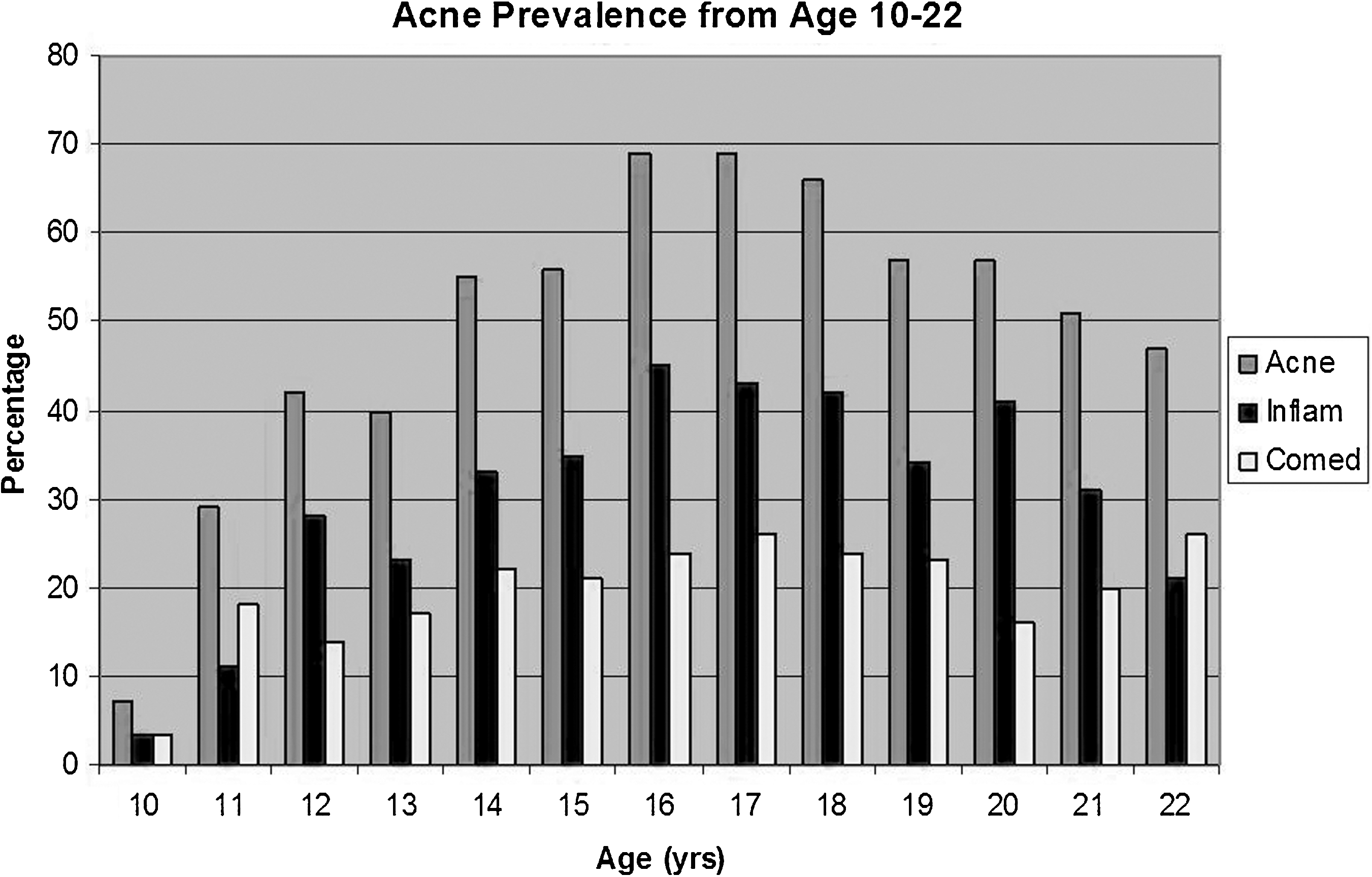
Prevalence of clinical acne and its subtypes, by age in women 10–22 years of age. Inflam, inflammatory acne; Comed, comedonal acne.
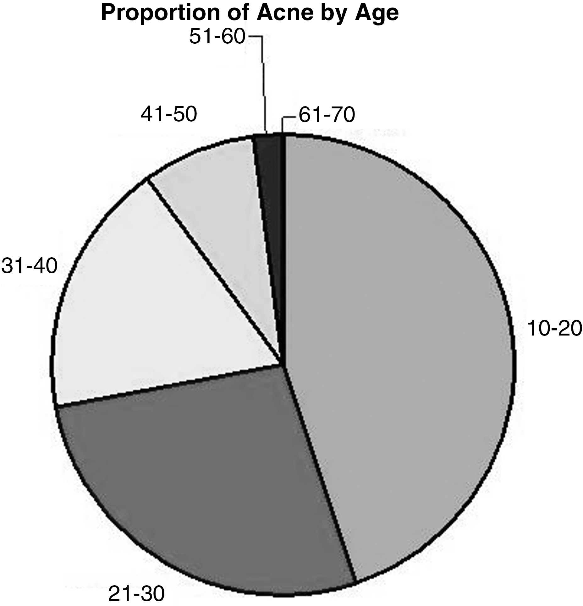
Proportion of acne by age group.
As expected, participants with acne (mean age 25 years, 95% confidence interval [CI] 24.4-25.9) were on average younger than those without acne (mean age 42 years, 95% CI 41.2-42.6, p≤0.001), and women with inflammatory acne were younger (mean 23.3 years, 95% CI 22.5-24.3) than those with comedonal acne (mean 27.2 years, 95% CI 26-28.3, p≤0.001). Severity of both inflammatory and comedonal acne was negatively predicted by age (F=129, 49, both p<0.0001). When women were separated by age-matched premenopausal and postmenopausal groups, postmenopausal women had significantly less clinical acne than premenopausal women (6.9% vs. 15.3%, respectively, chi-square=16, p=0.00006) (Fig. 6). As well, there was a negative relationship between acne severity and years menopausal (p<0.0001). When the relationship between acne and age was examined by menopausal status, age was negatively related to acne in premenopausal (p<0.0001) but not in postmenopausal women (p=0.4).
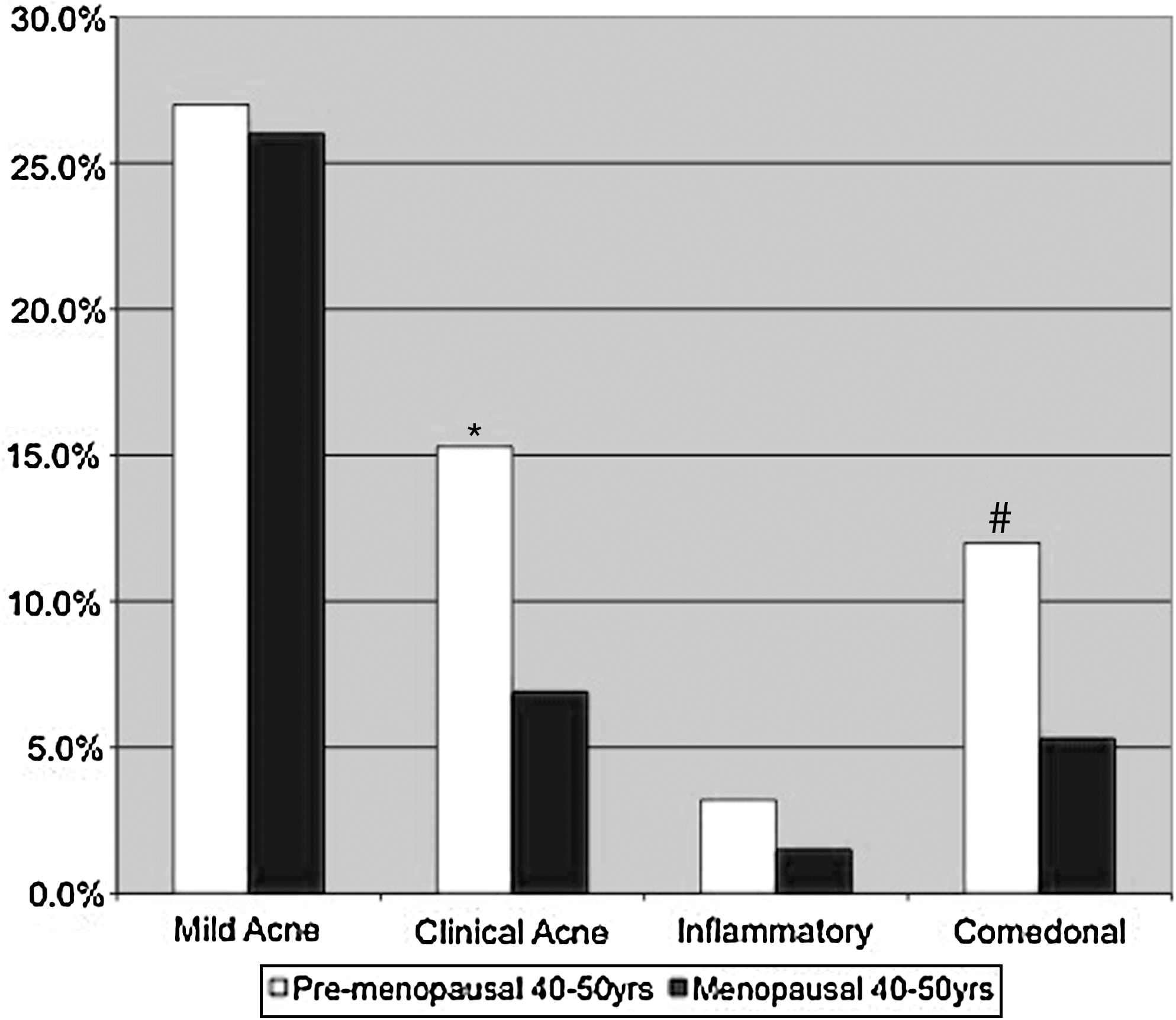
Mild acne, clinical acne, and subtypes in age-matched premenopausal and postmenopausal groups. *Premenopausal >menopausal; #premenopausal > menopausal: chi-square=13, p=0.0003.
Sequelae of acne
Of acne sequelae, atrophic and hypertrophic scarring were seen in 16% and 4.4% of women with acne, respectively, and hypopigmentation and hyperpigmentation were seen in 6% and 46%, respectively. Both forms of dyspigmentation and scarring were associated with clinical acne, but mild acne was associated only with atrophic scarring (t=−2.6, p≤0.01). Specifically, the inflammatory subtype of clinical acne was associated with hyperpigmentation, hypopigmentation (t=−17, −4.8, both p≤0.0001), and atrophic and hypertrophic scarring (t=−2.2, p≤0.02, t=−6.6, p≤0.0001). The comedonal subtype of clinical acne was associated with atrophic (t=−2.6, p≤0.009) and hypertrophic scarring (t=−2.7, p≤0.007). Both acne and its sequelae decrease in prevalence with age; however, dyspigmentation resolves, relatively, more than scarring (Fig. 7).
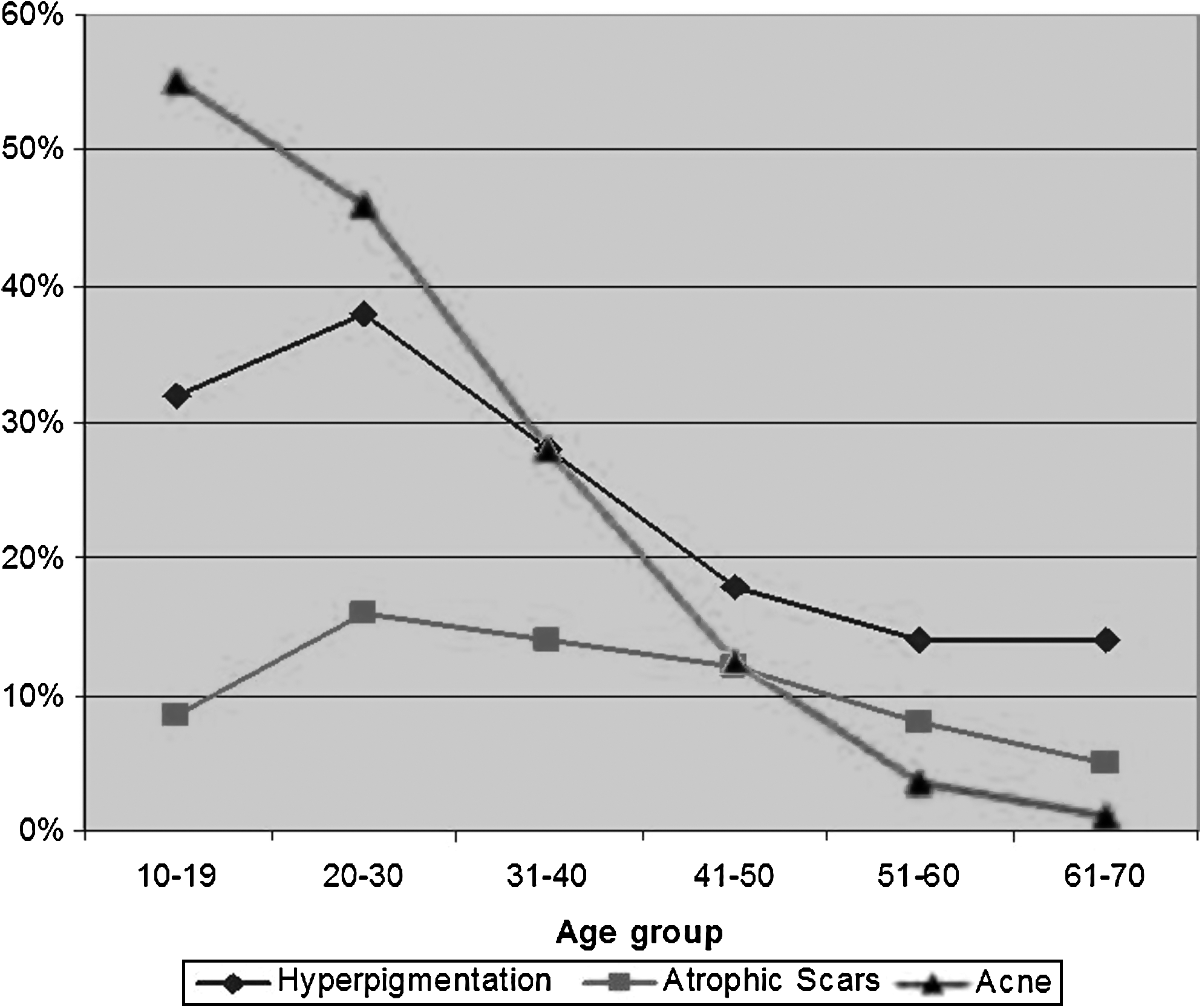
Acute sequlae: hyperpigmented macules, atrophic scars, and acne prevalence, by age group.
Acne and sebum
Sebum excretion was significantly higher in women with clinical acne (t=−6.2, p<0.00001) than in those without acne. When examined by acne subtype, sebum excretion was related to inflammatory (t=−6.2, p<0.0001) but not comedonal acne (t=−1, p<0.09) or mild acne (t=0.6, p=0.5). Additionally, there was a positive relationship between the severity of inflammatory acne and sebum excretion (F=10, p<0.0001). Women who reported having oily skin had somewhat higher sebum excretion rates, but this difference did not reach statistical significance (p=0.25).
Acne and hirsutism
More women with acne had hirsutism of the lip than those without acne (chi-square=10, p=0.001), a finding that was true for both acne subtypes but not for mild acne. Women with hirsutism of the upper lip had greater sebum production than those without hirsutism (t=−2, p<0.04) as well as larger pores (t=−2.1, p<0.03) and a greater BMI (t=−3.3, p<0.001). There were no age-related differences in hirsutism.
Acne and pore size
Scatter plot demonstrated that PCF increases with age, and age and PCF are positively related (Spearman's rho=0.3019, p<0.00001). In the entire sample, neither PCF nor clinical pore size was associated with acne. When the relationship between acne and pore size was examined by age group, both PCF and clinical pore size were positively related to clinical acne of both subtypes in women <40 years of age (p=0.01 for clinical acne, p=0.001 for comedonal, p=0.05 for inflammatory) but not in those >age 40 (p=0.08). Pore size was not related to mild acne in any age group.
Acne and behavioral or personal risk factors
For a subset of the sample, data were available on current smoking, alcohol, and caffeine habits. Of the 1083 women from Los Angeles, two thirds were nonsmokers, 18% were current smokers, and 15% were past smokers. More current smokers than nonsmokers or past smokers had acne (x=6.2, p=0.01, x=16, p<0.001), a finding that was true for both subtypes of clinical acne. There were no differences in acne prevalence based on alcohol or caffeine use.
Subjects with acne had higher BMI (mean BMI 25.6) than those without acne (mean BMI 23.9, t=6.5, p<0.0001). As well, acne was negatively related to years of menopause (p<0.0001) and years of hormone replacement therapy (HRT) (p<0.0001). Cumulative lifetime sun exposure, education, and income were not related to acne or its subtypes in this sample.
Logistic regressions were run with the following continuous variables, which were found to be related to acne in the general sample but which were possibly explained by the effects of age: BMI, sebum, PCF, years of menopause, years of HRT, and years of smoking. The significant factors relating to clinical acne were age (odds ratio [OR] 0.00, p<0.0001), sebum excretion (OR 20.08, p<0.002), PCF (OR 1,808.04, p<0.0001), and years of smoking (OR 14.8, p<0.006). When the regressions were repeated for acne subtypes, the significant factors relating to inflammatory acne were age (OR 0.00, p<0.0001), sebum excretion (OR 54.60, p<0.0001), and PCF (OR 90.02, p<0.0001), and the significant factors relating to comedonal acne were age (OR 0.01, p<0.0001), PCF (OR 148.41, p<0.0001), and years of smoking (OR 27.11, p<0.006). Logistic regressions were performed for the aforementioned age groups and differences were noted in factors associated with clinical acne in those greater and less than 40 years of age. Factors associated with clinical acne for subjects aged 10–40 were age (p=0.0001), sebum (p=0.003), and PCF (p=0.0001), whereas for those >age 40, associated factors were age (p=0.0001) and years of smoking (p=0.003). A logistic regression with acne, age, and years menopausal demonstrated an age-controlled relationship between acne and years of menopause (p=0.045); however, this relationship disappeared when sebum excretion was entered as a covariate (p=0.2). Table 2 describes the percentage of several risk factors by age group.
BMI, body mass index; HRT, hormone replacement therapy.
Discussion
Our results, based on clinical grading of photographs, demonstrate that clinical acne is common in a heterogeneous sample of adult women. The prevalence of acne in this sample confirms findings from previous self-report survey studies 5,9 that acne peaks in the teen years, but approximately one half of women in their 20s and roughly one third of women in their 30s have acne. Our results also indicate that mild acne, consisting of few comedones or papules, is prevalent, affecting roughly a quarter of women in all age groups, decreasing only slightly with age. Because of the cross-sectional nature of the study, we cannot distinguish persistent from adult-onset acne.
We separated clinical acne into inflammatory and comedonal subtypes to determine if there are factors unique to these different manifestations of acne. In this sample, the teenage spike in acne prevalence was primarily due to a sudden increase in inflammatory acne; women with comedonal acne were, on average, older than those with inflammatory acne. There are several potential explanations for this age-related difference in acne subtype. There may be a physiologic reduction in inflammation with increasing age that manifests as a shift from inflammatory to noninflammatory acne. Another possibility is that the age-related decline in sebum production itself decreases inflammation. Sebum is produced as adrenal androgens begin to rise at 10 years of age, peak in the early 20s, and then gradually decline from the mid-20s throughout life. 15 Recently, sebum has been implicated as proinflammatory, 16 a possibility supported by the positive relationship we observed between sebum production and inflammatory acne prevalence and severity. The age-controlled decrease in acne prevalence and severity seen after menopause suggests that ovarian hormones play some role in the pathogenesis of acne. After adding sebum as a covariate, years menopausal was no longer related to acne, suggesting that perhaps the postmenopausal decrease in ovarian androgen production and the resultant decrease in sebum excretion account for this observation.
The finding that inflammatory acne is associated with more sequelae, specifically more dyspigmentation, than comedonal acne is expected given the well-known effects of inflammation on the skin. That acne hyperpigmented macules and atrophic scars decrease with age is not surprising given the decrease in acne prevalence with age. Notably, acne itself decreased more dramatically with age than its sequelae, and atrophic scarring decreased only moderately with age, suggesting that the appearance of atrophic scars lessens as skin loosens with age. However, conclusions about the natural history of acne hyperpigmentation and scarring are limited by the cross-sectional nature of this study.
.Our results support associations between acne and both sebum production and hirsutism. Sebum production, hirsutism, and acne are known to be driven by hyperandrogenism, 17 and, therefore, our findings suggest that hyperandrogenism is an important etiologic factor of acne in this sample, although the lack of data on endocrine parameters prevents definitive conclusions. Whereas the relationship between clinical acne and sebum production was expected, the lack of relationship between sebum production and comedonal acne has not been reported before, as prior findings that individuals with acne have higher sebum excretion than those without 18 have not broken acne down into subtypes. Sebum is thought to be proinflammatory 16 and, therefore, is likely more essential a component of the inflammatory subtype of acne.
Although we found a positive association between hirsutism and BMI, supporting prior findings, 19 we did not find associations between BMI and acne or sebum production. One published study of BMI and acne in healthy children found that higher BMI was a risk factor for development of acne. 20 These authors found that Taiwanese children aged 6–11 with acne had higher average BMI (19.5) than those without acne (18.2) and that those with BMI≥95th percentile had more acne than children with BMI<18.5. Our sample was older, with no subjects aged 6–9 and only 2% aged 10 or 11, as well as heavier, and for those reasons, comparison of results is difficult. Our findings suggest that the classic Stein Leventhal polycystic ovarian syndrome (PCOS) phenotype of obesity, hirsutism, and acne does not account for the majority of acne in our sample, which is interesting in light of the increasing recent reports of nonobese PCOS. 21
Although the relationship between acne and enlarged pores is often mentioned, there are few published data to support this association, and one study failed to show a relationship between pore size and severity of acne. 22 Our observation that the relationship between clinical acne and pore size was specific to women <40 may be because large pores in younger women are most often associated with acne, whereas pore size increases as a function of age and is not necessarily associated with acne in older women.
The relationship between acne and genetic, behavioral, or environmental factors is of great interest but remains unclear because of conflicting data and the difficulty inherent in examining human behavior. The role of cigarette smoking in acne is unclear. Whereas some studies have shown no effect, 23 others have found an increased prevalence of acne among smokers, 24,25 and one study found that smoking, daily cigarette consumption, and duration of smoking appeared to be protective in the development of inflammatory acne in girls. 26 This study supports a relationship between smoking and clinical acne in women, as well as an age-controlled relationship between years of smoking and clinical and comedonal acne (both p<0.0001) but not inflammatory acne (p=0.3). When examined by age group, the effect of years of smoking is seen only in women over 40. This finding is interesting in light of the recent description of smoker's acne, microcomedones and macrocomedones with few inflammatory lesions in adult female smokers. 27 The authors explain that there are several pieces of evidence supporting a pathogenic role of smoking in comedonal acne, including the presence of keratinocyte nicotinic acetylcholine receptors that induce hyperkeratinization, the smoking-induced increase in oxidative stress, and increase in lipid peroxidation.
The most significant limitation of this study is the cross-sectional design, which prevents us from drawing conclusions about the progression of acne with age. As well, the restriction to female participants prevents the generalization of findings to acne in men. In addition, our definition of inflammatory acne has the potential to include a large number of comedonal lesions, as a woman with at least 3 papules is classified as having inflammatory acne regardless of the number of comedonal lesions. The photographic nature of the study imposes general limitations based on the quality of the image and the ability to perceive skin surface changes in a two-dimensional image. The restriction of the images to the left side of the face necessitates an assumption that acne will generally be symmetrical in nature. In addition, cases of subclinical acne, which can be assessed only with biopsy, were undoubtedly present in the populations examined, but the techniques used in this study were not sufficiently sensitive to define this group accurately. The goal of study recruitment was to collect an ethnically diverse sample of women from the general population. Original data collection was not conducted with this study in mind, however, and description of the study population is limited by the information gathered initially.
Conclusions
This study provides acne prevalence data over the life span for a large, heterogeneous group of women. These data suggest that although acne peaks in the teenage years, it remains a prevalent disease in women in their 20s, 30s, and 40s. Although inflammatory and noninflammatory acne were equally prevalent, there may be differences between the two subtypes in terms of pathogenesis and sequelae. There also may be age-related differences in factors associated with acne.
Footnotes
Acknowledgments
This study was funded in part by an unrestricted grant from Proctor & Gamble. A.C.P. received fellowship funding from the Doris Duke Clinical Research Foundation. A.B.K. became an investigator for Proctor & Gamble in May 2011.
Disclosure Statement
No competing financial interests exist.