




Abstract
Abstract
Torsion of the vermiform appendix is a rare cause of appendicitis that is clinically indistinguishable from the classical presentation. In this paper, we present the first report in the literature of an appendicular torsion associated with an undescended cecum. A 3-year-old male patient with persistent pain in the right hypochondrium and peritoneal irritation underwent diagnostic laparoscopy, finding necrosis of the vermiform appendix caused by torsion of its base associated with an undescended cecum located in an indurated area of the subhepatic region. A video-assisted transumbilical appendectomy was performed.
Introduction
Case Report
The patient was a 3-year-old male with abdominal colic of 18 hours located in the right flank with hyporexia and vomiting of gastric contents. At the time of the initial evaluation, there were no signs of peritoneal irritation. An abdominal ultrasound was performed without finding the appendix nor signs of inflammation in the right iliac fossa. The patient was reassessed after 18 hours of observation, finding abdominal distension, pain located in the right hypochondrium with a palpable mass, and signs of peritoneal irritation; an abdominal X-ray showed fluid levels. A complete blood count showed leukocytosis (18,500 wbc/mm3 with 95% neutrophils). A laparoscopic examination was decided because of clinical signs of an acute abdomen.
Surgical technique
We started with two 5-mm ports, one in the umbilical scar for 5 mm and 30-degree optics (Karl Storz GmbH & Co. KG, Tuttlingen, Germany) and another in the left iliac fossa. On abdominal examination, inflammatory liquid was found in the pelvic cavity and an indurated area in the right parietocolic and subhepatic spaces. We were unsuccessful in an attempt to separate the indurated area, so we placed another 5-mm port in the right iliac fossa. Lysis of adhesions of the omentum, small intestine, and cecum was done. Once the indurated area was separated, we found a necrotic subhepatic appendix, twisted three times counterclockwise, adhered to the abdominal wall of the right parietocolic and subhepatic spaces (Figs. 1 and 2). We also observed that the appendix had a narrow appendicular mesentery and a movable cecum without fixation. A video-assisted transumbilical appendectomy was performed, exteriorizing the appendix and the cecum through the umbilical scar (Fig. 3). The operative time was 55 minutes. Feeding was started on postoperative day 1, and the patient was discharged on the second day.

Arrow at the base of the necrotic appendix indicating counterclockwise torsion. The diagram shows the three turns.
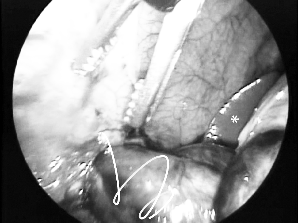
Diagram of the three turns of appendicular torsion. The asterisk marks the liver, showing the subhepatic location of the appendix and the cecum.
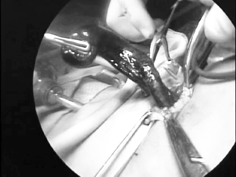
Extraction of the appendix through the umbilicus for video-assisted transumbilical appendectomy.
Discussion
Although torsion of the vermiform appendix has been described for 90 years, it is rare in pediatric patients, with 15 cases being reported previously.10,11 No specific age predominates, but the range found in the literature varies from 50 days 5 to adults. 12 Primary and secondary torsion of the vermiform appendix have been described. Among the primary causes are abnormalities of the mesentery, such as a narrow base, as occurred in our case, the absence of azygotic folds that normally laterally fix the appendix, and even abnormal peristaltic movements.3,4,11 Still, its etiology is uncertain. Benign tumor pathology such as mucocele, 13 malignant pathology such as cistadenoma, 11 or after intestinal invagination 10 or intestinal duplication 14 have been associated to secondary causes.
Abdominal ultrasound has been used to find the vermiform appendix, in the differential diagnosis of the most frequent causes of pain in the right iliac fossa,2,15 or to suggest the diagnosis of appendicular torsion. 16 In our case, the appendix was not found by ultrasound because it was located in the subhepatic area. Computerized tomography has been used for diagnosis in cases where the appendix was not found. 11
An undescended cecum has been reported in 6% of the population, predominantly in men. 9 Cecal malposition makes the clinical diagnosis of appendicitis and appendicular torsion difficult, since abdominal pain is not located in the right iliac fossa. The undescended cecum and its lack of fixation in this case contributed to the torsion of the vermiform appendix and the lack of classical signs of appendicitis.
Surgical treatment in pediatric patients is an appendectomy by the conventional approach or by laparoscopy.17,18 A complete laparoscopic appendectomy can also be performed. Our usual approach is the transumbilical video-assisted appendectomy. Since we do not have optics with an operating channel, we usually use two ports to perform a transumbilical video-assisted appendectomy. On this occasion, another port was necessary to separate the indurated area where the necrotic appendix was located. The mobile cecum made the transumbilical appendectomy easier.
Conclusion
In our patient, laparoscopic exploration led to the diagnosis of acute abdomen caused by appendicular torsion and an undescended mobile cecum. This case is the first report in the literature of this type of association.
Footnotes
Acknowledgements
The authors thank Sergio Lozano-Rodriguez, MD, for his help in translating the manuscript.
Disclosure Statement
No competing financial interests exist.