
Abstract
Abstract
Background:
The rat is increasingly being used in laparoscopic research. Laparoscopic microsurgical training is critical in order to develop new surgical indications in pediatric patients. This report evaluates laparoscopic splenectomy and nephrectomy in a rat model.
Materials and Methods:
A Wistar rat (weight between 250 and 400 g) was placed in the supine position. Inhaled 3% halothane anesthesia was administered. A Veress needle is inserted in the right-upper abdomen. After establishing a pneumoperitoneum of 3–4 mm Hg, a 2- or 5-mm trocar was placed, according to the procedure. A 2-mm 0-degree endoscope was used. Two aditional 2- or 5-mm trocars were then placed. Laparoscopic splenectomy involved two-handed dissection, intracorporeal ligation, and the division of gastrosplenic attachments and hilar and short gastric vessels. Laparoscopic nephrectomy was done by intracorporeal ligation and division of the renal vessels and the ureter after mobilization of the kidney.
Results:
Laparoscopic splenectomy was performed in 8 rats; laparoscopic nephrectomy was done in 4 rats. Operative time was 25–40 minutes for splenectomy and from 30 to 65 for nephrectomy. Postoperatively, 4 rats died from hemorrhage. No wound infections occurred at the port sites.
Conclusions:
Laparoscopic splenectomy and nephrectomy in an experimental rat model is technically feasible and may provide an excellent training model for pediatric minimally invasive surgery.
Introduction
Materials and Methods
The anesthesia technique was based on inhaled 3% halothane, using a facial mask. We used Wistar rats (weighing between 250 and 400 g). They were placed in the supine position, and, after shaving the abdominal wall, a Veress needle was inserted in the upper right abdomen. After establishing a pneumoperitoneum of 3–4 mm Hg, a 2- or 5-mm trocar was placed, according to the procedure. A 2-mm 0-degree endoscope was used and helped in the introduction of the two aditional trocars (Fig. 1).
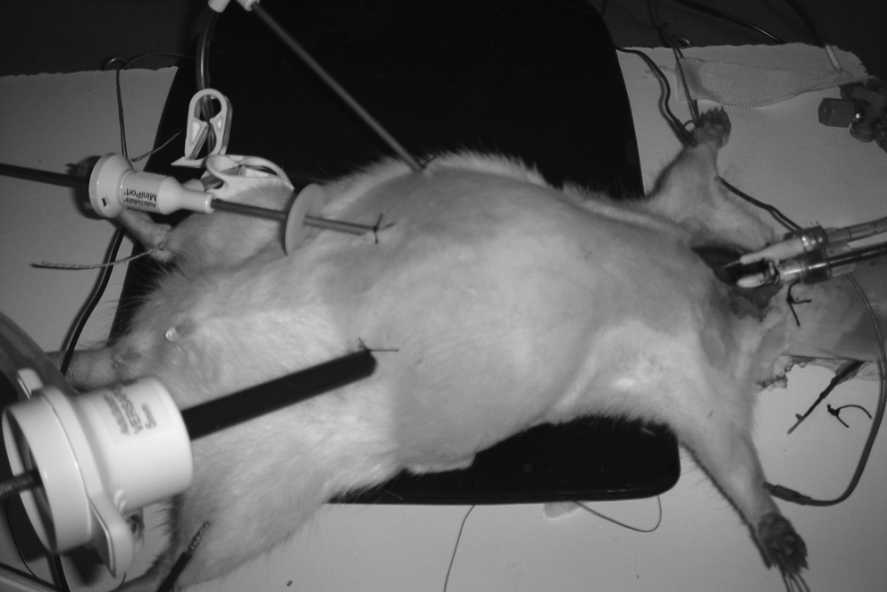
Rat position.
Operative procedures
Splenectomy
The procedure started with the retraction of the stomach and the division of the gastroesplenic ligaments. Hiliar vessels were dissected, and 5-mm clips were used for their ligation; eventually, small vessels could be coagulated. The spleen was removed through a port site.
Nephrectomy
A transperitoneal approach was used by retraction of the small bowel and colon with the dissection of the retroperitoneal fat around the kidney. The dissection of the renal vessels had to be careful and their ligation with silk ties or clips was possible.
Discussion
Advantages in laparoscopic procedures, such as shorter hospitalization, less postoperative pain, and no significant scars, are commonly accepted and produce an increase in surgical indications. Experimental models are necessary to study the effects of the laparoscopic techniques in a live organism. 4 Laparoscopic microsurgical training is critical in order to develop new surgical indications in pediatric patients. 5 The rat has been used for years in medical investigations and is now a feasible animal model for laparoscopic research. 6 It is easy to handle because of its small size, and the inhaled anesthesia is not difficult to perform. 7 The present report summarizes our experience with the rat as an animal model for laparoscopic splenectomy and nephrectomy. We consider that the surgeon must be familiarized with the rat anatomy and conditions of the pneumoperitoneum in this animal model. 8 The small size of the instruments is a surgical challenge in this type of procedure.
Conclusions
In conclusion, the rat is a feasible model for pediatric laparoscopic procedures due to its availability and low cost. It is important to study the effects of laparoscopic techniques in very low-weight live organisms and the development of training programs in minimally invasive surgery.
Footnotes
Disclosure Statement
No competing financial interests exist.