




Abstract
Abstract
Background:
Partial amniotic carbon dioxide insufflation (PACI) during fetoscopic interventions greatly improves visualization of intraamniotic contents. The purpose of this study was to assess any histologically discernable effects from this approach on the fetal brain after long-term survival in sheep.
Methods:
Six pregnant ewes between 63 and 92 days of gestation underwent PACI after fetoscopic intraamniotic access. Insufflation pressures ranged between 7 and 15 mm Hg (mean 11.7; median 12.5). Insufflation times ranged between 45 and 80 minutes (mean 55.8 minutes; median 52.5) and depended on the duration of various percutaneous fetoscopic maneuvers (e.g., posturing, fetal transesophageal electrocardiography, and chronic fetal vascular access) that were tested during these studies. After fetal spontaneous delivery between 147 and 150 days of gestation, 5 of the lambs were observed for abnormal neurological symptoms. The last ewe and her sheep were terminated at 133 days of gestation for humane reasons. All six brains were examined for hemorrhage, embolism, infarctions, inflammatory changes, and abnormal cortical maturation. An unoperated sibling was available as a control.
Results:
The 5 sheep that were spontaneously delivered exhibited no abnormal neurological findings. In all 6 sheep, PACI did not result in any histologically discernable damage to their brain in these long-term studies. Maternal and fetal complications were not observed during or after the approach.
Conclusion:
The application of PACI during minimally invasive fetoscopic interventions seems safe for the fetal brain. Due to the still limited clinical experience with PACI, continued assessment of its maternal and fetal risks as well as management are required.
Introduction
Before the clinical introduction of amniotic insufflation at our institution, we examined multiple maternal and fetal safety as well as management issues of this approach in sheep. Over the past decade, we have made many aspects of these animal studies regarding the institution, maintenance, and safety of amniotic insufflation with compressed air available in the literature.3–6 In this vein, the purpose of this manuscript is to present the results of macro- and microscopic brain studies that were performed in 6 sheep which had undergone fetoscopic procedures employing carbon dioxide for insufflation.
Animals and Methods
The studies were performed in six time-bred fetal sheep. The study protocol was approved by the local committee on animal research and was performed according to institutional and government guidelines for the provision of humane care to laboratory animals.
Age at fetoscopic intervention ranged between 63 and 92 days of gestation (median = 79.5 days; term = 145–150 days). Each ewe was positioned supine with a 15° left tilt, intubated, and ventilated with 0.5%–2% halothane in 50% oxygen. Placental transfer of the anesthetic gas provided fetal anesthesia.
Intraamniotic access to the fetus was achieved via one to three trocars that was/were percutaneously inserted into the amniotic cavity by an over-the-wire approach. The first trocar was placed under ultrasound guidance, whereas subsequent trocars were placed under fetoscopic guidance.
Via the trocar(s), the amniotic cavity was insufflated with carbon dioxide. Insufflation pressures ranged between 7 and 15 mm Hg (mean 11.7 mm Hg; median 12.5 mm Hg) and depended on the minimum pressure that was required by the insufflator to instill gas into the amniotic cavity. This pressure, which depends on the combined resistances of the abdominal wall, maternal intraabdominal organs, and the uterine smooth muscle tone to further uterine volume distension, has been defined as the opening pressure. 1 Then, amniotic fluid was partially removed and replaced with carbon dioxide at opening pressure until about half of the fetus lay exposed to carbon dioxide.
Insufflation times ranged between 45 and 80 minutes (mean 55.8 minutes; median 52.5 minutes) and depended on the duration of various percutaneous fetoscopic maneuvers (e.g., posturing, fetal transesophageal electrocardiography, and chronic fetal vascular access) that were tested during these studies.
During the operations, maternal electrocardiogram, blood pressure, and end-expiratory carbondioxide concentration were continuously monitored for detrimental cardiovascular effects on the ewe from PACI.
All 6 ewes and their fetuses recovered from surgery and continued gestation after fetal intervention. After fetal spontaneous delivery between 147 and 150 days of gestation, 5 of the lambs were examined for clinically evident neurological deficits presenting as movement disorders or the inability to establish a normal feeding pattern or other abnormal neurological symptoms or behavior. The last ewe and her sheep fetus were terminated at 133 days of gestation to avoid terminating the newborn sheep for humane reasons. All animals were sacrificed by intravenous administration of an overdose of potassium chloride after ketamine premedication and induction of general anesthesia.
Neuropathological evaluation
Immediately after their termination, the 5 lambs that had been spontaneously delivered and the fetus that had undergone postmortem Cesarean section were perfusion-fixed with 4% buffered formaldehyde and their brains were removed. In addition, an unoperated sibling that had been spontaneously delivered was available as a control. After submersion of the brains in 4% buffered formaldehyde for more than at least 2 weeks, they were subjected to neuropathological examination. After weighting and macroscopic inspection of the brains, samples of standardized areas (frontal and occipital poles of the cerebral cortex, hippocampus, basal ganglia, thalamus, cerebellum, and brain stem) were prepared for histological examination. Samples were paraffin-embedded, and sections (10 μm) were stained with hematoxylin-eosin. All brains were examined for bleeding complications, infarctions, embolic events, and acute inflammatory tissue reactions or abnormal cortical migration and maturation.
Results
Maternal findings during PACI
The application of PACI during fetoscopic interventions did not result in any detrimental cardiovascular effects on the ewe as assessed by arterial blood gas analyses, continuous maternal electrocardiogram, blood pressure, and end-expiratory carbondioxide concentration monitoring.
Fetal findings during and after PACI
All fetuses survived the application of PACI during fetoscopic interventions and continued gestation for periods of between 7 and 11 weeks. The 5 of them that were spontaneously delivered appeared to be neurologically normal after delivery, exhibiting typical postnatal movement and feeding patterns as well as normally interacting with their mother.
Neuropathological examination of the brains from the 6 fetuses that had undergone PACI and the control fetus yielded no abnormal macroscopical or histological findings. In particular, there was no evidence for intraventricular or parenchymal hemorrhage, embolic events, infarctions, acute inflammatory tissue reactions, or abnormalities of cortical maturation (Fig. 1).
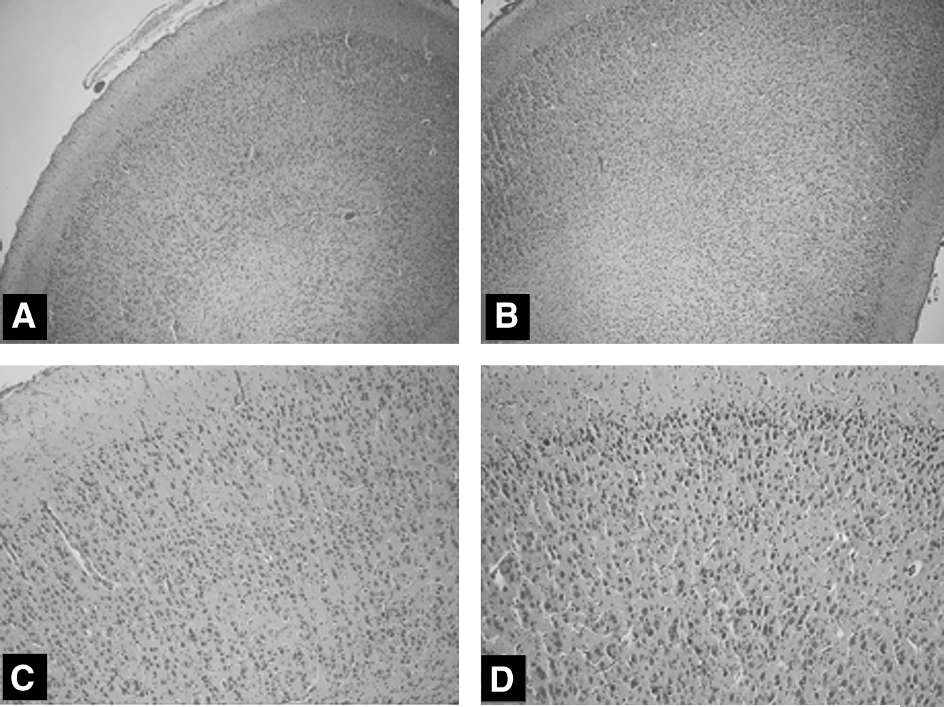
Cerebral cortex (
Discussion
PACI has been instituted over a wide range of gestational ages to improve visualization during technically difficult minimally invasive fetoscopic interventions in human fetuses affected by a variety of fetal malformations or disease states.1,2 Given the still vastly limited clinical experience with PACI during fetoscopic interventions in human fetuses, our study in a small group of sheep provides some further evidence that this approach can be performed without apparent detrimental effects on the fetal brain and maternal hemodynamics. Examining various brain regions particularly sensitive to vascular or hypoxic injury, we found neither macro- nor microscopic damage to this organ in all 6 study fetuses.
The risk for the occurrence of serious maternal or fetal complications during insufflation of the amniotic cavity with carbon dioxide or other gases in humans still remains unknown. Previous investigators observed that carbon dioxide insufflation of the amniotic cavity after maternal laparotomy and uterine exteriorization may result in fetal acidosis in sheep.7,8 In our own experience, uterine hyperdistension in sheep easily occurs when amniotic insufflation is performed after maternal laparatomy; it is further aggravated by uterine exteriorization and may largely impair fetoplacental blood flow by overstretching of placental vessels. This effect might have influenced the results of the previous studies by our colleagues as well as prompted their reluctance to incorporate PACI into their own clinical practice.
In contrast, after the 6 fully percutaneous fetoscopic procedures that used carbon dioxide as the insufflation gas in this sheep study and after 30 previous procedures which employed compressed air, 5 even amniotic insufflation pressures as high as 30 mm Hg were tolerated without apparent neurologic damage or macro- or microscopic brain injury when uterine hyperdistension was avoided. Likewise, during >50 fetoscopic procedures in human fetuses performed now at our center, no untoward hemodynamic or neurologic effects from PACI were observed in both pregnant women and fetuses. 1 Given the current limitations and potential risks of invasive fetal blood gas monitoring during fetoscopic surgery in human fetuses, these findings are reassuring that the risk to acquire cerebral damage from PACI seems low.
Footnotes
Acknowledgment
The development of the fetoscopic procedures described in this manuscript has been supported by educational and research grants by the German Research Society (Deutsche Forschungsgemeinschaft [DFG]) (Ko 1484/1-1; Ko 1484/2-1; Ko 1484/3-1; Ko 1484/3-2; Ko 1484/3-3).
Disclosure Statement
No competing financial interests exist.