
Abstract
Abstract
Introduction:
Transumbilical laparoscopically assisted appendectomy (TULAA) has been reported in the literature as an alternative to traditional three-port laparoscopic appendectomy (LA). Our study compares outcomes between LA and the one-trocar transumbilical technique in a single institution over a concurrent time frame for all cases of pediatric appendicitis.
Methods:
An Institutional Review Board–approved retrospective chart review of all appendectomies from July 2007 through June 2009 was performed. All appendectomies were performed either laparoscopically or transumbilically. One surgeon predominantly used the TULAA method, whereas the other 2 surgeons used strictly the LA method. No cases were converted to open. Categorization of specimens as normal, acute, or ruptured was based on pathology reports. Outcomes analyzed for each group included surgical duration, cost, length of stay, fever (>101.5F), wound infection, ileus, and postoperative abdominal-pelvic abscess.
Results:
A total of 131 appendectomies were performed by 3 surgeons, 83 were LA and 48 were TULAA. For all stages of appendicitis, outcomes differed significantly only for operating room cost, with the TULAA being significantly less expensive. All other outcomes were similar between the two techniques.
Conclusion:
Our study suggests that TULAA is a reasonable alternative to the standard minimally invasive technique for appendicitis in both acute and ruptured situations. All analyzed complications were similar between the groups, suggesting that TULAA is an acceptable surgical method in pediatric patients for all stages of appendicitis.
Introduction
Materials and Methods
We performed an Institutional Review Board–approved (exemption obtained) retrospective analysis of hospital and outpatient clinical records of 131 children who underwent appendectomy from July 2007 to June 2009. All the procedures were performed at a children's hospital by pediatric surgeons with the assistance of general surgery residents. Children with a clinical or radiographic diagnosis of acute appendicitis or early perforated appendicitis underwent immediate appendectomy. Cefoxitin was the preferred preoperative antibiotic. Children with a ruptured appendix and a walled-off abscess were treated with antibiotics and percutaneous drainage as needed, followed by interval appendectomy. The patients were not randomized to treatment group; 83 patients had traditional LA and 48 had one-trocar TULAA. Two of the 3 surgeons in the practice performed only LA and 1 surgeon preferentially performed TULAA, unless additional trocars were needed. If additional trocars were needed, these cases were classified as conversion to traditional LA.
Data were collected and analyzed using Microsoft Excel 2003 and SPSS 17 for Windows. Discrete variables are reported as number and percentages. Chi-square test was used for comparison of means. Fischer's two-tailed exact test was used to calculate P value. Continuous variables are reported as mean and standard deviation. Two-tailed Standard's test was used for calculation of significance. A P value of <0.05 was considered statistically significant.
Operative technique LA
LA was performed using three Step dilators (US Surgical, Norwalk, CT): umbilical (12 mm trocar), left lower quadrant (3 or 5 mm trocar), and suprapubic (3 or 5 mm trocar). The abdomen was entered by Veress technique at the umbilicus. An Endo-GIA stapler (US Surgical) or Endoloops (Ethicon, Cincinnati, OH) were used through the umbilical port to ligate and divide the appendix and mesoappendix. Endoloops were used in the smaller children due to limited intraperitoneal space. In a few cases, trocar placement varied slightly, based on surgeon judgment. The appendix was removed through the umbilical port.
Operative technique TULAA
TULAA was performed using one Step dilator (US Surgical) in an umbilical position (12 mm trocar) after access by Hasson technique using a 10-mm operative laparoscope (Richard Wolf Medical Instruments, Vernon Hills, IL). The appendix was observed, the adhesions of the appendix and cecum to the lateral abdominal wall, cecum, or small bowel were lysed using a 5-mm blunt grasper through the operative port of the working laparoscope. After mobilization, the appendix was grasped at the tip and drawn out through the umbilical incision (see Fig. 1). The mesoappendix was divided between ligatures, the base of the appendix ligated, and the mucosa of the appendix cauterized. Appendiceal stump and cecum were then returned to the abdominal cavity.
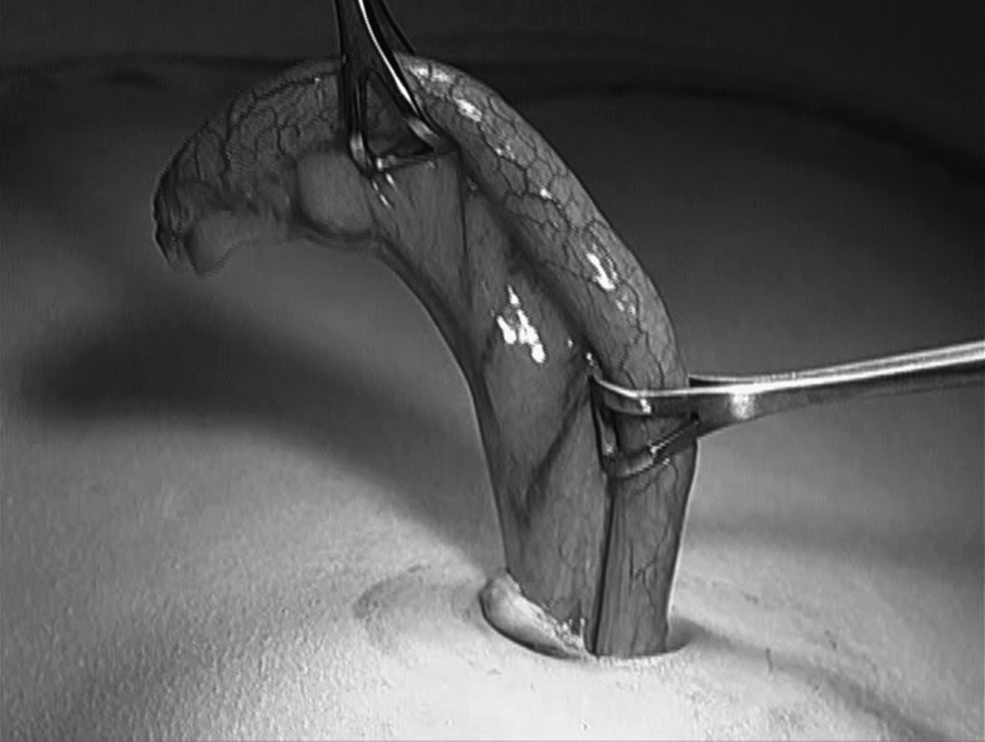
Intraoperative exterior image of extracted appendix.
Results
Data were divided into two groups for analysis based on the initial intent-to-treat procedure. Demographic information collected included age at operation and gender. Intraoperative data collected included duration of surgery, operating room (OR) cost, and conversion from TULAA to LA. Outcomes analyzed were length of hospital stay (LOS) and postoperative events, to include fever (>100.4F), all degrees of wound infection, ileus (defined as no return of bowel function within 2 days), and abdominal-pelvic abscess formation. Data were analyzed to include a comparison of the total number of appendectomies for the LA and TULAA technique, and also for each pathologic category of appendicitis (normal, acute, and ruptured).
A total of 131 appendectomies were performed. Eighty-three children underwent LA, and 48 underwent TULAA. No procedures were converted to open. Nine (18.8%) were converted from TULAA to LA. None of the normal appendices, 4 out 24 (16.7%) acute, and 5 out 11 (45.5%) ruptured appendices were converted to LA. All appendectomies performed by the TULAA surgeon were started using the TULAA technique and additional trocars were only added if necessary. In most instances, the reason for conversion with additional trocars was inability to appropriately observe intra-abdominal structures due to inflammation and dense adhesions. In one instance, additional trocars were needed due to an unexpected intraoperative finding (a large torsed omental cyst coursing into the right lower quadrant as the etiology of the patient's abdominal pain). For this study, conversion was not considered a complication. All appendix categories were based on pathologist examination, and not surgeon judgment. Of the 131 appendectomies, 81 (62%) were acute (57 LA and 24 TULAA), 25 (19%) were ruptured (14 LA and 11 TULAA), and 25 (19%) were categorized as normal (12 LA and 13 TULAA) (Table 1).
LA, laparoscopic appendectomy; TULAA, transumbilical laparoscopically assisted appendectomy.
For all categories of appendicitis, age and gender were similar between LA and TULAA groups. Mean duration of surgery, not including anesthetic time, in minutes was also similar between the groups (45 minutes LA, 46 minutes TULAA; P = 0.74). OR cost was significantly less for the TULAA group in total ($2053.47 LA and $1640.53 TULAA; P = 0.001) and for the acute and normal groups when analyzed separately. For the perforated appendices, mean OR cost was also lower for the TULAA group than for the LA group, but this did not reach statistical significance ($2243.50 LA and $2036.64 TULAA; P = 0.53) (Table 2).
OR, operating room; SD, standard deviation; USD, U.S. dollar.
Postoperative data analyzed included LOS and complications. Mean LOS was similar between groups (1.54 days LA and 2.66 days TULAA; P = 0.77). Although our LOS for acute appendicitis is ∼1 day, this includes all categories of appendicitis. One child in the TULAA group had chronic nephritic syndrome and required additional hospital days for fluid management after appendectomy, which elevated the LOS in days for the TULAA group, but without reaching statistical significance. Postoperative complications were analyzed for in total for LA and TULAA groups, as well as broken into pathology category of appendicitis within the LA and TULAA groups. When all postoperative events were analyzed, no significant differences were noted between the LA and TULAA groups. All incidences of postoperative fever, ileus, wound infection, and intra-abdominal abscess were similar between LA and TULAA for all stages of appendicitis (Table 3).
LOS, length of hospital stay.
Discussion
LA is now widely accepted as a treatment for pediatric appendicitis. Additionally, our institution previously reported in 2004 that laparoscopic removal of the appendix is feasible and safe in all stages of pediatric appendicitis. 10 In July 2007, the first author joined an established 2-surgeon practice at a major children's hospital where the 2 senior surgeons were performing traditional three-trocar LA. The junior surgeon brought a different technique for appendectomy to the practice and we sought to compare the two techniques. Operative costs and times, LOS, and postoperative complications have all been compared for LA versus OA, and have been evaluated using historical data to compare LA to TULAA. However, no study exists that compares these outcomes in a concurrent series of patients undergoing the techniques of LA versus TULAA in a single institution.
The negative appendectomy rate of 19% in our series is slightly higher than the 12% found in the large 1995 series reported by Pearl et al. 11 Similar to our previous report, we used pathology findings to categorize the stage of appendicitis in an attempt to remove surgeon bias from influencing the results. Thus, some patients who had intraoperative findings that appeared to be acute appendicitis to the surgeon were placed in the normal category based on pathology findings. Additionally, our group makes a concerted effort to diagnose appendicitis clinically, without the use of imaging studies unless the diagnosis appears questionable. With increasing evidence of radiation induced malignancy, evident after only a single computed tomography exposure we have attempted to minimize the use of this modality. 12 Nonetheless, our 19% is still within the published 5%–19% negative appendectomy rate.13–16
One of the potential downfalls of the TULAA technique is bringing the inflamed and infected appendix directly through the umbilical wound, potentially increasing the chance of wound cellulitis or abscess. In the TULAA group, the central portion of the umbilical wound is left open to heal, and cleaning with daily shower and twice daily half strength peroxide are begun on postoperative day 1. In the LA group, the wounds were sutured closed and covered with gauze and a Tegaderm (3M, St. Paul, MN) occlusive dressing, which was removed on the first postoperative visit, ∼14 days after operation. Wound infection rates in our study were similar between LA and TULAA groups in comparison of total numbers, as well as within the appendix pathology category breakdown. Our study demonstrates no significant increase in wound infections in the TULAA group than in the LA group.
Another potential concern of the TULAA technique is decreased overall observation of the peritoneal cavity and structures due to the limitations of mobilizing intra-abdominal contents with a single instrument and a zero-degree laparoscope. This could potentially leave contaminated areas unobserved, leading to increased rates of intra-abdominal abscess. Our study also dispels this possibility. When looked at in total between LA and TULAA, or based on pathologic category for each technique, the rate of intra-abdominal abscess was not significantly different between LA and TULAA groups.
Multiple studies, including our own, 10 have demonstrated the increased cost of LA over OA. The increased cost of laparoscopy, in all studies, including our own, can be attributed to the laparoscopic supplies necessary to remove the appendix. These include endoscopic stapling devices, Endoloops, disposable suction–irrigation devices, and multiple disposable laparoscopic trocars. The TULAA technique uses a single tie for the mesoappendix, a single suture to ligate the appendix, and a single disposable laparoscopic trocar. In addition, a re-usable suction–irrigation device or sterile 14 French nasogastric tube through the working port of the operative laparoscope is used for lavaging any contaminated areas of the peritoneal cavity. Our study demonstrates a significant decrease in cost of TULAA when compared to LA, similar to other TULAA articles. 8
Appendices in all stages of appendicitis were successfully removed using the TULAA technique, making it a potential operative intervention for all stages of appendicitis. Significant inflammation, phlegmon, or difficult appendiceal position (high in the retrocecal area) do make the TULAA technique more difficult. However, in over half (55.5%) of the perforated appendices treated by the TULAA technique, no additional trocars were required. If additional trocars are needed, the initial placement of the 12 mm port at the umbilical position does not compromise additional port placement, and will admit an endoscopic stapling device for ligation of the appendix and mesoappendix, as in the traditional LA technique.
The operating TULAA surgeon does not find it necessary to define an age cutoff for attempting the transumbilical technique and has, in fact, used this technique in teenagers with adult proportions. Rather, the patient's body habitus is a more significant factor when determining the ease with which the appendix can be exteriorized via the umbilical incision. Children with a thicker abdominal wall pose more of a challenge to extract the appendix, due to the fact that the abdominal wall is often as thick as the appendix is long. However, with an appropriately sized fascial incision, this should be possible. Occasionally, the mesoappendix may need to be divided sequentially, as opposed to using a single ligature at the mesoappendix base, to allow room for exteriorization through the fascia. Even the longer skin incision required in this instance can generally be kept hidden within the deeper umbilicus.
Finally, the cosmetic benefit of LA over OA cannot be disputed. This is important in children and especially in adolescent females, whose body image becomes an important part of their psychosocial development. The TULAA technique converts a surgery with minimal evidence of scar (LA) to a virtually scarless operation by placing the single incision within the umbilicus. While this really is a nonmeasurable benefit, we believe this to be an important surgical consideration for patient satisfaction, especially as single incision laparoscopic surgery techniques become more common.
TULAA may be used safely in all stages of pediatric appendicitis, without increases in operative time, LOS, or postoperative complications. Additionally, TULAA has the benefit of decreased operative cost, and an invisible scar resulting from surgery. In summary, TULAA is technically feasible, safe, and cheaper, and has improved cosmesis for all stages of pediatric appendicitis.
Footnotes
Disclosure Statement
No competing financial interests exist.