
Abstract
Abstract
Introduction:
We report the first pelvic kidney removal through the umbilicus using a scarless pure single-port technique in a young woman.
Patients and Methods:
A 27-year-old woman presented with uro-sepsis and acute renal failure secondary to a dilated, chronically infected, nonfunctioning left-sided pelvic kidney with ureteropelvic obstruction causing an obstruction to the right kidney. The acute episode was managed with bilateral ureteric stents and antibiotics. Definitive treatment involved removal of the diseased pelvic kidney through the umbilicus via a single-port access device (TriPor™; Olympus). A curved tissue grasper and extralong bariatric suction device were used along with standard straight laparoscopic instruments. In addition, a 10-mm flexible-tip video laparoendoscope (HD EndoEYE LTF-VH™; Olympus) and a robotic camera holder (FreeHand™; Prosurgics) were used to reduce external instrument clash.
Results:
The procedure was technically successful leaving the patient with a scarless abdomen. The operative time was 185 minutes, blood loss 100 mL, and length of stay 48 hours. There were no complications.
Conclusion:
Scarless transumbilical pelvic nephrectomy is technically feasible. The first reported clinical experience is discussed.
Introduction
LESS is a natural advance from traditional multiport laparoscopy and has the cosmetic advantage of either a single scar or no scar if performed transumbilically. The single-site approach has the advantage of a single incision, but there are significant ergonomic challenges. 7 External clashing of the camera head, hands, and operating instruments as well as internal clashing of instruments and shafts are the most limiting. Various adaptations of instruments, including flexible or articulating tip, varying length, and bent or flexible shafts, make single-port operating possible; however, despite these instruments it remains difficult to learn. Reconstructive procedures requiring advanced suturing skills or any procedure on obese patients remain a challenge and are seldom reported. Suturing through a single-port access device often needs the addition of an accessory 2–3 port. 8
We now report the first removal of a nonfunctioning pelvic kidney through the umbilicus using a scarless single-port LESS technique in a young woman.
Patients and Methods
A previously fit and well 27-year-old woman presented unwell with sepsis and acute renal failure requiring admission to the intensive care unit. After fluid resuscitation and intravenous antibiotics, cross-sectional imaging was performed. This revealed a chronically obstructed and infected left pelvic kidney secondary to an ureteropelvic junction obstruction. This was causing hydronephrosis on the right side due to local compression of the pelvic ureter (Fig. 1). A percutaneous nephrostomy was inserted into the right kidney. The creatinine improved from 350 to 148 μg/mL over 24 hours. Retrograde bilateral JJ stents were then inserted under general anesthesia with ∼750 mL of pus drained from the chronically infected left-sided pelvic kidney.

Noncontrast CT showing a left-sided pelvic kidney with a dilated renal pelvis causing compression to the right ureter.
After 3 months renography revealed the left pelvic kidney to contribute only 15% of overall renal function, and so repeated cross-sectional imaging including CT angiography was performed to plan the surgery. The right-sided ureteric stent remained in situ for the duration of the operation and perioperative period as a planned event in case of injury.
The operation was undertaken via a transperitoneal route using the TriPort™ access device. Modifications were made to previously reported techniques of laparoscopic removal of a pelvic kidney due to the ergonomic challenges of single-port surgery. These modifications included the use of a bent tissue-grasper, extralong slightly bent bariatric suction device, and the use of a robotic camera holder (FreeHand™). 9 In addition, to prevent internal instrument and camera clash, a 10-mm flexible-tip video laparoendoscope (HD EndoEYE LTF-VH™; Olympus) was used. The shaft of the telescope is directed away from the target and the flexible tip then re-deflected toward the point of interest (Fig. 2). This differs from the flexible cameras commonly used with single-port surgery, where the camera tip is straight and the handle is flexible to avoid external instrument clashing.
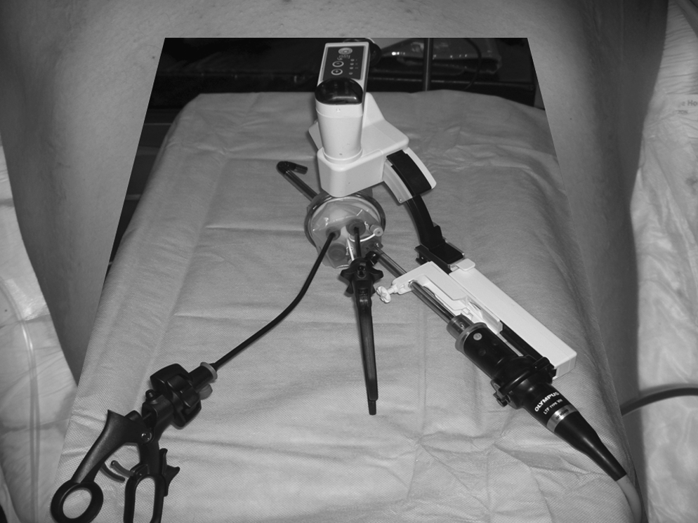
Schematic view of flexible-tip laparoendoscope supported and controlled by a robotic camera holder, a curved tissue grasper, and a traditional straight instrument with minimal external and internal potential for instrument clash.
The pelvic kidney was identified and the peritoneal attachments divided. The sigmoid colon was mobilized with the harmonic ACE scalpel and the kidney was skeletonised using a combination of harmonic ACE and blunt dissection (Fig. 3). Preoperative CT angiography had failed to identify a vascular pedicle and so the tissue laterally between the kidney and the internal iliac artery on the left was divided with the harmonic ACE without sutures or vascular clips. The ureter was partially divided; the stent was removed and then tied with 2/0 vicryl using an extracorporeal tie. The kidney was placed in a retrieval bag and mechanically morcellated with forceps to enable removal through the small midline fascial defect. A drain was inserted into the renal bed and the layers closed with 1/0 vicryl and 4/0 vicryl rapide. The right-sided ureteric stent was removed in the clinic with a flexible cystoscope under local anesthesia after 4 postoperative weeks.

Intraoperative view from flexible-tip laparoendoscope showing a small pelvic kidney and hugely dilated renal pelvis.
Results
The operation proceeded uneventfully with no complications. Operation time was 185 minutes and blood loss was ∼100 mL. Hospital discharge occurred on the second postoperative day. The histology of the morcellated specimen revealed chronic pyelonephritis secondary to a ureteropelvic junction obstruction. The immediate and 6-week postoperative cosmetic result was satisfactory to the patient (previous umbilical piercing not used for some years) (Fig. 4).
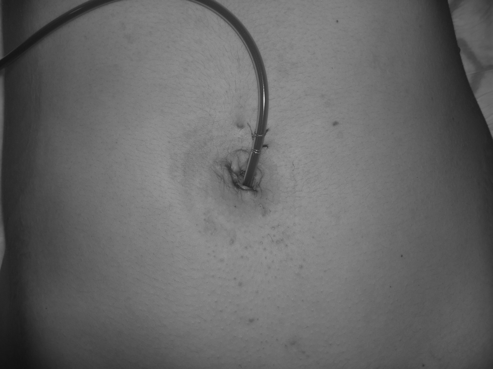
Immediate postoperative view (previous umbilical piercing).
Discussion
LESS scarless pelvic kidney nephrectomy is a safe and technically feasible alternative to traditional multiport laparoscopic pelvic kidney nephrectomy.
Failure of the mature kidney to reach its normal position is termed renal ectopia. Pelvic kidneys are defined as those found below the aortic bification. They are more common on the left and have an incidence on postmortem studies of between 1 in 2000–3000. 10 They are often hypoplastic and are prone to reflux and obstruction due to an anteriorly placed pelvis and malrotation; this can lead to impaired drainage of urine from a high insertion of the ureter or obstruction from an anomalous vessel. 11 Renal ectopia is associated with other congenital abnormalities, including bicornate or absent uterus, with or without vaginal abnormalities, including duplication or absence in females, 12 and undescended testes, urethral duplication, and hypospadius in males. 13
Minimally invasive pelvic kidney surgery has been previously performed and reported for all of the common conditions that effect kidneys. Due to the renal anatomy, anomalous vessels and adjacent structures these operations can be challenging. Traditional multiport laparoscopic pyeloplasty, 14 laparoscopic-assisted percutaneous nephrolithotomy (PCNL), 15 and nephrectomy, both transperitoneal 16 and retroperitoneal, 17 have all been reported with good surgical exposure and operative times comparable to those of laparoscopic operations in anatomically normal kidneys.
The recent trend toward LESS in urology is well known, and the largest single-centre experience of 100 ablative and reconstructive cases has been published. 18 LESS pelvic operations are reported less frequently, presumably related to the increased surgical challenge. Despite this, complex procedures, including colectomy, 19 hysterectomy, 20 and radical prostatectomy, 21 have all been reported, although usually single cases or small numbers only. The radical prostatectomy series had longer operation times and more complications than traditional multiport laparoscopy in the hands of the authors all of whom are experts in multiport laparoscopy with high-volume workloads; this may improve in time as the techniques and instruments are further refined. Long-term follow-up is required for any LESS procedure to report oncological and functional outcomes. Any move toward improving cosmetic results with a compromise in outcome needs to be known. Although randomized controlled trials in surgery are notoriously difficult to perform, it is possible to assess the advantages of LESS over traditional laparoscopy using surrogate markers of patient benefit such as length of stay, return to normal activities, wound complications, and operative narcotic use.
The LESS approach has the advantage of a single incision, but there are significant ergonomic challenges. Compared to standard multiport laparoscopy, where the ports are well spaced apart, in single-port surgery the ports are in proximity usually as part of a single-piece access device, for example, R-port and TriPort. This proximity leads to clashing of camera head, light lead, surgical instruments, telescope shaft, and hands both externally and internally. This can be frustrating for the operating surgeon and assistant, and may restrict movements required for accurate fine dissection and reconstruction with intracorporeal suturing. Many of the ergonomic challenges facing LESS can be overcome by modifying traditional laparoscopic techniques and equipment as detailed in Table 1. With time, advances in technology such as flexible-tip absorbable stapling devices and tissue glues may reduce the need for suture reconstruction.
In summary, LESS is an exciting and advancing area of laparoscopic urology and in this report we show that complex procedures on anatomical variants can be performed safely. With advances in access devices, for example, QuadPort™ advanced pelvic reconstructive procedures such as radical prostatectomy, may become the standard of care.
Disclosure Statement
No competing financial interests exist.