
Abstract
Abstract
Background:
The risk of intraabdominal contamination is a critical consideration during most natural orifice translumenal endoscopic surgery (NOTES) procedures.
Objective:
The objective of this study was to determine a safe and efficient pathway for the endoscope in a transgastric NOTES procedure.
Design and Settings:
A pilot experimental study in live pigs was performed.
Methods:
Five White Landrace pigs, weighing ∼30–35 kg, underwent the placement of a device consisting of an expandable sheath, the distal portion of which was composed of a fully covered self-expanding metal stent, and an introducer made with an outer catheter, a pushing catheter, and an inner, guiding catheter. The sheath was attached to the stent by suturing it in place. The initial gastric opening was made by means of a needle knife papilotome with electrocoagulation, aimed to the anterior gastric wall. Then, it was dilated with an endoscopic 1.8-cm balloon. The set was introduced over a Savary guidewire. After the set placement, the outer tube was slowly retrieved. Finally, the delivery system was removed from the pig, leaving the entire endoscopic port in place.
Results:
The expandable gastric port was placed without difficulty in all animals. Endoscope insertion into the expandable gastric port was very easily performed. The endoscope had a wide range of movement inside the peritoneal cavity. The gastric port sealed the gastric wall, avoiding gross contamination of the peritoneal cavity and maintaining the pneumoperitoneum without excessive inflation of the intestine.
Conclusions:
Use of a gastric port can minimize contamination of the peritoneal cavity due to the spillage of gastric contents during a transgastric NOTES procedure and can also facilitate performance of the procedure.
Introduction
This study describes a flexible and relatively sterile pathway from the mouth to the peritoneal cavity.
Materials and Methods
Five White Landrace pigs (weighing 30–35 kg) underwent the placement of an expandable gastric port (EGP). The procedures were performed with the animals under general anesthesia by endotracheal intubation. Halothane, fentanyl, and pancuronium were administered. The preanesthesia medication consisted of 2 mL of 1% acetylpromazine and 15 mg of midazolam.
The device, its placement, and its withdrawal
The EGP device consisted of a thin, expandable polyethylene sheath, the distal portion of which was composed of a fully covered self-expanding metal stent of 8 cm length and 18 mm diameter, and an introducer made up of an outer catheter, a pushing catheter, and an inner, guiding catheter (Fig. 1). The sheath was attached to the stent with sutures (Prototype; Cook Endoscopy, Winston-Salem, NC). For the last two animals, the distal part of the stent was attached to a steel wire with a design like a “crown.” This device had two threads attached, and when the threads were pulled inside a tube, the “crown” acquired an anchoring shape. The sheath was collapsible over a substantial portion of its length and was operable between collapsed and expanded configurations. Its length was suitable for forming a pathway for an endoscope to pass through (Fig. 2). The self-expanding stent was similar to the zigzag-shaped wire metal Gianturco stent. To fit the EGP, the stent was fully loaded into an outer catheter, and the proximal end of a prepositioned Savary-Gilliard dilator guidewire was placed through the guiding catheter. Under laparoscopic control, with a view of the anterior gastric wall, an endoscopy was performed. An initial opening in the gastric wall was created with a Needle Knife Papillotome (ECL-18 × 8; Cook Endoscopy) and dilated with a 1.8-cm balloon (ECL-18 × 8; Cook Endoscopy). The entire system was gently advanced over the guidewire until the stent portion reached the gastrotomy site (Fig. 3). The metallic stent and the entire clear sheath were slowly deployed by holding the inner catheter in place while withdrawing the outer catheter. Finally, the delivery system was removed from the pig, leaving the entire endoscopic port in place, with the “crown” opened and attached to the gastrotomy site, allowing visualization of the peritoneal cavity (Fig. 4). To remove the port, the “crown” was collapsed by tensioning the threads attached to it, and the entire system was pulled through the stomach and esophagus with the endoscope.

EGP for natural orifice translumenal endoscopic surgery. EGP, expandable gastric port.
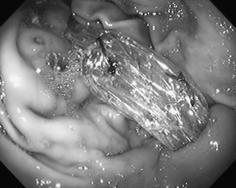
The EGP tip reaches the gastrotomy (endoscopic view).
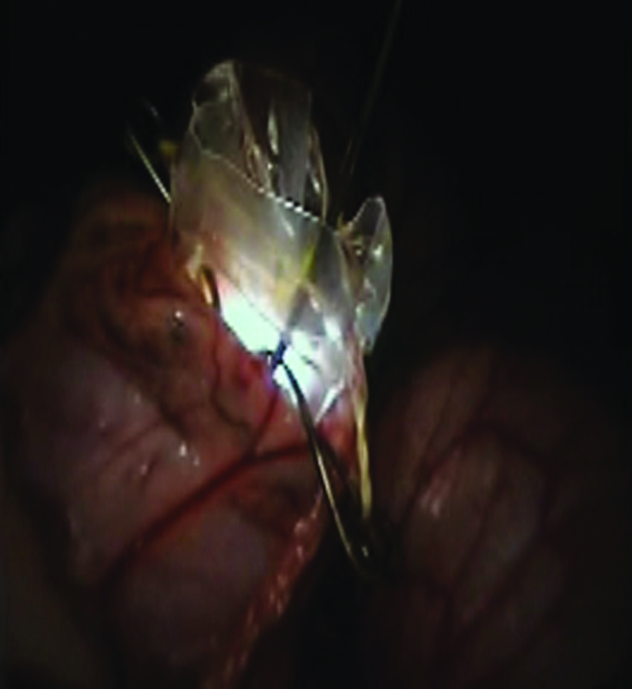
The endoscope reached the distal EGP end. The “crown” grabs the gastric wall (laparoscopic view).

Endoscopic view inside the EGP distal end.
Results
The EGPs were placed easily in most of the animals. The guidewire had to be pulled through a laparoscopic port inserted into the umbilicus during placement. After EGP placement, a conventional gastrointestinal endoscope was inserted easily through the device. The introduction was carried out slowly, with rotational movements, to decrease the friction against the plastic sheath. No difficulties were encountered during the insertion through the gastric port. The use of water or other lubricants was also important during this procedure. The EGP allowed wide movements of the endoscope inside the peritoneal cavity, without changing the position of the EGP, in three animals. In 2 animals, the port slipped into the gastric lumen during endoscopic management. Final evaluation of the cavity through laparoscopy showed absence of gastric contents, such as mucosal secretions and residual food found in the gastric lumen, suggesting that the port sealed the gastric wall and avoided gross contamination of the peritoneal cavity. Further, it seems that the EGP allowed for maintenance of the pneumoperitoneum without intense inflation of the intestine.
Discussion
One major concern regarding perforation of the gastrointestinal system is the contamination of the peritoneal cavity, which may cause peritonitis, infection, or adhesions. The EGP provides a flexible conduit for the endoscope to pass through (from the mouth to the peritoneal cavity) and a way to maintain a clean and relatively sterile pathway for the endoscope. Moreover, the use of a device tightly fitted to the gastric opening seems to avoid the continuous spillage of gastric contents into the peritoneum and allows for maintenance of the pneumoperitoneum without intense inflation of the intestine.
At present, procedures involving a transgastric route to access the peritoneal cavity are considered to involve contamination.3,4 Presently, endoscopes can be sterilized, and the gastrointestinal tract can be cleared with antimicrobiological agents. However, it is not absolutely clear whether preprocedural gastric lavage reduces the contamination of the peritoneal cavity. 5 Of course, such gastric preparation is necessary to ensure that no solid contaminants are present in the stomach, and delayed gastric emptying could be considered a contraindication for the approach.
It is important to emphasize that overtubes must be passed down through the mouth and, therefore, transport bacteria from the oropharyngeal cavity and esophagus to the stomach. The segment that will be in the peritoneal cavity and the tip of the overtube where the working endoscope will exit would also be contaminated. 6 For this reason, the use of prophylactic antibiotics and gastric antibiotic lavage has been recommended, although open and laparoscopic transgastric procedures usually do not require such precautions. 5
The EGP presented here has an advantage over traditional plastic overtubes, which have been also proposed for natural orifice translumenal endoscopic surgery (NOTES) procedures. 7 Traditional plastic overtubes are quite rigid and can restrict the ability to manipulate the endoscope as desired, especially when the endoscope must be curved, as has been demonstrated in several experiments prior to this study. Moreover, the results of band ligation procedures have shown that lesions occur at the edge of the overtube, suggesting that mucosal damage does not occur during insertion but rather during the procedure itself, possibly because of friction during respiratory movements. 2 The present study shows that the EGP does not interfere with intraabdominal procedures. However, the port slipped into the gastric lumen during endoscopic management in 2 animals. Improvement of the distal end of the EGP would avoid this inconvenient displacement.
The guidewire must also be improved before performing a full NOTES procedure. Namely, a stiffer Savary guidewire must be used, because the conventional Savary guidewire did not provide enough support for the EGP during insertion.
Conclusions
An EGP can minimize contamination of the peritoneal cavity due to the spillage of gastric contents during transgastric NOTES procedures and can also facilitate the performance of these procedures.
Footnotes
Acknowledgments
The authors thank Ms. Alvamar Helena de Campos Andrade Lamparelli for the manuscript revision and Mr. Vihar Surti, Senior Research Engineer, Cook Endoscopy, Winston-Salem, NC, for the development of the EGP set. The endoscope system and other devices were kindly provided for the study by Olympus Latinamerica, Miami, FL, and Cook Endoscopy, Winston Salem, NC.
Disclosure Statement
The following authors have no conflicts of interest or financial ties to disclose: Pablo R. de Siqueira, Horus A. Brasil, Daniel Moribe, and Marco Aurélio D'Assunção. Kiyoshi Hashiba acts as a consultant for Cook Endoscopy. The authors declare that this article has not been published or submitted for publication elsewhere. All authors have contributed significantly and are in agreement with the content of the manuscript.