
Abstract
Abstract
Objectives:
To report the first clinical experience with laparoendoscopic single-site (LESS) extraperitoneal aortic lymphadenectomy.
Materials and Methods:
A 33-year-old woman with biopsy proven locally advanced squamous cell carcinoma of the cervix was taken to the operating room for surgical staging. Preoperative imaging did not detect any aortic lymph node metastases. Informed consent for LESS extraperitoneal aortic lymphadenectomy was obtained. A 2 cm transverse incision was made on the left side midway between the iliac crest and inferior costal margin along the middle axillary line. The preperitoneal space was created and the TriportTM inserted. Using the Deflectable-Tip EndoEyeTM laparoscope and two straight instruments, the aortic lymphadenectomy was performed as defined by the disease-specific oncologic principles.
Results:
The procedure was completed in 125 minutes. There were no intraoperative or postoperative complications, and the blood loss was minimal (10 mL). The patient was discharged home on postoperative day number 1. LESS extraperitoneal aortic lymphadenectomy yielded 10 lymph nodes. Microscopic metastatic squamous cell carcinoma was detected in 1 out of the 10 lymph nodes. Her treatment plan was modified to extend the field of radiation to include the paraaortic lymphatic basins.
Conclusions:
LESS extraperitoneal aortic lymphadenectomy is feasible and safe, and provides a comprehensive assessment of aortic lymph nodes as defined by the disease-specific oncologic principles.
Introduction
Vasilev and McGonigle 7 and Dargent (Extraperitoneal aortic dissection, Award video presented at the meeting of the Society of Gynecologic Oncologists, Phoenix, 1997) are the pioneers of laparoscopic extraperitoneal approach to paraaortic lymph node dissection. The technique was further refined by Dargent et al., who reported their experience with transperitoneal, bilateral extraperitoneal, and left extraperitoneal paraaortic lymphadenectomy in cervical cancer. They found that the left approach is feasible without compromising the nodal yield and with significantly shorter operative time. 1 Further, Querleu et al. confirmed, in a pilot study, the utility of left laparoscopic extraperitoneal paraaortic lymphadenectomy as a tool to identify lymph node-positive cervical cancer patients who require extended-field radiation therapy. 8 The operative technique described by Querleu et al. is the most widely used. All current techniques utilize an initial umbilical intraperitoneal trocar to evaluate the abdominal cavity for evidence of intraperitoneal disease and three or four extraperitoneal trocars to perform the lymphadenectomy.
Recently, with the advancement of minimally invasive surgery, the concept of scarless surgery has gained interest. This lead to the development of natural orifice transluminal endoscopic surgery (NOTES). NOTES utilizes the natural body orifices for access; however, this approach has not been widely adopted due to several concerns about safety, consequences of poor closure of access sites (e.g., bladder, bowel, and stomach), and technical surgical difficulties. 9 Moreover, the frequent need for an additional transabdominal port with NOTES lead to a shift in interest to single-incision laparoscopic surgery as an alternative. Due to the several terms that existed to describe single-incision laparoscopy, the term laparoendoscopic single-site surgery (LESS) was introduced in a consensus statement in 2008. 10 In this report we present the first LESS extraperitoneal aortic lymphadenectomy.
Materials and Methods
Patient
A 33-year-old woman with biopsy-proven locally advanced squamous cell carcinoma of the cervix was taken to the operating room for surgical staging. Her medical and surgical histories were unremarkable. The body mass index was 22 kg/m2. Positron emission tomography revealed increased Fludeoxyglucose uptake in the area of the uterine cervix. There was no uptake in the paraaortic region. After detailed explanation of the risks, benefits, and alternatives of the surgery, the patient was consented for an examination under anesthesia, cystourethroscopy, proctosigmoidoscopy, and LESS extraperitoneal aortic lymphadenectomy. Preoperative prophylactic antibiotics were administered 30 minutes before incision. Sequential compression devices were applied to the lower extremities before induction of general anesthesia. Patient was placed in the dorsal supine lithotomy position with both arms abducted, and she was prepped and draped in a sterile fashion. We started with an examination under anesthesia and cystourethroscopy. Then, we performed LESS extraperitoneal aortic lymphadenectomy followed by proctosigmoidoscopy.
Operative technique
The surgical technique for LESS extraperitoneal aortic lymphadenectomy is as follows. Initially, standard intraperitoneal laparoscopy is performed via a 5-mm umbilical trocar inserted after pneumoperitoneum has been created. We use a 5-mm Deflectable-Tip EndoEyeTM laparoscope (Olympus Medical, Tokyo, Japan) (Fig. 1). The surgeon stands on the left side of the patient and the assistant on the right with two video monitors at the lower extremities of the patient. This allows the evaluation of the abdominal and pelvic cavities for any evidence of intraperitoneal involvement with metastatic disease. It also helps guiding a proper entry into the extraperitoneal space and positioning of the multi-channel port during the next stage of the procedure.
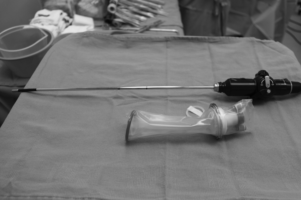
The Deflectable-Tip EndoEye laparoscope (Olympus Medical, Tokyo, Japan) and Triport (Advanced Surgical Concepts, Dublin, Ireland).
A 2-cm transverse incision is made on the left side midway between the iliac crest and inferior costal margin along the middle axillary line. The incision is carried down to the underlying layers of external oblique, internal oblique, and transversus abdominis muscles. The muscle fibers are separated bluntly along their course until the underlying peritoneum is reached. Care must be taken to avoid disrupting the peritoneum. The forefinger is then introduced through the incision and is used to separate the peritoneum from the overlying muscles of the abdominal wall under laparoscopic monitoring as described by Querleu et al. 8 The extraperitoneal space may also be developed using a balloon trocar. Subsequently, the TriportTM (Advanced Surgical Concepts, Dublin, Ireland) (Fig. 2) with one 12-mm, two 5-mm ports, and two insufflation channels is introduced through the incision in such a way that the internal ring rests in the extraperitoneal space previously developed by the forefinger. Once proper placement is confirmed, the Deflectable-Tip EndoEye laparoscope is removed from the umbilical trocar and brought through the Triport. The abdomen is deflated and extraperitoneal space is insufflated with carbon dioxide up to 15 mm Hg. The assistant then moves to the patient's left side and the right monitor is moved to over the patient's right shoulder. A Harmonic ACE® (Ethicon Endo-Surgery, Cincinnati, OH) and a grasping blunt forceps are introduced through the remaining two ports of the Triport and are used to perform the lymphadenectomy. The extraperitoneal space is developed bluntly off the psoas and medially until the left common iliac artery and ureter are identified. The development of the extraperitoneal surgical space and the margins of the paraaortic lymphadenectomy follow the steps described by Querleu et al. 8 The psoas muscle is freed cephalad up to the fascia of the left kidney. The space between the ureter and the common iliac artery is developed in such a way that the iliac and lumber segments of the ureter stay attached to the overlying lifted peritoneal sac. The dissection is then carried along the lateral and anterior aspects of the common iliac artery caudally to the level of its bifurcation and cephalad up to the level of aortic bifurcation and left renal vessels. Care must be taken to identify, isolate, and preserve the inferior mesenteric artery. At this point, the left common iliac and aortic lymph nodes can be dissected from the bifurcation of the common iliac artery caudally to the renal vessels cephalad.
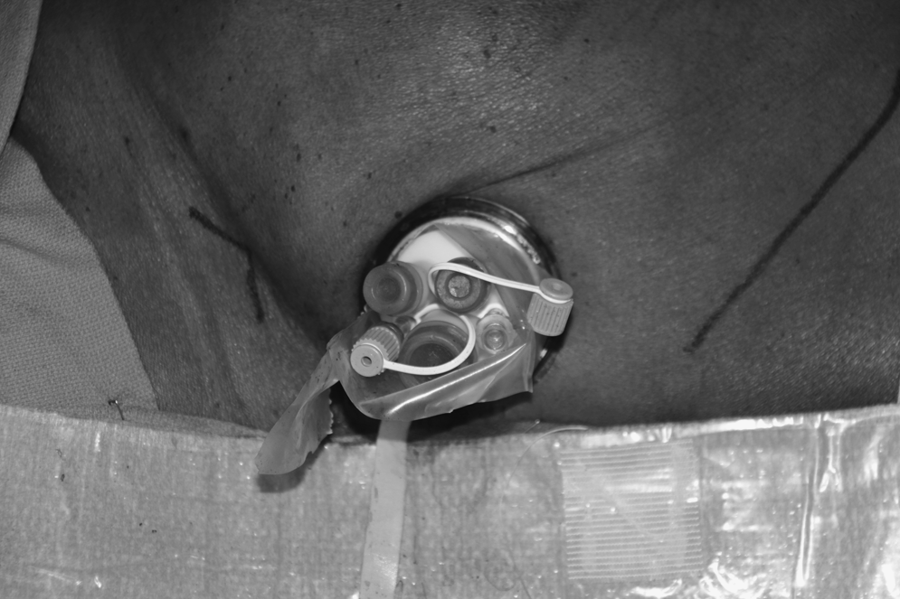
The Triport position for laparoendoscopic single-site extraperitoneal paraaortic lymphadenectomy.
Following this, we continue elevating the peritoneal sac over the sacral promontory, bifurcation of the aorta and inferior aspect of the vena cava. The right common iliac artery is identified and followed caudally to its bifurcation. Then, the right ureter is identified and lifted up together with the overlying peritoneal sac, thus separating it from the underlying iliac vessels and psoas muscle. At this point, the right lateral common iliac nodes, precaval nodes, and presacral nodes are dissected off. This completes the paraaortic nodal dissection. Hemostasis is ensured under low pressure. The upper limit of lymph node dissection is labeled using a 5-mm laparoscopic vascular clip. Dissected lymph nodes are removed through the Triport after uncapping the external portion. Once the specimen is removed the cap is latched on and the extraperitoneal space re-expanded. To prevent lymphocyst formation, a 3-cm opening in the peritoneal sac is routinely made. The Triport is removed and the incision is closed using 3-0 polyglactin to re-approximate the muscle layers and 4-0 poliglecaprone in a subcuticular fashion on the skin.
Results
The procedure was completed in 125 minutes. There was no evidence of intraperitoneal involvement with metastatic disease upon standard laparoscopy via the 5-mm umbilical trocar. Cystourethroscopy and proctosigmoidoscopy ruled out tumor extension to bladder or rectal mucosa. There were no intraoperative or postoperative complications and the blood loss was minimal (∼ 10 mL). The patient was discharged home on postoperative day number 1. LESS extraperitoneal aortic lymphadenectomy yielded 10 lymph nodes. Microscopic metastatic squamous cell carcinoma was detected in 1 out of the 10 lymph nodes. Her treatment plan was modified to extend the field of radiation to include the paraaortic lymphatic basins.
Discussion
During the last 10 years, the use of laparoscopic surgery for treatment and staging of gynecologic malignancies has gained wide acceptance. With the evolution of laparoscopic surgery, the idea of scarless surgery gained interest. The standard laparoscopic extraperitoneal aortic lymphadenectomy requires three or four trocars, in addition to the umbilical trocar, according to the techniques described by Querleu et al. 8 This can lead to poorer cosmetic results, increased pain, and potentially increased risk of serious injury to underlying organs with each trocar insertion. In our initial case, the procedure was completed with a Triport, thus decreasing the number of extraperitoneal trocars from three or four to one. The comprehensive assessment of aortic lymph nodes as defined by the disease-specific oncologic principles were respected. Estimated blood loss was 10 mL. The patient was discharged home on postoperative day number 1 and did not have any intraoperative or postoperative complications. She was able to start her definitive chemoradiation on time without any delays related to the surgical intervention. As a result of the presence of metastatic disease in 1 out of 10 paraaortic nodes, the radiation field was extended to encompass the paraaortic lymphatic basins.
The Triport is, by design, a wound protector that may decrease or eliminate the incidence of wound site metastasis. In addition, it can be adjusted to fit different abdominal wall thicknesses, thus expanding its utility. It provides a good seal, allowing proper expansion of the extraperitoneal space without any significant gas leakage. However, it also suffers from several limitations. The Triport introducer has a sharp tip, which may perforate the peritoneum during insertion. We do not use the introducer that is prepackaged with the Triport, and we manually insert and position the internal ring of the port in the preperitoneal space. Manual insertion, however, can be challenging especially in patients with thick abdominal wall and the Triport may benefit from some design modifications that would optimize its use for extraperitoneal procedures. A blunt tip introducer that can split the muscle fibers during entry without disrupting the peritoneum may facilitate proper positioning of the Triport in the preperitoneal space. The QuadportTM (Advanced Surgical Concepts) with its larger internal ring may provide an easier manual insertion mechanism. During this initial experience, we utilized straight laparoscopic instruments. The use of Deflectable-Tip EndoEye laparoscope minimized instrument clashing and is essential when straight laparoscopic instruments are used. However, precurved instruments that take into consideration the distances and angles needed for this type of procedures may allow the use of a straight laparoscope, minimize instrument clashing, and improve the performance.
Conclusions
LESS extraperitoneal aortic lymphadenectomy is feasible and safe, and provides a comprehensive assessment of aortic lymph nodes as defined by the disease-specific oncologic principles. The procedure can be further improved by refining the port and instruments to address the specific requirements of laparoscopic extraperitoneal surgery. Further clinical studies are necessary to define the oncologic outcomes and potential benefits of LESS extraperitoneal aortic lymphadenectomy.