
Abstract
Abstract
Background:
Gynecomastia is a benign clinical condition that is the unilateral or bilateral enlargement of the male breast, but sometimes it causes serious psychological and physical stress in young men and brings about phobia of malignancy in elderly men. Minimally invasive and functional therapy represents an inevitable trend in breast surgery. We investigated the feasibility and safety of vacuum-assisted biopsy device in treating gynecomastia.
Methods:
From January 2006 to January 2010, 20 male patients with gynecomastia were treated by an 8-gauge vacuum-assisted biopsy device. The average age was 24.7 years (range, 18–47 years).
Results:
The operation was successfully performed in all 20 patients with a mean operating time of 51 minutes and a hospital stay of 4 days. Postoperative complications included 1 case of hematoma, but no nipple necrosis, local skin necrosis, or skin buttonhole occurred. No other operation-related complications were observed. Satisfactory chest contour was gained in all cases without any abnormality, skin redundancy, or recurrence during the follow-up of 6–48 months.
Conclusions:
Treatment of gynecomastia by the Mammotome device is distinctive, practicable in manipulation, safe, and can achieve excellent cosmetic results. The Mammotome procedure simply represents another novel treatment option for gynecomastia.
Introduction
Materials and Methods
Patients
From January 2006 to January 2010, 20 male patients with Simon's grade II and III gynecomastia (Fig. 1), 6 who were requesting surgery, underwent ultrasound-guided Mammotome excisional surgery. These patients aged 18–47 years (mean 24.7 years) and with 2–21 years of gynecomastia progression. Ten patients were preoperatively treated with tamoxifen. Both historical and physical examinations are frequently sufficient to make an appropriate diagnosis. Ultrasound exam was performed in all the cases. The longest lesion diameter ranged from 4 to 8.5 cm (mean, 6.4 cm). Eleven patients were having bilateral gynecomastia.
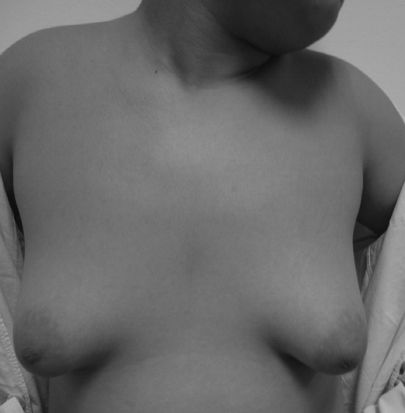
Preoperative anteroposterior view of the patient with Simon's grade III gynecomastia.
Surgical technique
The procedure was performed after the patient had been given a detailed explanation and an informed consent had been obtained. The patients were operated on by the skilled surgeon with special interest in breast surgery.The Mammotome procedure was performed in a supine position with the ipsilateral arm raised above the head. Real-time ultrasound guidance was performed using a Philips iU22 system (Philips Medical Systems, Andover, MA). Color Doppler ultrasound was performed in all cases to evaluate lesion vascularization and avoid major vessels during the excisional procedure. The edge of the breast disk was marked on the skin with indelible ink (Fig. 2). Local anesthetic, consisting of 0.75% lidocaine containing a 1:1000 mixture of epinephrine (used for the breast tissues to minimize bleeding), was applied in all the patients. One hundred milliliters of physiological salt solution containing a 1:1000 mixture of epinephrine was injected subcutaneously along the edge of the breast disk.

The marked resection range was removed with guidance of ultrasound using vacuum-assisted biopsy device.
Once local anesthetic was administered, a 5-mm skin incision was made in the lateral breastfold, through which the 8-gauge Mammotome device (Mammotome EX, Johnson and Johnson Company) was passed and positioned beneath the breast disk for real-time ultrasound's guidance. Resection of the breast tissues can then be systematically performed under constant ultrasound control until completion. At the end of the procedure, all hematomas were suctioned out, and a further 100 mL of physiological salt solution containing a 1:1000 mixture of epinephrine was infused into the cavity. All the patients did not receive liposuction. The extent of procedure depends on both the amount of breast tissue to be removed and the degree of skin redundancy. A continuous negative pressure drainage tube was placed to continue the suction and chest strap hemostasis.
The follow-up time was 6–48 months. Patients' opinion and level of satisfaction was obtained and graded as poor, average, good, or excellent. A scoring system was used by the operating surgeon to grade the cosmetic outcome between 0 and 10 (Patient satisfaction score was checked after procedure in all patients, the definition of satisfaction score was not satisfied [less 5], satisfied [5–8], and very satisfied [9–10]). All complications were recorded.
Results
Twenty male patients underwent Mammotome excision of their gynecomastia. During operation under local anesthesia, all patients tolerated the procedure. The weight of gland resected in the 31 breasts was 68–160 g, with a mean of 93 g. The mean operating time was 51 minutes (range, 40–73 minutes)and a hospital stay of 4 days (Fig. 3). Postoperative complications included 1 case of hematoma, but no nipple necrosis, local skin necrosis, residual gynecomastia, or skin buttonhole detected. No other operation-related complications were observed. Satisfactory chest contour was gained in all cases without any abnormality, skin redundancy, or recurrence during the follow-up of 6–48 months (average, 26.8). All patients were satisfied (scores of 8) or very satisfied (scores of 9 or 10). The surgeon's scoring mean was 8.7. All lesions were histologically benign.

Four days after Mammotome excision (postoperative lateral view of the patient with gynecomastia).
Discussion
Gynecomastia describes a benign development of the mammary gland in men. The incidence of malignancy or abnormal pathology associated with gynecomastia tissue in the adolescent man is extremely low.6,7 Patients with idiopathic gynecomastia do not respond to medical treatment, so they need surgical treatment. Patients who have long-standing symptomatic gynecomastia or whose medical therapy is not successful, who are experiencing psychological or emotional distress, or who have severe mastalgia are potential candidates for surgery. Gynecomastia can cause severe emotional phobia for breast cancer in elderly men and physical distress in young men. Treatment of gynecomastia depends on the underlying cause. 8 Treatment may be either medical or surgical but should be individualized (e.g., reassurance, medical treatments, or surgery).9–16 If it is drug induced, it may regress once the medication is stopped. Due to limited experience and unknown long-term side effects, trials of medical therapy should be limited to only 6 months. When medical treatment fails, the surgical procedure is the choice.
The aim of surgical treatment is to achieve a normal appearance of the masculine thorax, with the smallest possible scar. Surgical technique will mainly depend on the degree of the gynecomastia, the distribution, and the proportion of the different components (fat and parenchyma) of the breast. Traditionally, open surgery such as subcutaneous mastectomy is enough to remove the breast tissue through a periareolar or inframammary incision. Unfortunately, this operation can leave a scaphoid defect in the soft tissue of the chest wall. Results are cosmetically unsatisfactory in up to 50% of patients.4,5 Liposuction can correct the abnormal and excessive collections of fatty tissue and is regarded by many investigators as one of the most effective treatments for gynecomastia, as it is associated with few adverse sequelae for its rare association with adverse sequelae. However, patients with a firm or fibrous breast disk still require open surgery. Studies by Petty et al. reveal that arthroscopic mastectomy was safe and effective in treating most grades of gynecomastia, with excellent cosmetic results. 17 Endoscope-assisted subcutaneous mastectomy was performed successfully in 125 breasts of 65 patients with gynecomastia, but postoperative complications only included 2 cases of partial nipple necrosis and 1 case of subcutaneous hydrops. 10 Endoscopic subcutaneous mastectomy had three small incisions.
The Mammotome procedures can be performed in the outpatient setting under a local anesthetic, and we now routinely perform Mammotome excision for all benign breast lesions, providing an excellent cosmetic result with little morbidity. 18 The 8-gauge Mammotome device was advanced through the 5mm incision in the lateral breastfold to excise the breast disk and fatty tissues. Ultrasound guidance was performed to avoid residual gynecomastia. The merits of Mammotome procedure in the treatment of gynecomastia, in terms of cosmetic maintenance and safety, were fully demonstrated in this study. Hematoma is a potential complication related to this procedure, because of the use of an 8-gauge probe and suction system. Bleeding can be avoided by injection of 100 mL of physiological salt solution containing a 1:1000 mixture of epinephrine (surrounding the breast disk) before the procedure and by sufficient compression immediately after the procedure. The Mammotome procedure is a minimally invasive technique for the complete removal of gynecomastia.12,13 All cases reported excellent satisfaction, and there were no severe complications. It may become the method of choice for the surgical treatment of gynecomastia.14,15 The normal male chest contour can be restored by the Mammotome device.
Conclusion
Vacuum-assisted biopsy device excision was an effective surgical option, especially for glandular gynecomastia. It may be one of the useful and safe methods for correction of gynecomastia.
Footnotes
Acknowledgment
This work was supported by China Postdoctoral Science Foundation (No. 20080431408; No. 201003759).
Disclosure Statement
No competing financial interests exist. All patients provided written informed consent for the study, and the study was approved by the Jinan Military General Hospital Research Ethics Board.