
Abstract
Abstract
Introduction:
Laparoscopic procedures for children with urological problems are common under the present conditions. Laparoscopic surgery is associated with complications such as port site hernia that are not associated with open surgery. Drain site hernia is one variety of port site hernia.
Subjects and Methods:
We did a retrospective analysis for the development of port site hernias among pediatric patients who underwent laparoscopic procedures. We also analyzed the various methods of prevention.
Results:
Among the 148 children who underwent laparoscopic procedures, 5 (3.4%) had a port site hernia in the early postoperative period. All of them were drain site hernias with early presentations, and the content was omentum. Three patients had reduction under sedation. Two patients needed laparoscopy: one for the reduction into the preperitoneal space and the other for the nonreducible hernia due to omental edema. All the patients had an uneventful recovery.
Conclusions:
Port site hernia is an uncommon complication in children undergoing laparoscopy. The drain site is the predominant location of port site hernia. Sedation during drain removal and judicious use of drain may help to decrease such occurrences.
Introduction
Subjects and Methods
We reviewed the case records at our institution of all the children who underwent laparoscopic procedures over the past 15 years (1995–2011). All the patients had a 10-mm port inserted for the camera. Two or three secondary ports were inserted depending upon the procedure performed. The port sites were not stretched at the time of port insertion. All ports that were 10 mm or larger had fascial closure. Fascial closure was not done for 5-mm port sites. Drains were placed through the 5-mm port site at the flank. A 12-French tube drain was used in all patients. The drain was removed when the drainage was less than 20 mL/day. The incidence of port site hernia, management, causes of herniation, and the preventive aspects were analyzed.
Results
Between 1995 and 2011, in total, 148 laparoscopic procedures were performed among the patients in the pediatric age group (Table 1). Ages ranged from 3 months to 15 years. Procedures included orchidopexy, orchidectomy, pyeloplasty, pyelolithotomy, nephrectomy, heminephrectomy, and ureteric reimplantation. Laparoscopic orchidopexy was the commonest surgery performed. All patients except those who underwent orchidopexy had a drain inserted. The drain was placed through one of the port sites (5 mm) in the flank.
Port site herniation occurred in 5 children. All these children were below the age of 5 years. Herniation occurred between postoperative Days 3 and 5, during drain removal. In all the cases the hernia was through the drain site, and the content in the hernia was omentum. There was no obvious infection at the drain site.
No patient had herniation through the 10-mm port. All of the 5 patients had a hernia through the 5-mm port site through which the drain was inserted. Herniation occurred on the day of drain removal. None had intestinal herniation.
Three children were managed without repeat laparoscopy. Omentum was pushed back gently into the peritoneal cavity, and the drain site was closed with tight application of plaster. Two patients needed repeat laparoscopy under anesthesia. Both procedures were after laparoscopic pyeloplasty.
In one of the patients the omental herniation was noticed 6 hours after the drain was removed. There was minimal edema of the omentum. On attempted reduction, instead of returning to the peritoneal cavity, the omentum partially entered into the preperitoneal space. Diagnostic laparoscopy was done through the 10-mm port. Omentum was protruding through the drain site. It was reduced into the peritoneal cavity using a grasper, and the port site was closed under laparoscopic guidance.
The other child was discharged following drain removal on postoperative Day 5. The patient presented after 24 hours with a mass protruding through the drain site (Fig. 1). This child also needed a repeat laparoscopy. The redundant omentum was excised after ligation as it was edematous, and the remainder was returned to the peritoneal cavity. A 5-mm port was introduced through the same site to confirm complete reduction of the hernia. The child recovered well and was discharged on the second day.
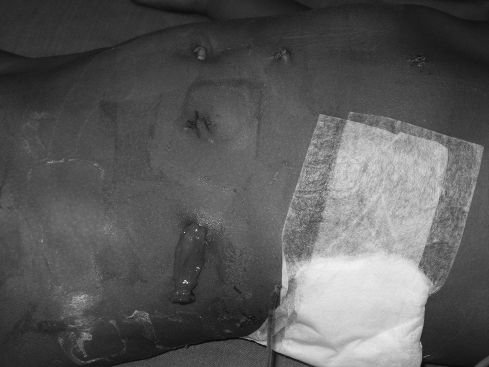
Drain site hernia with edematous omentum protruding out.
All the children are being followed up. None of the children had developed any recurrence of hernia, either in the same site or at other port sites, at a mean follow-up of 108 months.
Discussion
Port site hernia is one of the complications of laparoscopy needing surgical intervention. Port site hernia was first reported in 1968 by Fear 4 in gynecological laparoscopy. Since then many such cases have been reported. Komuta et al. 5 presented a case report of drain site hernia after laparoscopy. Drain site hernias following open surgery have been reported. 6 With the increase in pediatric laparoscopy, many port site hernias are being reported now.7,8 Our report focuses on the occurrence of port/drain site hernias following laparoscopic pediatric urological procedures.
Drain site hernias differ from other types of hernias that occur due to congenital or acquired weakness in the fascia. In the port site where the drain is placed, the fascia is not closed in any of the patients. This forms a persistent and potential fascial defect. In our series of 148 cases we had 5 patients with drain site hernia. This translates to about 3.4% of children undergoing laparoscopy. This is comparable to the incidence of port site hernia in other studies.2,7
The abdominal wall fascia and muscles are thinner in children. With increased intraabdominal pressure (during crying), herniation of the peritoneal contents can occur. 1 In adults the muscle bulk and the tough fasciae might prevent herniation. If the child is prevented from straining during drain removal (with sedation), the possibility of immediate herniation can be reduced.
Patients in the pediatric age group need observation for about 24 hours after drain removal to look for herniation. Reduction of hernia without sedation may not be possible in very young children. In one child, the omentum could not be reduced into the peritoneal cavity and was instead reduced into the preperitoneal space. However, this was recognized immediately, and reduction was done under sedation.
The other observation was regarding the reduction of hernia after a significant time lag. If there is a delay in presentation, the content of the hernia can become considerably edematous and incarcerated. This would make reduction difficult. However, in our series of children, the bowel was not involved. So if there is a delay in recognition, diagnostic laparoscopy with the patient under anesthesia would be necessary. The edematous omentum can be excised between ligatures for easy reduction.
Drain site hernias in children can be prevented by removing the drain with the patient under sedation and strapping the port site. Tube drains may be irrigated prior to removal. Suction drains may better be avoided, as there is a possibility of omentum or bowel adhering to the drain. 9 Avoiding the use of drains such as Penrose drains with few large holes and instead using drains with multiple small holes or spiral cuts (Vario drain) or a corrugated drain could decrease the possibility of omentum entering the drain.
Placement of fascial closure sutures on the muscle fascia of port sites (particularly drain sites) at the time of surgery (incorporating all the layers of the abdominal wall fascia at the port site) may allow for closure of the drain site at the time of drain removal (the technique of delayed port closure). Such approach may potentially prevent occurrence of the drain site hernia. It is important to note that even 3-mm port site fascial defects can lead to herniation. 10 Additionally, in laparoscopic pyeloplasty, the drain may be placed in a retroperitoneal position through a separate incision, and the colon can be repositioned on top of the drain as illustrated in Figure 2. Another possible preventive measure would be to perform removal of all ports after complete carbon dioxide deflation in order to avoid the possibility of the omentum being pushed out through the port site by the gas flow and pressure. 11 Finally, judicious use of drains limited to a selected group of patients would also reduce the incidence of drain site.

Drain in the retroperitoneum, prior to closure of the mesocolon after transmesocolic pyeloplasty.
Conclusions
Port site hernia is an uncommon complication in children undergoing laparoscopy. Port sites through which drains are inserted are more prone to develop herniation than other port sites. Sedation of the patient during drain removal and selective use of drain may help to decrease such complications.
Footnotes
Disclosure Statement
No competing financial interests exist.