
Abstract
Abstract
Use of secured independent tools (SIT) is changing the laparoscopy paradigm, which involves the use of instruments inside the abdominal cavity that are operated via a port that is larger in diameter than the instrument itself. However, in SIT instead of ports we used filaments or cables. Here we describe a modified SIT for use in the introduction of sutures or cables inside the peritoneum. Cables or sutures are passed through a tunnel made by an intravenous catheter and then exteriorized via a 12-mm port for tying, plugging (attaching), or connecting to different types of devices such as an endoscopic bulldog, alligator clamps, lights, and micromotors. These devices are introduced inside the abdomen and remotely operated with cables or filaments. The use of SIT is not limited to laparoscopy; it was successfully used in clinical experiences of single-port and single-incision laparoscopy and could facilitate natural orifice surgery. The technique offers a good force for traction, retraction, and mobilization. In addition, it has transmission capabilities for cameras and may facilitate the placement of wired microrobotics.
Introduction
We know that all layers of the abdomen are, in part, responsible for pain, the skin for cosmetic results, the fascia for hernias, and the peritoneum for adhesion formation. It is in our opinion that a controlled needle entrance through the abdominal wall could be less traumatic than a large anchor, screw, or suture to the peritoneum and fascia. Use of SIT has been previously done with preconnected devices introduced via a main port and exteriorizing the tail or tails using a suture passer or other means of exteriorization. 1
Here we are presenting the same principle with a different option, where intravenous catheters serve as a tunnel that allow for the passage of sutures or cables. The suture or cables are withdrawn via a main port. The device is connected outside the main port and introduced into the abdomen.
Surgical Technique
Our experiment was first conducted with simulators. We used a laparoscopic trainer (LapTrainer with Simuvision, Simulab Corp., Seattle, WA), fluorinated ethylene propylene intravenous catheters (Jelco® [Smiths Medical] and Angiocath™ [BD]), alligator clamps, mini-lights and micromotors, one 12-mm port and one 5-mm port, cables, and 2-0 polypropylene filaments 75 cm in length. The 12-mm port has a cannula with an open and close switch or a removable trap to allow for the withdrawal of devices and specimens. We are describing the use of the alligator clamps as an example for all other SIT setups (Fig. 1). SIT initial clinical experiences with alligator clamps were performed with good results by Tsin et al. 1 in 4 cases of single-port Nissen fundoplications and by Dominguez et al. 2 in 4 cases of single-incision cholecystectomies.
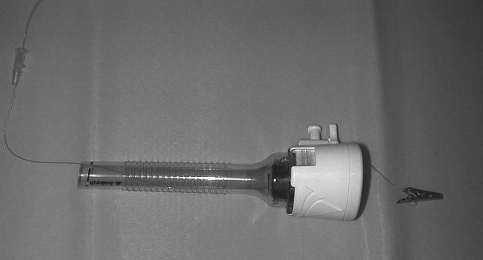
Intravenous catheter, 2-0 polypropylene filament, 12-mm cannula, and an alligator clamp.
The 18-gauge intravenous catheter was placed percutaneously under laparoscopic surveillance until the peritoneal cavity was entered (Fig. 2). The inner needle was removed to free the tunnel made by the sleeve of the catheter. The filament was threaded through the intravenous catheter, and the distal end was held to prevent slippage into the abdomen. The filament was identified with the endoscope as it entered the cavity. The end inside the abdomen was grasped and withdrawn until it exited via the operative port. Outside the port, the filament was tied to the orifice suited at the longer end of the alligator clamp (Fig. 3). The clamp was introduced inside the peritoneum backwards by a gentle pull of the filament or cable at the end of the catheter. We followed the device's entry into the abdomen with an endoscope until it was parked in the anterior abdominal wall. The same maneuver was done with an endoscopic bulldog clamp. When using a 12-mm port we follow the alligator clamp until the long end touched the intraperitoneal end of the intravenous catheter. This maneuver helps to keep the alligator steady in a desirable position for grasping. We use a 10-mm-diameter right-angle endoscopic clamp with a rotating ratchet handle; the angle clamp is directed to grasp both alligator ends.

Clinical experience. The intravenous catheter is visualized inside the abdomen.

Clinical experience. The alligator clamp is tied.
The clamp is applied as distal as possible at the alligator's end to achieve the maximum opening of the jaws. The alligator is held, directed, and placed in position for grasping the target. When the position point is reached the angle clamp grasp is released to allow the closing of the alligator jaws. For removal, the filament is pulled taut to align and steady the alligator's ends, and then the same maneuver described to open the jaws is repeated.
To withdraw the alligator we grasp the long end of the alligator and continue with the extraction. On some occasions, releasing the alligators or bulldog clamp is not needed because they are removed together with the target; for these extractions the cannula trap must be open or removed to allow passage. Extraction of specimens with diameters larger than 12 mm requires different techniques.
Two filaments are used as two tails for alligator clamps or endoscopic bulldogs, and two cables are used to plug lights or micromotors. The cable filaments come from strategically placed intravenous catheters in the abdominal wall in order to park the device in the anterior peritoneum or to provide side-to-side movements for traction, retraction, and mobilization. The introduction and removal of lights and micromotors are similar to the techniques used for the alligator clamps.
The devices were removed via the 12-mm port, and cable filaments were released from the devices. Cables or filaments were removed after the device was disconnected. Using the alligator clamp via a 6-mm working channel of an operative laparoscope was previously published.1,2 Our clinical experience was done using operative laparoscopes in Nissen fundoplications, where the alligator clamp was placed at the central tendon of the diaphragm as a liver retractor. In cholecystectomies the alligator clamp was used as a grasper for traction, retraction, exposure, and mobilization (Fig. 4). The use of the endoscopic bulldog clamp as an organ retractor has been done before, using a cinch to hook to the parietal peritoneum. The SIT technique works by tying a filament to the spring end of the bulldog clamp. 3 In the event of needing to change instruments, the instrument is withdrawn and detached from the existing cable filaments, and a different instrument is attached and introduced as previously described. Clamps, lights, and micromotors performed well.

Clinical experience. The alligator clamp is used to mobilize the gallbladder.
Discussion
This article focuses on the deployment of filaments or cables to hold tools and to power engines and lights through the anterior abdominal wall without specifics on the devices.
There are many ways to perform traction, retraction, and exposure in laparoscopy besides the usual secondary port.1–8 Leashes, marionettes, and reins perform well yet require a needle passage through the target.5–7 The SIT alligators or bulldog clamps achieved all of these functions without the needle entering the target.
The pneumoperitoneum has a minor escape when using an 18-gauge intravenous catheter sleeve; this is diminished with the presence of the filament inside the sleeve. Furthermore, in most cases the sleeve is removed and pulled outside of the abdominal wall until it exits the free end of the filament. Removing the sleeve allows for the abdominal wall track to close over the filament-cable, thereby further reducing the leakage of CO2. Sometimes the complete removal of the sleeve at the free end of a cable is not possible in some connections. When such situations occur, the sleeve is pulled and remains threaded to the cable outside the skin.
The use of micro- or mini-laparoscopy ports to pass cables or filaments will not defeat the principles of SIT as far as the diameter of a port or ports remain smaller than the instruments being used. Dávila et al. 9 used intravenous catheters for the introduction of sutures in single-port laparoscopic cholecystectomy, colangiography, gastrostomy, Nissen fundoplication, and hysterectomy. They also used the intravenous catheter for the introduction of suture passers, snares, and micro-laparoscopy graspers.
Independent lights, cameras, or engines inside the abdomen are not yet used in standard laparoscopic surgery. The progress in miniaturization of instruments, lights, and cameras combined with the affordability of micromotors and the recent advances in micro-optic fiber transmissions brought about several experimental works with such technologies.10–14 SIT could facilitate the clinical implementation of different types of the above-mentioned tools, including microrobotics.
The approach here described aims to reduce skin, fascia, and peritoneal damage caused during peritoneoscopy, with the goals of improving cosmetic results and decreasing pelvic and abdominal pain, hospital stay, infections, hernias, and peritoneal postsurgical adhesions.
Footnotes
Disclosure Statement
The authors certify that there is no actual or potential conflict of interest in relation to this article, and they reveal no financial interests or connections, direct or indirect, or other situations that might raise the question of bias in the work reported or the conclusions, implications, or opinions stated—including pertinent commercial or other sources of funding for the individual author(s) or for the associated department(s) or organization(s), personal relationships, or direct academic competition.