
Abstract
Abstract
Aim:
This study aimed to develop a novel procedure for esophagoesophageal anastomosis for natural orifice translumenal endoscopic surgery (NOTES).
Materials and Methods:
An ex vivo feasibility study was performed in eight porcine models. The procedure was as follows: (1) A BraceBar™ (Olympus Medical Systems Corp., Tokyo, Japan), a double T-bar suturing device, was placed endoscopically at the blind end of the upper esophagus (UE). (2) The blind end was incised, and the scope was advanced out of the esophagus. (3) A balloon catheter was inserted into the lower esophagus (LE). (4) The catheter and a thread on the BraceBar were withdrawn so that the end of the UE was inverted, and the LE was pulled into the UE. (5) After the catheter was removed, a short tube was placed inside the duplicated part of the esophagus via the transgastric route. (6) A double ligature was performed using a ligating device over the tube. A liquid leak test was performed after the procedure.
Results:
All steps in this procedure were technically successful under the endoscopic visualization without any assistance from outside of the esophagus. The median time of this procedure was 31 (23–66) minutes. The median internal pressure of the UE was 122 (82–142) mm Hg when the anastomosed esophagus was separated into two specimens during the leak test.
Conclusions:
Translumenal esophagoesophageal anastomosis was feasible. The duration of the procedure was short, and the anastomoses appear to have sufficient strength for use in clinical practice. An in vivo survival study is needed to confirm the safety and reliability of this NOTES procedure.
Introduction
We previously reported a laparoscopic gastric pull-up using NOTES techniques and succeeded in translumenal esophagoesophageal anastomosis in acute nonsurvival experiments. 5 However, that procedure was complicated and associated with risk; therefore, a simpler and safer method is needed. The aim of this study is to develop a novel procedure for esophagoesophageal anastomosis for NOTES.
Materials and Methods
The study protocol was approved by the Animal Care and Use Committee of The University of Tokyo and Japan NOTES. An ex vivo feasibility study was performed in eight porcine models. Eight esophagi and whole stomachs were harvested from 100-kg pigs, and each esophagus was transected at the middle and closed at both ends with interrupted sutures. A TOP Overtube (model 16632; Top Corp., Tokyo, Japan) was introduced into the upper esophagus (UE) and mounted in a phantom of the human upper body. The lower esophagus (LE) and whole stomach was set in the phantom (Fig. 1).
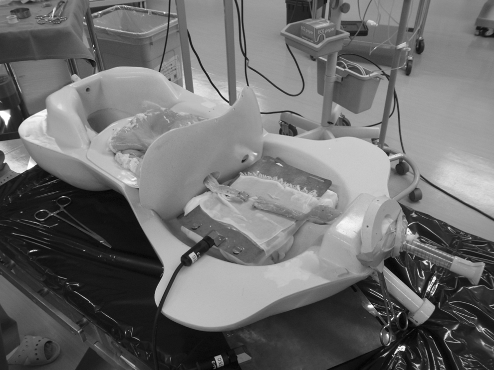
Experimental setting.
Surgical procedure
A two-channel endoscope (model GIF-2TQ260M; Olympus Medical Systems Corp., Tokyo) was introduced into the upper esophagus, and a BraceBar™ (Olympus Medical Systems Corp.), a prototype of the double T-bar suturing device originally developed to establish endoscopic full-thickness closure of large gastric perforations, was placed at the blind end. This device consisted of an applicator, which was a needle catheter with a slidable outer sheath (Fig. 2a) and a suture unit composed of a bifurcated nylon suture with three T-tags, two of which were fixed at the distal ends of the separated nylon threads, with the remaining tag placed at a position proximal to both (Fig. 2b). The suture unit was loaded inside the applicator in advance (Fig. 3a). The applicator was then advanced through the scope, and all layers of the blind end were penetrated by the needle, after which the first T-tag and thread were released out of the applicator blindly behind the intestinal wall (Fig. 3b). The applicator was withdrawn into the lumen, leaving the T-tag and thread in place. Next, another puncture was made on the other side of the blind end at a position equidistant from the center as the first puncture, and the second T-tag was released in the same manner as the first. After the needle was pulled back into the lumen, the outer sheath was advanced, and the proximal tag, which could slide over the thread, was pushed to cinch the pair of T-tags loosely (Fig. 3c).

The BraceBar, a prototype of a double tissue anchoring device, consisting of two parts:
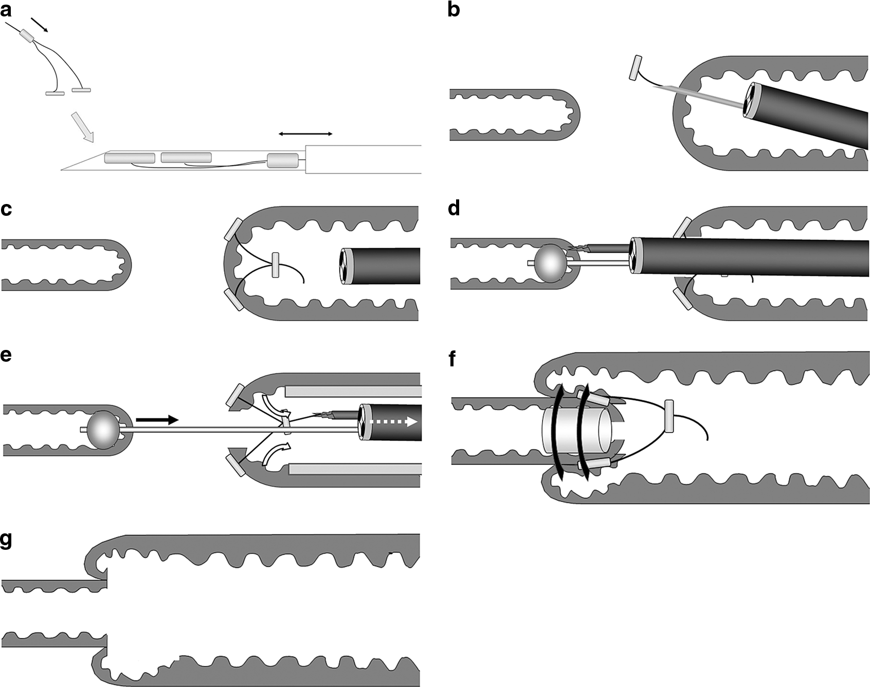
Schema of translumenal esophagoesophageal anastomosis.
The esophageal wall between the T-tags was incised, and the scope was advanced out of the esophagus, after which the LE was identified. The blind end of the LE was grasped, and a small hole was made at its center. A balloon catheter (Multi3V™; Olympus Medical Systems Corp.) was inserted into the LE through the hole, and the balloon was expanded with water (Fig. 3d). The scope was withdrawn back into the UE, leaving the balloon catheter in place, and the thread on the BraceBar was grasped by a forceps. After the tip of the overtube was advanced up to 2–3 cm proximal from the blind end, the scope, together with the catheter and the forceps, was pulled so that the edge of the UE was inverted, and the LE was dragged into the UE (Fig. 3e). The catheter was removed, and a short (approximately 3 cm) 16 French silicon tube was placed inside the duplicated part of the LE via the transgastric route. Finally, a double ligature over the transanastomotic tube was performed using a ligating device (Olympus Medical Systems Corp.) with the expectation that a wide opening would be created after the ligated tissue became necrotic and dropped off together with the tube (Fig. 3f and g).
Liquid leak test
The following experimental system was set up to evaluate the strength of the anastomosis. An extension tube was inserted into the UE through the oral opening and ligated tightly. The other end of the tube was connected to a TruWave® disposable pressure transducer (Edwards Lifesciences LLC, Irvine, CA), and the internal pressure of the UE was displayed in real time on a monitor. A small hole was made on the side wall of the UE, and another extension tube, which was connected to a syringe, was inserted and ligated firmly with double purse-string sutures. The LE was clamped with two intestinal forceps just proximal to the cardia. Indigocarmin aqueous solution in a syringe was manually infused into the UE, and the state of the specimens and the pressure values were recorded. Leakage pressure was defined as the value at the time when the esophageal duplication was released and the anastomosed esophagus was separated into two specimens, or when the solution was seen leaking from the anastomotic part.
Results
The procedure was technically successful under endoscopic visualization without any manipulations from outside of the esophagus in all attempts except one, in which the ligating device broke down. Endoscopic views and the appearance of the exterior of the esophagus during the procedure are shown in Figure 4. The overtube played an important role in inverting the end of the UE, and advancing the overtube close to the blind end was an essential process. The median time of this procedure was 31 (23–66) minutes (Table 1).
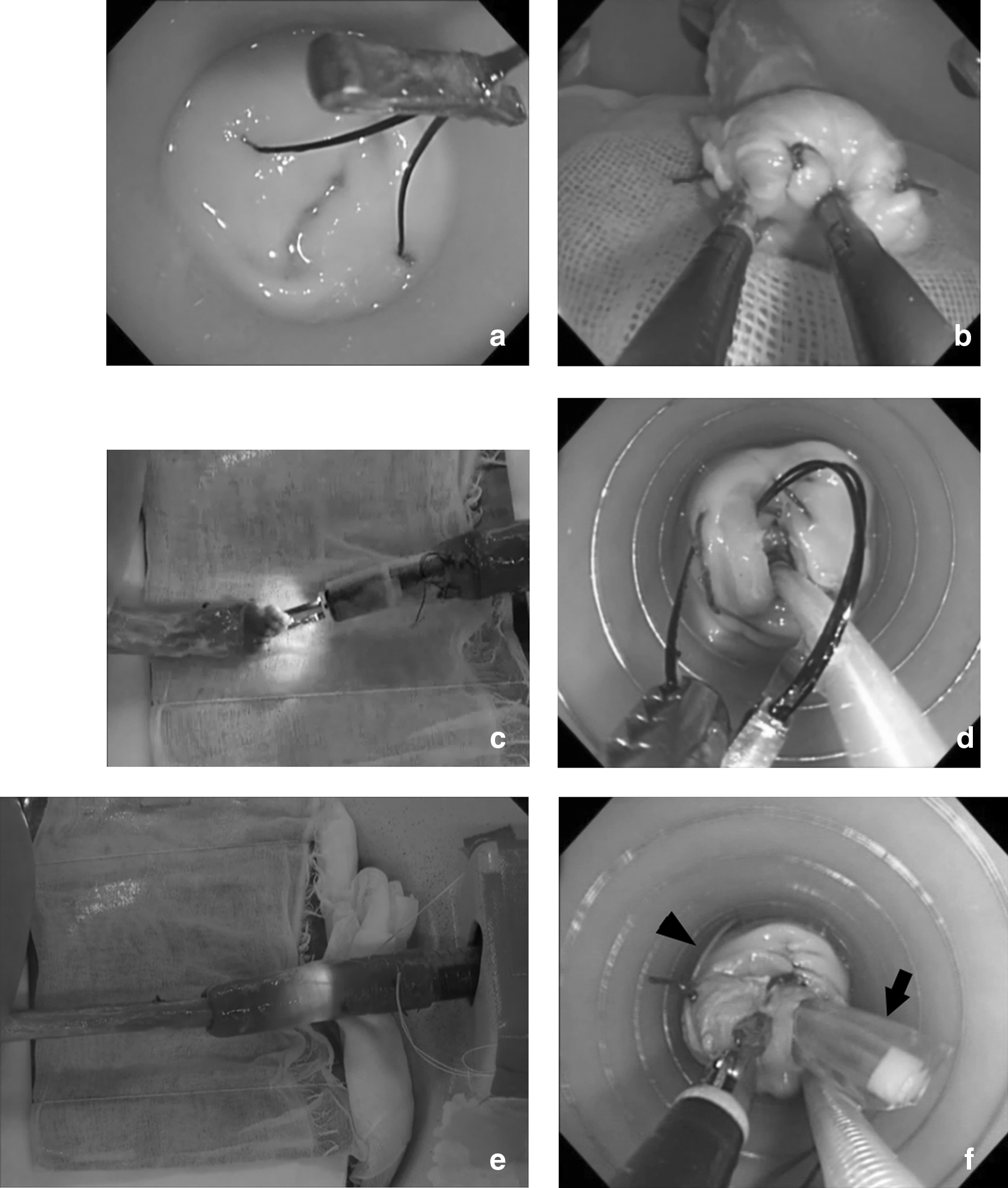
Endoscopic views and outer appearances during the procedure.
Dislocation of the transanastomotic tube occurred, and leakage pressure could not be measured correctly. Pressure values are shown in parentheses as reference.
Leakage from needle holes was observed.
The leak test was performed in all seven specimens for which the procedure was completed. Table 1 shows results of the liquid leak test. The internal pressure of the UE was adequately measured in four of the seven specimens (Cases 3, 4, 6, and 8), and the median value was 122 (82–142) mm Hg. In these cases, as the solution was infused, both the UE and LE were expanded, which indicated patency of the anastomosis (Fig. 5). Finally, the LE was pushed out of the UE, as if an intussusception was reduced by a high-pressure enema, and the anastomosed esophagus was separated into two specimens. In contrast, there was no expansion of the LE during infusion of the solution in two of the three specimens in which the leakage pressure was not measured correctly. The reason was that the transanastomotic tube slipped from the duplicated part when we transferred the anastomosed esophagus from the phantom of the human upper body to the experimental setting of the leak test. As a result, the patency of the anastomosis was lost (Cases 1 and 2). The internal pressure of one of those specimens reached the maximum limit (300 mm Hg) without separating the esophagus into two segments (Case 1). In the other case, the internal pressure was 248 mm Hg when the LE was pushed out of the UE (Case 2). Leakage from the needle holes was observed in one (Case 7).

State of the specimen during the liquid leak test.
Discussion
Many studies to develop methods for gastrotomy or colotomy closure for NOTES have been performed in both ex vivo and in vivo settings. However, only a few studies have aimed to establish a method for bowel anastomosis for NOTES. Rolanda et al. 6 reported peroral esophageal segmentectomy and anastomosis, but a single transthoracic trocar was used to assist the procedure. Kantsevoy et al. 7 and Bergstorm et al. 8 described endoscopic gastrojejunostomy, but these procedures were performed in the stomach. The stomach provides a large working space, which enables easy manipulation of the scope and various instruments. The target of our research is esophagoesophageal anastomosis, which presents difficulty in handling the scope or instruments because of the limited space. Our previous method achieved translumenal esophagoesophageal anastomosis but was complicated, and the blind puncture of the upper esophagus presented risks of injury. 5 Subsequently we devised a new procedure and herein report its feasibility. The minimum duration of the procedure was 23 minutes, a time sufficiently short for clinical practice. Moreover, with more experience and with more cases, the time could be shortened to less than 20 minutes.
We performed the liquid leak test to evaluate the strength of the anastomosis. Hookey et al. 9 reported that a liquid leak was detected at the site of hand-sewn gastrotomy closure at a pressure of 80.8 mm Hg. Sodergren et al. 10 noted leakage of water dyed blue from a colotomy site that was closed with interrupted sutures at a pressure of 23 mm Hg. Of course, in these studies leak tests were not performed for the esophagus, and the procedures examined were not anastomoses. However, our result showing a pressure of 122 mm Hg was much higher than in those studies described above and indicates that the anastomosis formed by our procedure was sufficiently strong to be used in clinical practice.
This study has several limitations. To begin with, in this study the experiments were ex vivo. Considering future clinical application, we admit concern about how to dissect the UE and LE. Our previous report described that dissection of the postmediastinum could be easily performed, 5 and we believe that dissection of the LE is possible, even though challenging.
We performed an in vivo experiment on a 40-kg pig to confirm the feasibility of dissection of the UE (authors' unpublished data). First, the LE was transected through a right thoracotomy, taking care not to injure the upper and the middle mediastinum. Next, a BraceBar was placed at the end of the UE. Then, a thread of the BraceBar was grasped endoscopically, and the endoscope was withdrawn. The outer appearance of the esophagus was observed under direct vision through the thoracotomy. As a result, the edge of the esophagus was inverted, and the fold was moved from the lower mediastinum through the upper mediastinum to the outside of the thorax at the end of the procedure. This meant that dissection of the UE, including the area between the trachea and the esophagus, was feasible just by traction of the BraceBar in the porcine model.
In addition, we have yet to confirm that the tissue ligated by the ligating device becomes necrotic and drops off together with the short transanastomotic tube. If these phenomena do not occur and only the tube drops off, leaving behind living ligated tissue, our procedure may result in a kind of papillary formation. That this could be a concern was indicated in Cases 1 and 2, where displacement of the tube caused loss of anastomotic patency. Animal survival experiments are needed in future.
Our long-term aim is to use this technique in neonates or infants with esophageal atresia. However, the endoscope and instruments used in the current study were those developed for adults and thus were too large to be used in infants. Therefore, in this study we had to use specimens larger than the esophagus of neonates or infants.
Despite these limitations, our results showed the feasibility of translumenal esophagoesophageal anastomosis. Moreover, the current procedure was simpler and safer than our previous one. 5 This method might after further study become available in general surgery such as colorectal resection and could contribute to widening the indication for NOTES in the future. Although a transgastric maneuver to insert the tube was used in this study, our final goal is to accomplish the anastomosis only by the oral approach. An improved procedure is under development.
Footnotes
Acknowledgments
This study was supported by grant 23592626 from the Ministry of Education, Culture, Sports, Science and Technology of Japan.
Disclosure Statement
No competing financial interests exist.