
Abstract
Abstract
Introduction:
In the United States, the prevalence of myasthenia gravis (MG) is approximately 14–20 per 100,000. One treatment option involves a thymectomy, which can lead to remission of symptoms. The amount of thymic tissue removed is correlated with a better outcome for patients. Thus, it is critical that the procedure used when performing a thymectomy maximize the resection of thymic tissue. Robotic-assisted thoracoscopic thymectomy provides a minimally invasive platform that avoids the mortality and morbidity of a median sternotomy while providing better visualization and a more delicate dissection than is available in a standard thoracoscopic procedure.
Patients and Methods:
Following Institutional Review Board approval, in total, 9 patients who underwent robotic thymectomy were reviewed. Intraoperative statistics such as operative time and blood loss were reviewed from operative records. Postoperative outcomes such as hospital stay, discharge medications, and complications were reviewed from hospital charts. Lastly, disease response was evaluated in consultation with a pediatric neurologist who specializes in MG.
Results:
Age at operation ranged from 2 to 15 years of age (average, 9.4 years). A majority of patients had an MGFA classification of II or greater (n=5). All patients were on pyridostigmine preoperatively, and 7 of 9 (77%) were taking prednisone. Mean operative time was 160.1±6.1 minutes. Average postoperative hospital stay was 1.1±0.3 days. One patient had a documented persistent pneumothorax on postoperative Day 1, which was treated with nasal cannula oxygen for an additional day. There were no additional operative complications, and all patients were discharged home on acetaminophen with codeine for pain control. Eight of 9 patients had improvement in MG symptoms after the procedure.
Conclusions:
Robotic-assisted thoracoscopic thymectomy is a safe and effective operation for children with MG. Robotic assistance allows for articulating instruments, three-dimensional visualization, and minimal blood loss. These factors may allow for a more complete resection compared with a standard thoracoscopic thymectomy.
Introduction
Historically, a thymectomy was performed via a median sternotomy, and a majority of the morbidity of the procedure was associated with the incision. More recently, video-assisted thoracoscopic surgery (VATS) has been used for thymectomies. Compared with median sternotomy, this minimally invasive procedure reduces postoperative pain and the chance for infection, leads to a shorter hospital stay, less tissue injury and blood loss, and better cosmetic results, while maintaining a very low postoperative mortality.5,6 However, studies have suggested that VATS thymectomies are not as successful in removing all thymus tissue. 7 This may be due to technical limitations in visualization and restriction of access to the superior-most portion of the thymus as well as the contralateral thymus in many patients.
Robot-assisted thymectomy is a relatively new procedure with proven feasibility and success in adult patients.8,9 It offers the aforementioned benefits of VATS, but also gives a three-dimensional view and has the technical advantages of articulating arms that provide greater access to the superior portion of the thymus and the contralateral side.9–11 A retrospective study has demonstrated a more complete resection in robot-assisted patients compared with VATS thymectomy and, accordingly, a significantly improved rate of complete stable remission. 11 This may be especially true for thymectomies performed in pediatric patients with MG, as the larger thymus gland in a smaller chest poses unique anatomical challenges that may affect outcomes. Studies have also shown more favorable clinical outcomes when a thymectomy is performed on younger patients and when patients are in the earlier stages of disease.12–14 Taken together, these are reasons to focus a study on robot-assisted thymectomy in pediatric patients.
We hypothesize that a thymectomy performed with robot assistance will lead to a more complete removal of thymic tissue with a good clinical outcome, minimal postoperative discomfort, a short hospital stay, and low complication rate.
Patients and Methods
Following Institutional Review Board approval, the charts of 9 patients who underwent robotic thymectomy at one of two institutions were reviewed. All procedures were performed by the same surgeon (D.L.). Intraoperative statistics such as operative time and blood loss were reviewed from operative records. Postoperative outcomes such as hospital stay, discharge medications, and complications were reviewed from hospital charts. Lastly, disease response was evaluated in consultation with a pediatric neurologist who specializes in MG (J.T.). All data are shown as mean±SEM values.
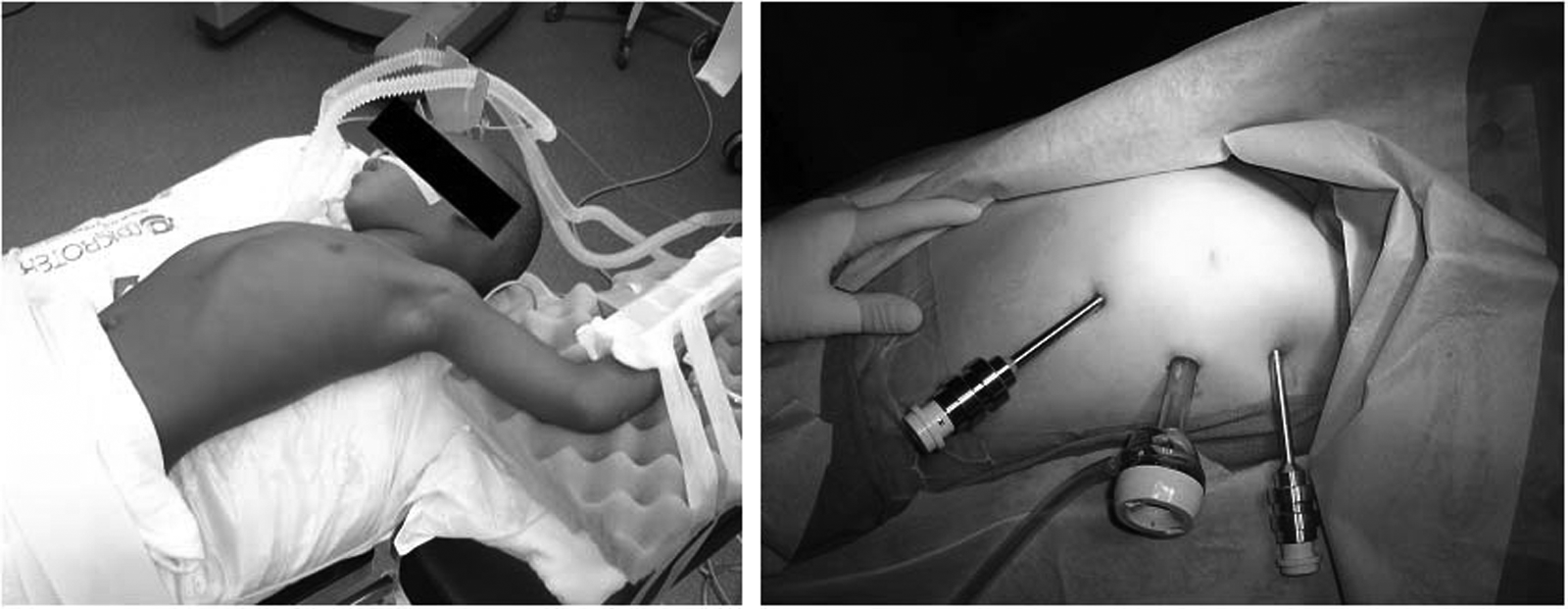
All operative procedures were performed in the same manner. Right lung ventilation was established by mainstem intubation, when possible. A low-pressure pneumothorax (4 mm Hg) was used to improve exposure in all cases. All thymectomies were performed from a left-sided approach (Fig. 1). A standard 5-mm thoracoscope was initially placed through a 5-mm trocar in the fourth intercostal space in a midaxillary line, which was then used to visualize the placement of the remaining two ports under direct visualization. A robotic 5-mm trocar was placed in the 7th intercostal space in midclavicular line, followed by an 8.5- or 12-mm camera port in between the working ports in the 6th intercostal space in the anterior axillary line. The standard 5-mm trocar was then changed out to a robotic 5-mm trocar. An assistant port was not needed for any of the procedures.
The da Vinci® surgical robot (Intuitive Surgical, Inc., Sunnyvale, CA) was used to perform a total thymectomy with resection en bloc of all thymic tissue. Once the robot was docked, a Maryland grasper was placed through the left working port and a hook cautery in the right. Dissection was started at the left inferolateral portion of the thymic gland medial to the phrenic nerve and continued cranially. The left lateral approach provided excellent visualization of the thymic veins that drain into the left innominate vein, which were ligated with clips (first case only) or taken with Bovie coagulation in continuity. The contralateral (right)-side dissection was undertaken in an inferomedial to superolateral fashion, taking great care to preserve the right phrenic nerve. Three-dimensional visualization as well as articulating arms greatly facilitated this portion of the dissection compared with standard thoracoscopic techniques. Dissection was continued into the neck to include the upper horns of the gland. Once again, robotic assistance provided superior visualization and access to the superior mediastinum, facilitating the most difficult portion of the dissection without disrupting other mediastinal structures. Once the dissection was completed, the robot was undocked, and the specimen was removed with an endocatch bag through the central camera port site. The lung was allowed to reinflate, and air in the pleural space was evacuated prior to closure of the incisions in layers.
Results
In total, 9 patients were reviewed. All patients had a robotic thymectomy performed between November 2007 and March 2011. Patients were seen at a routine postoperative visit approximately 14 days after the operation and were subsequently followed by a pediatric neurologist.
Female-to-male ratio was 2:1. The mean age at operation was 9.4 years and ranged from 2 to 15 years (Table 1). All patients presented with ophthalmologic symptoms (ptosis and diplopia), and diagnosis was confirmed with a edrophonium chloride (Tensilon®; ENLON) stimulation test. Prior to operation, all patients were treated with pyridostigmine, and 7 of 9 (77%) were taking prednisone. Mean time from diagnosis to operation was 14.4±7.3 months. All patients were classified by the Myasthenia Gravis Foundation of America (MGFA) classification system as determined by a neurologist (J.T.) based on degree of symptoms. 5 One patient had MGFA class I disease (11%), 3 patients had class II disease (30%), 4 patients had class III disease (44%), and 1 patient had class IV disease (11%).
Most patients (7 of 9) fell into Myasthenia Gravis Foundation of America (MGFA) class II or III (intermediate disease). All patients (9 of 9) were on pyridostigmine, and 78% (7 of 9) were on prednisone at the time of operation.
Mean±SEM value.
All procedures were performed with assistance from surgical housestaff and operating room staff specially trained in robotics. Average operative time was 160.1±6.1 minutes. Operative time declined progressively with a greater number of procedures performed (Table 2 and Fig. 2), with the initial procedure taking 190 minutes and the last procedure performed taking 136 minutes. Estimated blood loss was less than 25 mL for all patients, and no patients required blood transfusions intraoperatively or postoperatively. An average amount of thymic tissue was 23.6±3.8 g. A majority of patients had a pathologic diagnosis of thymic hyperplasia (7 of 9); the remaining 2 patients had pathologically normal thymus tissue. The first patient had a tube thoracostomy placed, which was removed on postoperative Day 1. The remainder of the patients had the air evacuated from their pleural cavity via a suction catheter at operation and did not have chest tubes placed. One patient had a small residual pneumothorax visualized on chest x-ray on postoperative Day 1. This patient was managed conservatively with nasal cannula oxygen supplementation, and the patient was discharged home on postoperative Day 2. There were no additional major or minor postoperative complications. Average hospital stay was 1.11±0.11 days. All patients were discharged with acetaminophen with codeine for pain control and were seen at approximately 2 weeks postoperatively by the operative surgeon.

Operative time for the 9 myasthenia gravis patients. Operative time declined as the number of cases increased, with an average operative time of 160.1±6.1 minutes.
One patient had a residual pneumothorax on postoperative Day 1, which was managed with observation.
Mean±SEM value.
After thymectomy, all patients returned to their neurologist (J.T.) for continued follow-up and treatment of their MG. Average time of follow-up was 22.5±13.3 months (Table 3). Four patients are asymptomatic but remain on pyridostigmine (DeFillipi score of 2), and 4 patients have had an improvement in symptoms since thymectomy (DeFillipi score of 3). 15 One patient had a persistence of symptoms with respiratory fatigue and a hospital admission for intravenous immunoglobulin infusion. Of note is that this patient had the most symptomatic disease (MGFA class IV) prior to thymectomy and had multiple admissions preoperatively for respiratory failure. Overall a symptomatic improvement was seen in 8 of 9 (88%) patients who underwent robot-assisted thoracoscopic thymectomy. Average time to return to normal activities was 1 week, with most patients reporting a return to normal activites of less than 3 days postoperatively.
Eighty-nine percent (8 of 9) patients had an improvement in disease status after thymectomy.
Mean±SEM value.
Discussion
Thymectomy is indicated for juvenile MG in children with moderate or severe generalized disease, and complete stable remission is achieved in about two-thirds of patients in 3 years. 16 Long-term administration of steroids and immunosuppressant medications has a number of significant side effects in children, including decreased growth velocity, diabetes, hyperlipidemia, central obesity, immunocompromised state, and pathologic bone fractures. In addition, the greatest benefit from thymectomy in disease remission is seen early in the disease course. 17 Therefore, we advocate for early thymectomy in children directed by disease onset and severity. The decreased morbidity and improved cosmesis associated with a minimally invasive approach remove some of the barriers for parents and providers in proceeding with surgery at a young age.
Surgery with robotic assistance has demonstrable advantages, including three-dimensional visualization and articulating instruments. Use of the robot also has some disadvantages, most notably the costs associated with purchasing and maintaining a unit as well as the time to dock and set up the robotic arms. Current trends appear to suggest that pediatric robotic surgery is useful and indicated in a select group of procedures and a select group of patients. 10 In our institution, the cost of the robot is shared by adult surgical services. Having pediatric surgeons specially trained in robotic surgery within a primarily adult hospital system allows for the use of robotic technology at a relatively low overall cost. Similarly, having an operating room staff that is trained and experienced in robot docking decreases overall operative time as demonstrated by decreasing operative times seen in our study. Based on this case series, we conclude that a thymectomy for MG in children is a procedure in which the robot can be safely used and the technical advantage of robotic assistance is not associated with a significant increase in cost or operative time. This is largely owing to the anatomic constraints of performing a minimally invasive thymectomy in a child and the generally accepted principle that in MG the optimal operation removes the entirety of thymic tissue. Technical advantages afforded by the use of the robot are particularly useful in the dissection of the superior thymus and the contralateral (right) side, which are areas of difficulty in a standard thymectomy.
Our results suggest that this procedure can be performed with minimal morbidity in children as young as 2 years of age. Previous studies in adults have demonstrated an improved rate of remission in robotic thoracoscopic thymectomies compared with nonrobotic thoracoscopic thymectomies for MG. 11 A case series of several children with MG who had a thoracoscopic thymectomy had to have an additional resection due to retained thymus. 18 To date no large-scale studies have been conducted in children comparing the two techniques. Although our study size is small and time to follow-up is limited, our results seem to suggest that a robotic thoracoscopic thymectomy provides a more complete resection leading to increased rates of remission. Additional study is warranted with more patients and for a longer follow-up period.
Footnotes
Disclosure Statement
No competing financial interests exist.