
Abstract
Abstract
Objective:
A thyroid isthmus nodule is a relatively rare condition. A small number of patients will present with thyroid mass isolated at the thyroid isthmus, which can cause discomfort in swallowing and cosmetic problems. Thus, some patients choose to have these nodules excised. The surgical removal of the thyroid isthmus mass is usually accomplished through an external incision of the neck. However, this procedure inevitably results in a neck scar.
Patient and Methods:
We report a case of an 18-year-old woman with a thyroid isthmus mass. We implemented a modified approach for the removal of the thyroid isthmus mass by using a frenotomy incision of the mouth, accompanied by an endoscope system.
Results:
A modified approach for the removal of the thyroid isthmus mass was used on the patient. The total operating time was 70 minutes. The patient continues to be free of any diseases 12 months after the excision.
Conclusions:
Resection of the thyroid isthmus mass can be performed by an intraoral endoscope-assisted approach through a frenotomy incision of the mouth. We described the detailed procedures for an endoscope-assisted transoral thyroid isthmus mass excision using a frenotomy incision.
Introduction
Surgical excisions are the treatment of choice for symptomatic or clinically apparent thyroid isthmus nodules. However, observation of an asymptomatic or nonclinically apparent thyroid isthmus nodule is another treatment choice, unless these thyroid isthmus nodules are confirmed as carcinomas.
Considering that most patients who undergo surgical resection are women, it is desirable to develop a surgical technique for the removal of thyroid isthmus nodules without producing external scarring of the neck. Thanks to the advancements in medical technology, we were able to develop a modified approach where a frenotomy incision of the mouth was endoscope-assisted.2–4 This modified method allowed for the reduction of risks in damaging the mouth floor structure because we used an anatomical dehiscence from the frenotomy incision.
Additionally, this surgical procedure was a form of translumenal endoscopic surgery through a natural orifice of the human body (natural orifice translumenal endoscopic surgery [NOTES®; American Society for Gastrointestinal Endoscopy (Oak Brook, IL) and Society of American Gastrointestinal and Endoscopic Surgeons (Los Angeles, CA)]). We developed this technique while taking into consideration that it could also be applied to transoral thyroid surgery. In this report, we describe this new technique in detail, as well as the surgical outcome.
Patient and Methods
An 18-year-old female patient was admitted with the chief complaint of a mass at the anterior part of her neck, which was detected 12 months before her visit. The size, since its initial detection, had gradually increased, and the patient experienced discomfort during swallowing due to the sensation of the lump in her throat. During palpation, a painless, movable, soft mass, 1.0 cm in diameter, was located between both thyroid glands at the thyroid isthmus. Laryngoscopic examination did not reveal any protruding masses at the base of the tongue. The medical history of the patient was unremarkable, and she had no history of thyroid disease.
The neck computed tomography imaging of the thyroid verified that the thyroid was at its normal location. With contrast enhancement, there was a calcified solid mass at the thyroid isthmus, approximately 1.0×1.0 cm in size (Fig. 1). Computed tomographic scans suggested a thyroid nodule in the thyroid isthmus, but aspiration cytology failed because the mass was so hard that the fine needle could not puncture the calcified mass. The thyroid function tested normal. The patient was a young woman, and she did not want a surgical scar to remain in the neck area. Therefore, the surgery was performed through a transoral approach using an endoscope.
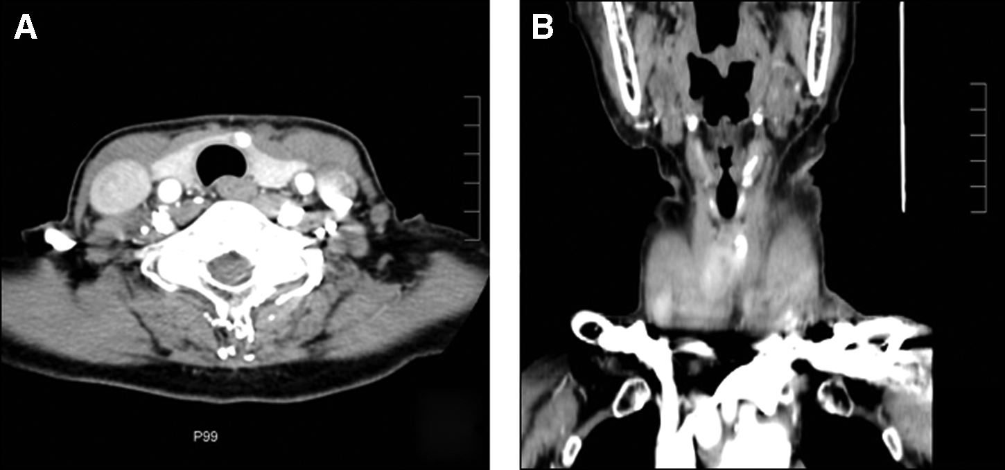
Computed tomography scan confirmed the 1.0-cm-sized mass was that of a thyroid isthmus calcified mass:
With regard to the operation, with the patient under general anesthesia, the mouth was opened, and the tongue was held in the upper position. An approximately 3-cm incision was made in the oral mucosa, which included the frenulum. After careful dissection of the soft tissue at the floor of the mouth, we found the genioglossus muscles. We separated the muscles at the midline and retracted them bilaterally. With the aid of an endoscope (rigid, 10 mm, 0°; Olympus, Tokyo, Japan), after the retraction of the genioglossus muscles, we found the hyoid bone.
We transected the hyoid bone with an Osteo-Punch™ Rongeur (1 mm, 40°; Koros USA, Inc., Moorpark, CA). Then, the hyoid bone was retracted bilaterally; from there we then went downward and were able to identify the thyroid isthmus, both thyroid lobes, trachea, and inferior thyroid vein (Fig. 2).

After these procedures, a thyroid isthmus mass was found at the middle of the thyroid isthmus. The mass was easily resected from the adjacent tissue with ultrasonic scissors (Harmonic® scalpel 300; Ethicon, a Johnson & Johnson Company, Cincinnati, OH) (Fig. 2D). In the operating room, we checked the frozen biopsy specimen, and the results indicated a benign calcified goiter. Afterward, the operation field was irrigated. A drain was inserted through the floor of the mouth, and it was sutured to the edge of the lower teeth (Fig. 3). Then, the genioglossus muscles were sutured with 4-0 Vicryl® (Ethicon), followed by the suturing of the opened oral mucosa. The total operation time was approximately 70 minutes. The drain was removed on postoperative Day 2. We encouraged frequent oral gargling with 0.02% chlorhexidine and allowed for a normal diet on postoperative Day 2. The patient was discharged on postoperative Day 5.
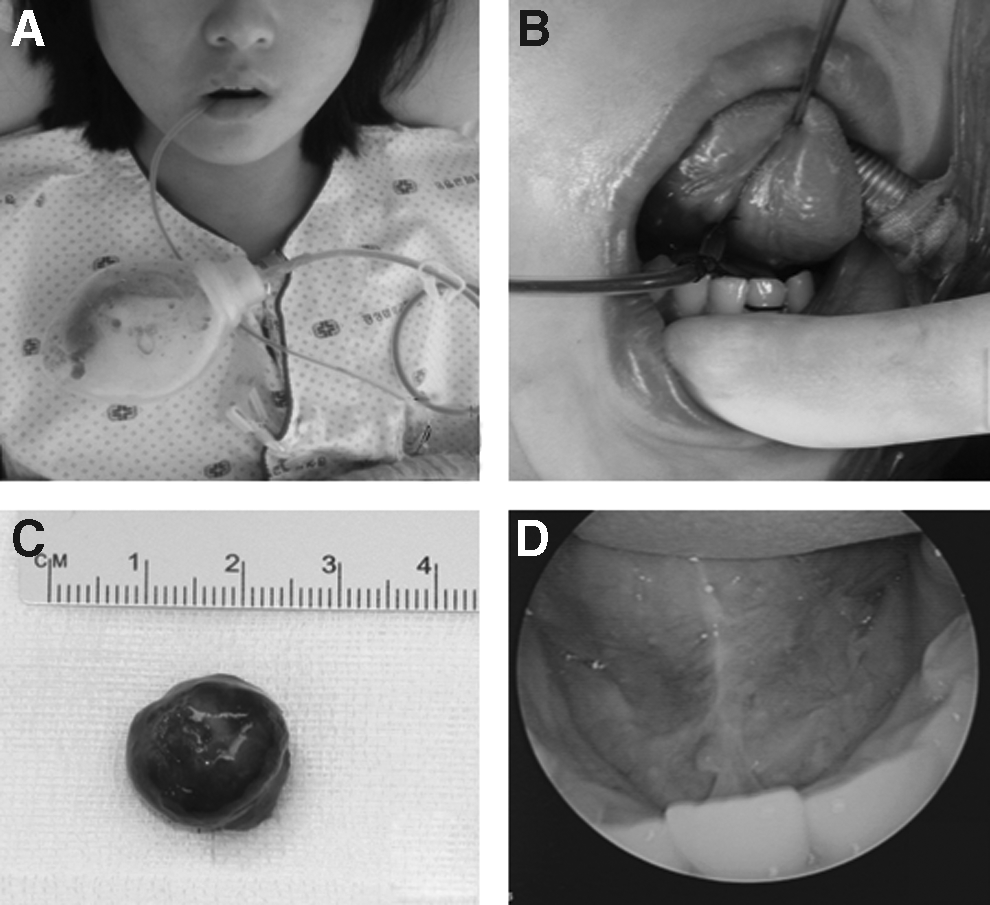
Twelve months have passed since the operation, and there have been no special findings of complications in thyroid function, articulation, and tongue movement. The tongue scar was seen as just a linear mark (Fig. 3). The patient is currently under follow-up observations at our outpatient clinic. She was quite satisfied with the surgical outcome. The mass was confirmed as a calcified nodular goiter through pathological examination.
Discussion
The thyroid isthmus is the central part of the thyroid gland that connects the lobes and lies directly anterior to the trachea. 5 When nodules arise from the isthmus or the pyramidal lobe, it may be adequate to perform surgical excisions. The treatment for thyroid isthmus masses depends on the location, size, and presence of symptoms or complications.6–8 Therefore, an accurate diagnosis can greatly reduce post-treatment complications.
In the neck, a thyroid isthmus mass is primarily a cosmetic problem, whereas secondary ulcerations, hemorrhages, and malignant degenerations become major problems. The treatment for an isolated thyroid isthmus mass that occurs at the midline of the neck, above the trachea, is surgical excision. However, this operation requires a transcervical approach, which results in external scarring of the neck.2–4 Considering the embryological development of the thyroid, it seemed obvious to us that an entirely transoral access to the thyroid region could be established. 9 During its descent, the thyroid gland, which originates from the bottom of the tongue, is connected to the tongue through the thyroglossal duct. The thyroglossal duct is located behind the strap muscles of the neck and the hyoid bone and either may persist postpartum or result in the so-called pyramidal lobe in up to 30% of cases. 9
Therefore, many surgeons strive to achieve scar-free surgery using the oral cavity. However, there are many obstacles to this approach. In contrast to the external surgical removal of a thyroid isthmus mass, transoral removal must pass through the hyoid bone and requires a long surgical pathway. The hyoid bone is the biggest obstacle in the pathway from the mouth to the thyroid.
In previous studies, the authors reported the safe excision of dermoid cysts, thyroglossal duct cysts, and ectopic thyroid tissues through the cutting of the hyoid bone with a frenotomy incision.2–4 Following this naturally predetermined access alongside the former thyroglossal duct, we were able to develop a new surgical approach to the thyroid region. This approach showed that a transoral hyoid bone excision was technically feasible, which allowed us to reach the upper side of the thyroid gland without major tissue damage or bleeding.
This surgical approach passed through a midline incision at the floor of the mouth and used the natural midline dehiscence present between the genioglossus muscles.2–4,10 This area has been shown to be a relatively avascular dissection plane. When the space between the genioglossus muscles was spread out using a tractor, the mylohyoid muscle was reached. When we traced the posterior line of the mylohyoid muscle, the hyoid bone was reached.
When we reached the hyoid bone, we resected the middle portion of the hyoid bone (0.5 cm in width). We then went downward and were able to identify the thyroid isthmus and both thyroid lobes. After identification, we were able to find the thyroid isthmus mass and resected the mass located between both thyroids. The patient was quite satisfied with the surgical outcome.
Translumenal endoscopic interventions, via so-called natural orifices, are considered to be refinements of minimally invasive procedures.9,11 These new interventional techniques are gaining increasing interest because they allow for surgical treatments without incision of the skin. These methods are not only technically possible, but they could also provide an appropriate surgical field, and thus it is believed that these methods can be applied to patients with thyroid isthmus masses that have manifested as central midline neck masses. The satisfaction level of our patient, after the operation, was good, and significant swelling or bleeding was not observed. Because neck exploration creates the possibilities of turning from an aseptic operation to a potentially infectious surgical intervention through the spread of pathogenic germs from the oral microflora, infections after operation must be managed carefully. Therefore, we used antibiotics and an oral gargle, and the drain was removed 2 days after the operation. Follow-up observations were carried out for 12 months after the operation, and there were no noteworthy recurrences or complication patterns observed.
Conclusions
This report suggests a new surgical method to approach the thyroid through the floor of the mouth, thus allowing for the possibility of resecting thyroid isthmus masses through an intraoral approach.
Footnotes
Acknowledgment
This research was supported by the Basic Science Research Program through the National Research Foundation of Korea (NRF) funded by the Ministry of Science, ICT, and Future Planning (2013R1A1A1012542).
Disclosure Statement
No competing financial interests exist.