
Abstract
Abstract
Objective:
To report a minimally invasive and reproducible technique that greatly facilitates the identification of the stricture during laparoendoscopic single-site ureteroureterostomy (LESS-UU) for benign proximal and middle ureteral strictures, using the intraoperative retrograde ureteroscopy-assisted technique.
Patients and Methods:
Between April 2011 and January 2013, 13 patients with a benign proximal or middle ureteral stricture underwent LESS-UU at our institution. A combination of diuretic renal scans, antegrade/retrograde ureteropyelography and/or computed tomography, and stent placement or exchange was preoperatively performed to assess all patients. The intraoperative retrograde ureteroscopy-assisted technique was used to identify the exact position of the stricture and place the stenting during LESS-UU.
Results:
Intraoperative retrograde ureteroscopy was successfully performed in all cases. The mean operative time was 156 minutes (range, 125–190 minutes), and the estimated blood loss was 80 mL (range, 20–160 mL). The mean hospital stay was 5 days (range, 4–7 days). One patient required conversion to open surgery because of the severe adhesions surrounding the stricture that resulted in failure to progress. Urine leakage occurred in 1 patient postoperatively and was successfully treated by conservative management. Postoperative fever occurred in another patient, who was treated with a dose of oral antibiotics. No major intraoperative or postoperative complication occurred. Clinical and radiographic success was achieved in 100% (13/13) of patients during a mean follow-up of 13.1 months (range, 9–27 months).
Conclusions:
LESS-UU is feasible and safe for repairing benign proximal and middle ureteral stricture. The intraoperative retrograde ureteroscopy-assisted technique during LESS-UU is useful for localizing the stricture.
Introduction
U
In the present study, we report a minimally invasive and reproducible technique that greatly facilitates the identification of the stricture during LESS-UU for benign proximal and middle ureteral strictures, using the intraoperative retrograde ureteroscopy-assisted technique.
Patients and Methods
Patients
Between April 2011 and January 2013, 13 patients with a benign proximal or middle ureteral stricture underwent LESS-UU at our institution. This study obtained informed consent from the patients, and ethics approval was granted from the ethics committee at Xiangya Hospital, Central South University, Changsha, Hunan Province, China.
Demographic and preoperative data were collected. The patients included six men and seven women, with an average age of 35.8 years (range, 31–48 years). Their average body mass index was 26.9 kg/m2 (range, 22.1–30.8 kg/m2). All patients presented with mild to moderate flank pain. Indications for LESS-UU included 9 patients with a refractory ureteral stricture after ureteroscopy for urolithiasis (treated by ureteroscopic lithotripsy using a holmium laser or ultrasonic lithotripsy), 3 patients with iatrogenic ureteral trauma during hysterectomy, and 1 patient with an idiopathic stricture refractory to conservative treatment. There were eight proximal and four middle ureteral pathologies. Three patients presented with percutaneous nephrostomy tubes placed by outside institutions at surgery. A combination of diuretic renal scans, antegrade/retrograde ureteropyelography and/or computed tomography, and stent placement or exchange was preoperatively performed to assess all patients at our institution.
Operative technique
Under general endotracheal anesthesia, patients were placed in a hyperextensive lithotomy position with the trunk in a 45° lateral decubitus position. The abdomen and pelvis were prepared separately. A 2-cm periumbilical transverse incision was made at the umbilicus. An ASC TriPort™ (Advanced Surgical Concepts, Bray, Ireland) was inserted through the incision, using the method described by Rane and Rao 7 (Fig. 1). Pneumoperitoneum was obtained by carbon dioxide insufflation at 12–14 mm Hg of intraabdominal pressure. A rigid 5-mm, 30° digital video-laparoscope was used in all 13 cases. In general, standard laparoscopic instrumentation was used for most dissection and retraction maneuvers. Prebent instruments were selectively used to facilitate retraction.
An ASC TriPort was inserted through the periumbilical transverse incision.
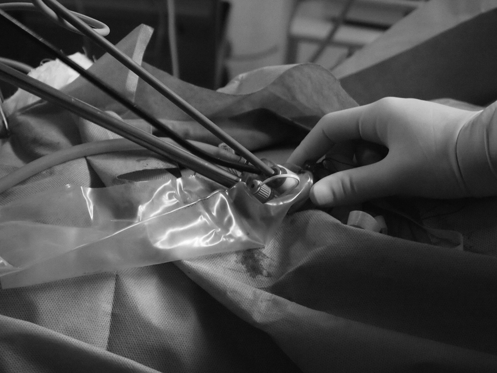
In our experience, confirmation of a normal part of the ureter before dissecting toward the affected tissue has been the optimal approach for the identification and dissection of a ureteral stricture. Therefore, after the line of Toldt was incised and the retroperitoneum was dissected, the iliac vessels and the middle ureter (proximal ureteral stricture) or the lower pole of the kidney and the proximal ureter (middle ureteral stricture) were first identified. Then a rigid ureteroscope was introduced and advanced to the site of the stricture (Fig. 2). The tip of the ureteroscope could be clearly seen under laparoscopy after the light source of the laparoscope was dimmed. Then distal-to-proximal or proximal-to-distal ureterolysis was performed. The healthy ureter should be adequately dissected to facilitate tension-free ureteroureteral anastomosis. Care must be taken to preserve the periureteral blood supply.
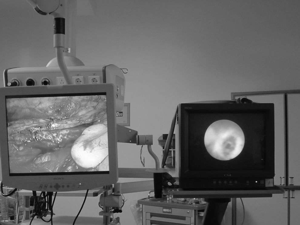
A rigid ureteroscope was introduced and advanced to the site of the stricture.
UUs were started by spatulating the normal distal ureter below the distal aspect of the stricture for a length of 1 cm. The proximal segment of the ureter was longitudinally incised laterally through the stricture until at least 1 cm of normal ureter was visualized. Then the diseased segment was excised. Ureteroureteral anastomosis was performed in an interrupted fashion with 4-0 polyglactin 910 (Vicryl™; Ethicon, Somerville, NJ) sutures. A 6 French double-J stent was placed in the retrograde manner under ureteroscopy. The irrigant was aspirated by the suction probe through the laparoscopic port, and a drain was placed. A Foley catheter was placed to maintain bladder emptying.
The drain was removed if there was no increase in output and it was less than 10 mL postoperatively. The Foley catheter was removed 5 or 7 days postoperatively, and the ureteral stent was removed in 4–6 weeks. Diuretic renal scintigraphy and renal ultrasonography were performed 3 months postoperatively and at 6-month intervals thereafter. Overall success was defined as the resolution of symptoms and resolution or improvement of radiographic obstruction at the most recent follow-up examination.
Results
Intraoperative retrograde ureteroscopy was successfully performed in all case. The mean operative time was 156 minutes (range, 125–190 minutes), and the estimated blood loss was 80 mL (range, 20–160 mL). The mean hospital stay was 5 days (range, 4–7 days). One patient required conversion to open surgery because of the severe adhesions surrounding the stricture that resulted in failure to progress. Urine leakage occurred in 1 patient postoperatively and was successfully treated by conservative management. Postoperative fever occurred in another patient, who was treated with a dose of oral antibiotics. No major intraoperative or postoperative complication occurred. Clinical and radiographic success was achieved in 100% (13/13) of patients during a mean follow-up of 13.1 months (range, 9–27 months).
Discussion
Laparoscopic ureteroureterostomy has been reported in the treatment of ureteral ectopia, 8 retrocaval ureter, 9 iatrogenic injury during gynecological surgery, 10 and benign strictures. 11 LESS has recently emerged as a potentially less invasive alternative to conventional laparoscopy, with improved cosmetic outcomes and possibly reduced morbidity. In our opinion, LESS could be more suitable for construction procedures or ablative procedures in which the specimen is relatively small because further enlargement of the incision for retrieving the specimen was not needed. Based on our increasing experience of LESS, we have expanded the indication in urologic surgery, including pyeloplasty 12 and UU. 5 In this study, we report our initial experience with and the short-term outcome of LESS-UU for repairing benign proximal and middle ureteral strictures, using the intraoperative retrograde ureteroscopy-assisted technique.
Difficulty or uncertainty in identifying the site of the stricture or determining the length of resection poses a technically challenging situation for the urologist. Several reports have reported methods to resolve this problem. Baldie et al. 3 reported that normal saline can be injected intraoperatively through the ureteral catheter to establish the hydroureter and facilitate identification of the stricture location and length during robotic laparoscopic UU. The mean surgery time was 258.6 minutes (range, 146–450 minutes). The mean hospital stay was 2.5 days (range, 1–8 days). The mean estimated blood loss was 171 mL (range, 30–500 mL). The mean follow-up was 6.4 months (range, 2–18 months). Open conversion was needed in 1 patient for anastomosis completion. All cases were clinically and radiographically successful at the last follow-up examination. Intraoperatively, normal saline injection to establish the hydroureter may be limited by failure in the ureteral catheter through the stricture. The wide adhesions surrounding the affected ureter may also present an ambiguous plane to identify the location of the stricture. In addition, previous placement of the ureteral catheter may increase the operative time.
Lee et al. 1 reported a novel method via indocyanine green visualization under near-infrared light to intraoperatively localize ureteral strictures during robot-assisted UU. Mean operative time was 171.3±52.4 minutes, and mean estimated blood loss was 175.0±146.5 mL. There were no immediate or delayed adverse effects attributable to intraureteral indocyanine green administration. Gallbladder laceration during trocar placement occurred in 1 patient with severe perihepatic adhesions, who required robotic cholecystectomy. No postoperative complication occurred. Mean follow-up was 5.9±1.5 months, and all cases were clinically and radiographically successful at last follow-up. This novel method presented a good outcome. However, it may be limited by the expensive device and intraoperatively complex manipulation, which may influence the widespread applicability, especially in developing countries.
Buffi et al. 4 reported that a flexible ureteroscope was used to transilluminate ureteral strictures successfully in 3 of 5 reported patients during robot-assisted UU. The median operative time was 124 minutes (range, 115–139 minutes), median hospital stay was 3 days (range, 2–5 days), and the median follow-up was 8 months (range, 6–20 months). There were no intraoperative complications and one postoperative complication. No ureteral stricture recurrence was found.
In our series, we used a rigid ureteroscope intraoperatively to identify the location of ureteral strictures. This report was the first to describe a precise technique to localize ureteral strictures during LESS-UU. Our technique poses some advantages. First, it may further increase cosmetic outcomes and reduce morbidity compared with traditional laparascopic UU and robotic laparoscopic UU. Second, the remaining rigid ureteroscope could increase the tactile feedback and protect the ureter from excessive dissecting forces. Third, intraoperatively, retrograde stenting placement under ureteroscopy could be also easily performed, with no need of additional position change. That may contribute to reducing the operative time. In addition, a rigid ureteroscope can be easily obtained in developing countries, and thus this technique may have widespread applicability.
Our study has some limitations. This technique was limited by the localization of only the lower margin of the stricture. The use of the two-camera system, allowing visualization of laparoscopy and ureteroscopy at the same time, was required. In addition, the present study was limited by its retrospective design, short follow-up, and small sample size. However, our study highlights the feasibility and the observed benefits of the intraoperative retrograde ureteroscopy-assisted technique during LESS-UU.
Conclusions
LESS-UU is feasible and safe for repairing benign proximal and middle ureteral strictures. The intraoperative retrograde ureteroscopy-assisted technique during LESS-UU is useful for localizing the stricture.
Footnotes
Disclosure Statement
No competing financial interests exist.