
Abstract
Abstract
Background:
Transanal endorectal pull-through for Hirschsprung's disease (HD) is a relatively safe and feasible procedure in neonates and infants. However, overstretching on the anal sphincter and mesentery of the sigmoid colon might cause potential risk of impaired defecation function. Single-incision laparoscopic endorectal pull-through (SILEP) is technically feasible and safe in HD patients, offering a better cosmetic result. However, it is stressful for the surgeon in view of its low manipulability and poor visualization causing clashing of instruments, especially in older children or patients with long-segment aganglionosis. We developed an age- and type-appropriate technique of hybrid SILEP (H-SILEP) using a trocarless instrument via another abdominal stab incision to obtain further improvement of SILEP in selected HD patients.
Patients and Methods:
Between August 2010 and July 2013, 36 patients (24 boys and 12 girls, with a mean age of 3.9 months) with HD underwent H-SILEP. Patient age, gender, transitional zone, operative time, blood loss, and intraoperative and postoperative complications, as well as short- and long-term results, were assessed.
Results:
Ten patients had transitional zone in the rectum, 17 patients in the sigmoid colon, and 9 patients in the descending colon. Neither additional ports nor conversion to laparotomy was required in these 36 patients. The mean operative time was 116 minutes. There was no major intraoperative complication. Perianal excoriation was the main early postoperative complication, which occurred in 9 patients. No anastomotic leak occurred. Postoperative enterocolitis occurred in 2 patients. There was no recurrent constipation. Follow-up for 6 months to 3 years in all patients showed an excellent cosmetic result.
Conclusions:
Our procedure is feasible and safe, and it is technically less challenging to perform H-SILEP in selected HD patients. Moreover, it is better to use additional instruments for ergonometric reasons.
Introduction
R
We have developed an age-appropriate and type-appropriate technique of hybrid SILEP (H-SILEP) using two trocars instead of three trocars at the umbilicus and inserting a trocarless 3-mm grasping forceps via a stab incision to the left side of the abdomen to obtain further improvement of SILEP in selected HD patients.
Patients and Methods
Patients
Between August 2010 and July 2013, 36 patients with HD underwent H-SILEP. There were 24 boys and 12 girls, with a mean age of 3.9 months. These patients were diagnosed with HD by anorectal manometry, suction rectal biopsy, and barium enema in our department. Patients with total colon aganglionosis, who had had previous colostomy, or who were lost to follow-up were excluded from this retrospective study. The diagnosis of HD was confirmed by intraoperative frozen section biopsy specimens and postoperative pathology results. Preoperatively, colon irrigations were performed for 2–5 days. Patients were started on preoperative intravenous antibiotics 1 or 2 days before the operation and continued for 3 days after surgery.
Before the parents signed the consent form for surgery, they were informed that the procedure was minimally invasive and that the procedure would be carried out via a small incision at the umbilicus ring and one stab incision in the left abdominal wall. Parents were told that there was a chance of needing additional trocars or conversion to an open technique. Advantages and disadvantages were reviewed to ensure that the parents were fully educated about the procedure. This study was approved by the Ethics Committee of Huazhong University of Science and Technology (Wuhan, China).
Techniques
Under general anesthesia, the patient was placed supine, transversely across the end of the operating table, with the foot elevated by 30°. The monitor was positioned at the patient's feet. The operating surgeon stood at the head of the table, and the assistant surgeon stood on the patient's left. A single transumbilical vertical incision was made. Two 5-mm trocars were inserted into the peritoneal cavity at the horizontal ends of the umbilical incision. After capnoperitoneum was established, a 30° straight camera was inserted in one of the ports, and either a grasping forceps or an ultrasonic scalpel was inserted in the other port. Then, through a stab incision on the left abdominal wall, a trocarless 3-mm grasping forceps was inserted (Fig. 1).

Two 5-mm trocar ports were placed into the abdomen through small incisions on the umbilicus ring, and a trocarless 3-mm grasping forceps was inserted through a stab incision on the left abdominal wall.
After an overall view was obtained, two or three seromuscular leveling biopsy specimens of the rectum and colon were obtained and sent for rapid frozen section to pathology, to identify the transitional zone by the presence or absence of ganglion cells in the submucosal nerve plexus. The rectum and sigmoid colon were mobilized 5 cm proximal to the normal colon by elevating or tenting the mesentery using a 3-mm grasping forceps, and dissection with the ultrasonic scalpel was used until the colonic pedicle was long enough to reach deep into the pelvis without tension. The dissection was continued to the peritoneal reflection of the rectum.
Transanally, we used the modified Soave's procedure 6 to complete the operation, where the aganglionic colon and 5 cm of the normal colon were resected, and coloanal anastomosis was performed. The umbilical fascia was closed by using 2-0 polyglactin 910 (Vicryl®; Ethicon, Somerville, NJ) suture. The skin at the umbilicus and the stab incision was coated with skin glue.
Statistical analysis
Follow-up was started 1 month after surgery and then once every 1–3 months. The age at operation, sex, transition zone location, operative blood loss, operative time, postoperative hospital stay, intraoperative and postoperative complications, and defecation frequency were analyzed. The results were expressed as the mean±standard deviation or as a percentage.
Results
The general characteristics for age at operation, sex, and transition zone location in this study are shown in Table 1. There were 24 boys and 12 girls, with a mean age of 3.6 months. There were 10 patients with the transitional zone in the rectum, 17 patients in the sigmoid colon, and 9 patients in the descending colon.
H-SILEP, hybrid single-incision laparoscopic endorectal pull-through.
The mean operative time was 116±15 minutes (range, 70–150 minutes). Neither additional ports nor conversion to open approach was required, even for the 9 patients with transitional zone located in the descending colon. There was no major intraoperative complication, with insignificant blood loss of 4.5±1.0 mL. The main early postoperative complication, which occurred in 9 patients, was perianal excoriation. No anastomotic leak occurred. Postoperative enterocolitis occurred in 2 patients, which was treated by a course of antibiotics, total parenteral nutrition, and colon irrigation during rehospitalization. There was no recurrent constipation. The defecation frequencies gradually improved to 2±1 times per day 3 months postoperatively. The operative data and complications are shown in Table 1.
The wound appearance at 1 week postoperatively and the barely visible scar on follow-up, 4 and 8 months postoperatively, respectively, showed excellent cosmetic results in all patients (Fig. 2).
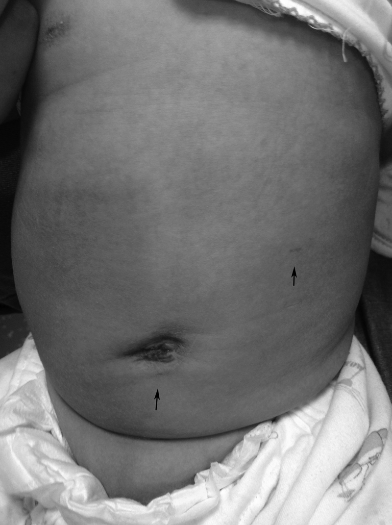
The appearance of the abdomen 4 weeks postoperatively.
Discussion
In the past, patients diagnosed with HD were committed to a multistage sequence of operations, including the creation of a colostomy and an open surgical pull-through procedure, followed by a colostomy takedown. Since introduction of laparoscopy in the early 1990s, the surgical management of HD has seen much progress, which has encouraged surgeons to attempt more complex laparoscopic procedures to reduce complications and promote early resumption of gastrointestinal function. 3
Nowadays, we are using minimally invasive techniques, like laparoscopic-assisted transanal endorectal pull-through and transanal-endorectal pull-through, described by De la Torre-Mondragón and Ortega-Salgado 1 and Gobran et al., 7 conventional laparoscopic endorectal pull-through (CLEP), described by Georgeson and Robertson, 3 and SILEP, described by Muensterer et al. 2 These procedures have widely recognized benefits, including decreased postoperative pain, 8 improved cosmetics, and decreased convalescence, and have become the standard treatment for HD in many centers around the world.4,5,9 Nevertheless, still today we have not been able to completely rule out the open approach.
SILEP is technically feasible and safe in selected HD patients, offering better cosmetic results in comparison with CLEP. However, greater technical challenges are encountered by surgeons, especially in older children or patients with long-segment aganglionosis, in view of its low manipulability and poor visualization, which is related to the instrument triangulation, coaxial alignment of instruments, and the single site of the working ports at the umbilicus, causing internal and external collision of laparoscopic instruments. 3
Considering the odds of SILEP,5,10 it became a new challenge to improve the SILEP by using conventional laparoscopic instruments. After a large amount of accumulated experience of CLEP11–13 and SILEP 5 with HD, we came up with a hybrid version (H-SILEP), a combination of both techniques. The parents of the patients received an explanation about this age- and type-appropriate new technique, and advantages and disadvantages were discussed before they choose H-SILEP over the SILEP and CLEP techniques. 5 Age-appropriate referred to the young age of the patients (mean, 3.9±2.1 months) at the time of surgery, and type-appropriate referred to the available techniques for treating short- and long-segment HD in our hospital. The H-SILEP procedure was completed using standard laparoscopic instrumentation. Special expensive instruments, such as the TriPort™ (Advance Surgical Concepts, Bray, Ireland) system, and curved instruments were inappropriate for this procedure.14,15
The H-SILEP approach provided better intraabdominal exposure, greater degrees of freedom, and greater in-line endoscope viewing. Minimal clashing between the laparoscope and the instrument was achieved by their interchange between the two 5-mm trocars located at the umbilicus, and the different axis alignment between the instruments located at the umbilicus and the 3-mm grasping forceps at a separated site allowed easier dissection, better triangulation, and more comfortable manipulations. Laparoscopic dissection made the transanal manipulation become much simpler. Only the rectal mucosa was dissected, but the mesentery of the colon was not freed through the anus. Thus, overstretching of the sphincter and mesentery of the sigmoid colon was also reduced.
In experienced hands, the learning curve was not steep, and the operating time significantly decreased after 3 cases, with a mean operative time of 116±15 minutes. The estimated blood loss of 4.5±1.0 mL was insignificant, with no intraoperative complication. With a mean hospital stay of 7±1.0 days, we can conclude that the patients recovered rather quickly from surgery, which consequently decreases the convalescence period. The early postoperative complication of perianal excoriation was not a major threat to the recovery of the 9 patients in which it occurred. Whereas postoperative enterocolitis is a serious complication, fortunately it occurred in only 2 cases and was successfully treated. Peristalsis of the bowel was confirmed by the passage of flatus in less than 24 hours postoperatively, and the frequency of defecation gradually improved to 2±1 times per day 3 months postoperatively without constipation, suggesting that the goal of the operation was achieved.
All the 36 patients were successfully operated on; neither extra additional trocars nor conversion to the open procedure was needed. We had 9 cases with the transitional zone located in the descending colon, where dissection was done to a higher level. Although it is wise to use an additional trocar for ergonometric reasons, the operation underwent smoothly, and no additional instruments were required.
During the past 5 years, single-incision laparoscopy has become increasingly popular among pediatric surgeons to achieve a scarless appearance in regard to cosmetic, self-esteem, self-rating, and socialization skills of the patients.8,9,16,17 From our follow-up of our patients, we obtained excellent cosmetic results in all patients who underwent H-SILEP, with almost no visible scarring, and no parents complained about the scar (Fig. 2).
The limitations of this study are mainly due to its retrospective nature and relatively small number of patients. But, this research has demonstrated that H-SILEP has some advantages, can be routinely used in selected cases of HD with the transitional zone up to the descending colon, and is appropriate to the age group of the patients. Continuous improvement of techniques and increased experience might expand the curative spectrum of this procedure.
One of the latest novel techniques consists of the totally transanal laparoendoscopic single-site pull-through colectomy, 18 a combination of minimally invasive laparoendoscopic single-site surgery with the scarless concept of natural orifice translumenal endoscopic surgery, which allows resection of long-segment aganglionosis without an abdominal incision. It has been proven to be safe, effective, and feasible in 3 patients. It offers some advantages: minimal trauma; no abdominal incisions, thus diminishing the postoperative pain and leaving no scars; and a shorter hospital stay. However, it has a greater learning curve with the same disadvantages of single-incision laparoscopic surgery (especially for long-segment aganglionosis): clashing and crossing of the instruments, poor visualization, low manipulability, overstretching of the adjacent structures, and the usage of an expensive TriPort instrument. Similar to our technique, it is a one-stage operation and novel technique, but we think that this technique should be practiced on a large number of patients, in order to provide more benefit regarding clinical outcomes.
Our results demonstrate that the H-SILEP approach is feasible and safe, and it is technically less challenging and has an excellent cosmetic result. Moreover, it is better to use additional instruments for ergonometric reasons.
Footnotes
Disclosure Statement
No competing financial interests exist.