
Abstract
Abstract
Objectives:
This study evaluates the safety and efficacy of thoracoscopic lobectomy in infants and children.
Materials and Methods:
From January 1994 to November 2013, 347 patients underwent video-assisted thoracoscopic lobe resection at two institutions. All procedures were performed by or under the direct guidance of a single surgeon. Patients' ages ranged from 1 day to 18 years, and weights ranged from 2.8 to 78 kg. Preoperative diagnosis included sequestration/congenital pulmonary airway malformation (n=306), severe bronchiectasis (n=24), congenital lobar emphysema (n=13), and malignancy (n=4).
Results:
Of the 347 procedures, 342 were completed thoracoscopically. Operative times ranged from 35 minutes to 240 minutes (average, 115 minutes). Average operative time when a trainee was the primary surgeon was 160 minutes. There were 81 upper, 25 middle, and 241 lower lobe resections. There were four intraoperative complications (1.1%) requiring conversion to an open thoracotomy. The postoperative complication rate was 3.3%, and 3 patients required re-exploration for a prolonged air leak. Hospital length of stay (LOS) ranged from 1 to 16 days (average). In patients <5 kg and <3 months of age, the average operative time was 90 minutes, and the LOS was 2.1 days.
Conclusions:
Thoracoscopic lung resection is a safe and efficacious technique. With proper mentoring it is an exportable technique, which can be performed by pediatric surgical trainees. The procedures are safe and effective even when performed in the first 3 months of life. Early resection avoids the risk of later infection and malignancy.
Introduction
T
However, thoracoscopic lobectomy can be one of the most technically demanding procedures performed by a pediatric surgeon. The ability to first correctly identify vital structures to both the affected lobe and those going to areas needing to be preserved, and then safely secure the large pulmonary vessels, and a general lack of adequate lung case volume for most pediatric surgical trainees make these procedures even more difficult to adopt. In order to address these issues, we have developed a standardized approach to perform thoracoscopic lobectomy and applied these techniques in training fellows and junior staff. The goal is to improve surgical outcomes and the surgeon's comfort and confidence in performing these complex cases.
Materials and Methods
Subjects
From January 1994 to November 2013, 347 patients underwent video-assisted thoracoscopic lobe resection at the Rocky Mountain Hospital for Children in Denver, CO, and the Morgan Stanley Children's Hospital in New York, NY. All procedures were performed by or under the direct guidance of a single surgeon. Patients' ages ranged from 1 day to 18 years, and weights ranged from 2.8 to 78 kg. Preoperative diagnosis included sequestration/congenital pulmonary airway malformation (n=306), severe bronchiectasis (n=24), congenital lobar emphysema (n=13), and malignancy (n=4). All patients had preoperative computed tomography scans, which documented the disease and helped with the preoperative decision-making. An additional 24 patients underwent anatomical segmentectomy and are not included in this series.
Technique
The basics of the technique have been previously described, 2 but the most important points are emphasized here. The procedures were performed with the patient in a lateral decubitus position and in most cases single-lung ventilation, obtained by mainstem intubation of the contralateral side. In cases where single-lung ventilation could not be achieved, CO2 insufflation alone was used.
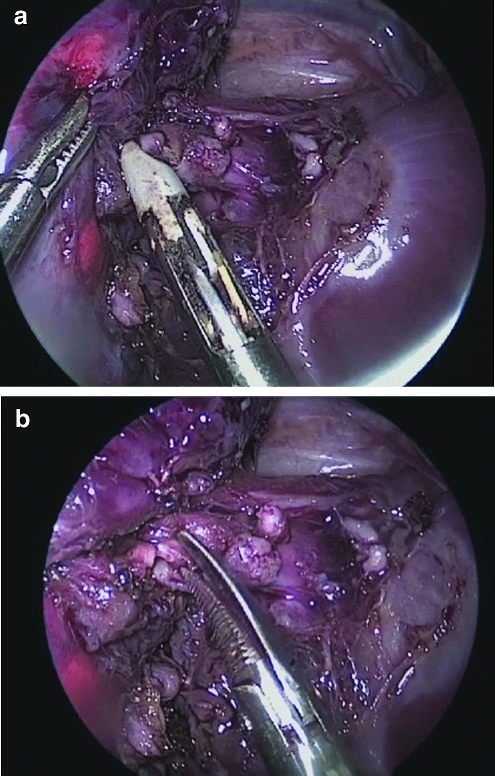
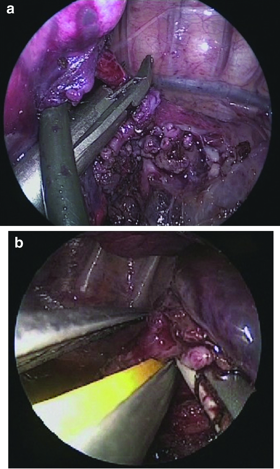
Three to four valved ports, ranging from 3 to 5 mm, were used. In the majority of cases, the LigaSure™ device (Covidien Energy Devices, Boulder, CO), a bipolar sealing device that came in a 5-mm curved dissector design, was the primary mode of vessel ligation. In more recent cases, a 3-mm vessel sealer (JustRight Surgical, Boulder) was used. These devices were also was used to seal and divide the lung parenchyma in cases of an incomplete fissure. The vessels were managed by obtaining adequate length of the vessel to create two seals approximately 3–5 mm apart and then dividing the vessel between them (Fig. 1). In larger patients (generally those over 15–20 kg), an endoscopic stapler was used to secure some or all of the major pulmonary vessels. Occasionally endoscopic clips and suture ligation were also used. The bronchus to the affected lobe was dissected in a similar fashion and isolated. In infants under 10 kg, endoscopic clips were primarily used to seal the bronchus (Fig. 2a), and in larger patients a 12-mm endoscopic stapler was used. More recently a 5-mm endoscopic stapler (JustRight Surgical) has been used in patients under 10 kg to seal and divide the bronchus (Fig. 2b).
The room is set up to facilitate an anterior approach. The surgeon and assistant are at the patient's front with the monitor at the patient's back. First, the chest is insufflated with CO2 using a Veress needle to help collapse the lung and avoid injury of the parenchyma with a trocar. Three trocars are used in almost all cases. Occasionally a fourth trocar is placed if added retraction is necessary. The first port is placed in the mid- to anterior axillary line in the fifth or sixth interspace to determine the position of the major fissure and evaluate the lung parenchyma. Position of the fissure should dictate the placement of the other ports. The working ports are placed in the anterior axillary line above and below the camera port (Fig. 3).
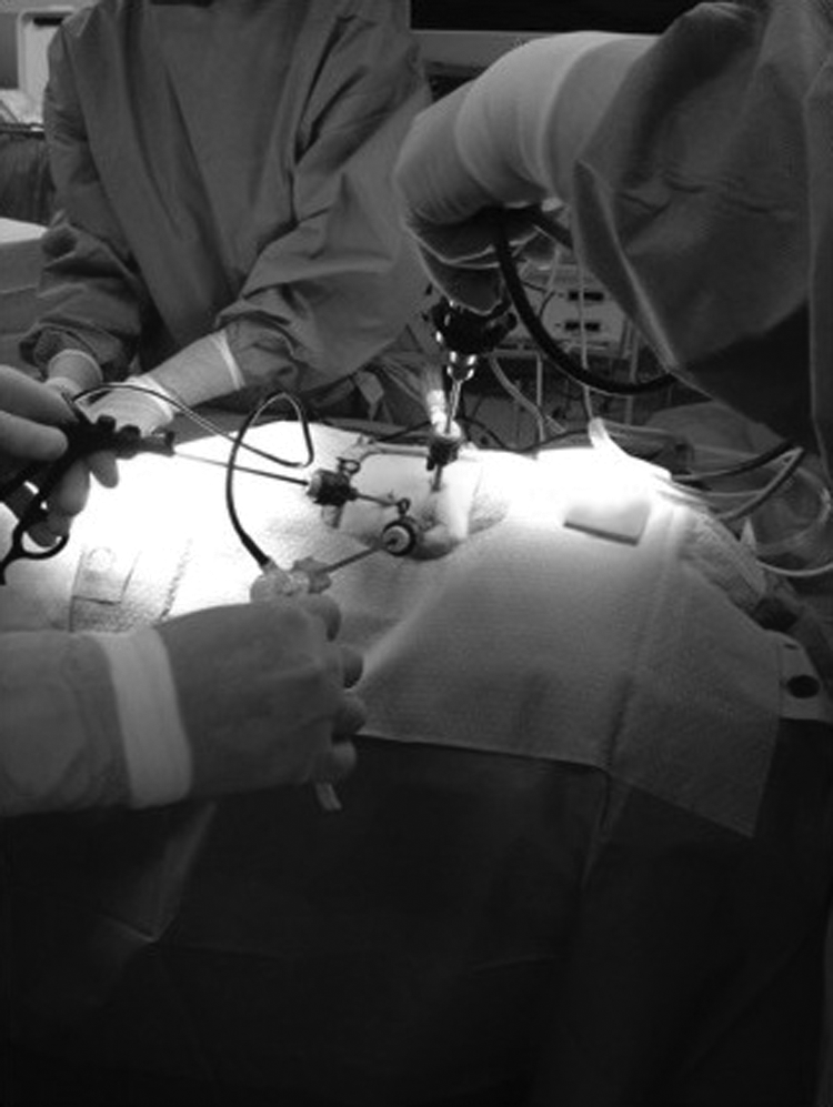
Trocar placement for a right-sided lobectomy. The camera port is in the mid-axillary line one interspace below and anterior to the tip of the scapula.
The branches of pulmonary artery to the affected lobe were sealed and divided first, often at the segmental level, then the pulmonary vein, and lastly the bronchus. If there were large space-occupying cysts, these were first “collapsed” using the vessel sealer. The cystic areas of the lung were grasped, and energy was applied (Fig. 4). This caused the cyst to decompress and collapse, creating more intrathoracic space and improving the surgeon's ability to manipulate the lung and identify important structures. For the last 2 years, in lower lobectomies, the bronchus was divided prior to the vein because this yielded excellent exposure of the trunk of the inferior pulmonary vein, facilitating isolation and safe division. A chest tube was left in all cases of lobar resection.
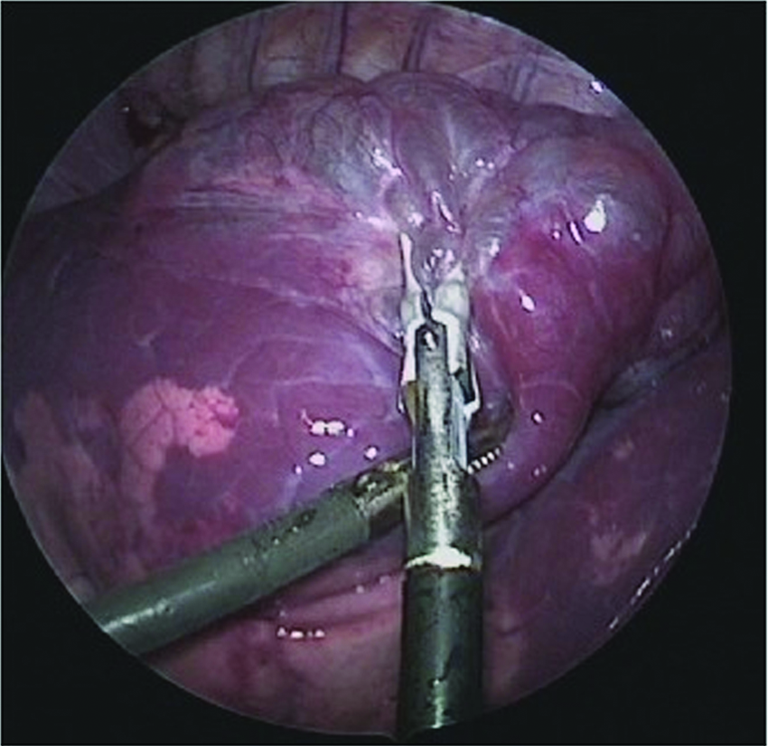
Decompressing the larger cysts in a right lower lobe congenital pulmonary airway malformation using the 3-mm sealer.
Results
Of the 347 procedures, 342 were completed thoracoscopically. Operative times ranged from 35 minutes to 240 minutes (average, 115 minutes). Average operative time when a trainee was the primary surgeon was 160 minutes (n=63), versus 95 minutes when the senior surgeon was performing the procedure. There were 81 upper, 25 middle, and 241 lower lobe resections. There were four intraoperative complications (1.1%) requiring conversion to an open thoracotomy. Three of these were secondary to bleeding, and one was to repair a compromised bronchus to an upper lobe following a lower lobectomy. Only one of these, a bleeding vessel to a pulmonary sequestration, occurred in the last 15 years of the study period. The postoperative complication rate was 3.3%, and 3 patients (0.8%) required re-exploration for a prolonged air leak. In 2 cases a small accessory bronchus was found and sutured closed. In the third, no leak was identified, and the patient had no air leak postoperatively. Hospital length of stay (LOS) ranged from 1 to 16 days with a mean of 3.2 days. In patients <5 kg and <3 months of age, the average operative time was 90 minutes. The postoperative complication rate was 2.6%, and the LOS was 2.1 days.
Discussion
Thoracoscopic lobectomy in children for congenital cystic lung disease is now an accepted and well-described technique.3–7 Most authors agree on the relative merits of a thoracoscopic approach, including less pain, shorter hospital stay, and decreased long-term morbidity, including chest wall deformity, shoulder girdle weakness, and scoliosis. 8 Despite this general consensus, the adoption of this technique and surgeons' comfort with the approach remain relatively low. We believe there are three main reasons for this. First, the average trainee in general surgery and pediatric surgery fellowship receives very little open or endoscopic thoracic training. Experience with lung biopsy, empyema, and mediastinal masses is usually adequate, but exposure to complex lung resections maybe limited. This lack of volume results in a decreased familiarity with pulmonary anatomy. Using a thoracoscopic approach further compounds this, as the surgeon can no longer put his or her hand in the chest cavity to palpate the structures and identify the anatomy.
We have tried to address this in a number of ways. First, a senior surgeon with significant thoracic experience (over 500 lobectomies done open and via thoraocoscopy) is present in every case. As the majority of these cases are semi-elective, scheduling is not an issue. This allows the greatest amount of experience to be present in every case, ensuring the best possible outcome for the patient as well as the greatest amount of teaching points to be related to every trainee. In cases performed by the trainee, the senior surgeon is able to interpret difficult or unusual anatomy improving the flow of the case. One of the most difficult aspects of these cases is when the fissure is incomplete and the pulmonary vessels are not readily visible. We have found that using the tissue-sealing technology to dissect and divide the parenchyma of an incomplete fissure is the safest way to approach this. The fissure is approached layer by layer until the pulmonary artery is visualized (Fig. 5). Using the vessel sealer results in limited bleeding and air leak. The senior surgeon's experience with this technique has helped facilitate the junior surgeons' comfort level with approaching and completing this task safely. Each of the junior surgeons involved in the study has been able to successfully complete a thoracoscopic lobectomy in his or her new job, and many are seen as a resource for their institution.

Using the tissue sealer to dissect through and open the incomplete fissure layer by layer until the pulmonary artery is exposed.
The second issue has been standardizing an anterior approach. During an open thoracotomy the surgeon is generally positioned at the patient's back. For thoracoscopic lobectomies the surgeon and assistant are positioned at the patient's front. This is especially important in smaller patients, as there is more room from the chest wall to the mediastinum, where the pulmonary vessels arise. This added space was even more important because of the relatively large nature of the sealing and stapling technology available during the majority of this study. In many cases the jaws of the instruments barely fit into the thoracic cavity, and this could make the manipulation and firing the devices difficult.
The anatomic relationships for each lobe using this anterior approach are critical. During mentoring of these procedures, three-dimensional relationships of the vessels and bronchi to each lobe, which cannot always be seen in the two-dimensional view of the scope, are stressed and discussed to further the junior surgeons understanding and comfort with the anatomy. This constant dialogue is a well-known training tool but becomes even more important in relatively rare cases where the specific anatomy often cannot be seen or palpated.
The third major issue was standardizing the management of the pulmonary vessels. Early in our experience we learned that thoracoscopic suture ligation of each individual vessel was difficult and time consuming. The small working space, difficulty in achieving traction and countertraction to obtain adequate vessel length while suturing, and the technical demands of tying a secure knot made this process laborious. We did not favor endoscopic clips for most vessels because of the risk of dislodging them during the extensive tissue manipulation necessary during a lobectomy.
Therefore, early on in our experience we adopted vessel sealing as a way to safely mange the pulmonary vessels. This approach has been shown to be a proven modality in adult lobectomy. 9 The initial 5-mm sealing device used could manage a vessel up to 7 mm in diameter and was an adequate tissue dissector. The 3-mm sealing device now available can seal vessels up to 5 mm and works well as a dissector, especially in the smaller chest cavities of infants. It is more than adequate for most pulmonary vessels in children under 10 kg and for segmental branches in larger children. A key to using vessel-sealing technology effectively is to make proximal and distal seals on the vessel approximately 3–5 mm apart. Using scissors, a partial cut is made to determine that the seals are secure and that there is no bleeding once the lumen is entered. Once the vessel is partially divided and no bleeding is seen, the vessel can be completely divided. The benefit of the technique described is the opportunity to reseal the vessel before the vessel retracts and control is lost.
Because of the relatively large nature of the pulmonary vessels and the limited space in the chest cavity, it takes very little blood to obscure the operative field and force conversion to an open thoracotomy. For this reason we have avoided any energy devices that seal and divide the vessel in one step, because if the seal fails the ability to salvage the situation is minimal. We have had only one conversion to open for bleeding since adopting this technique. That was a clear technical complication by the surgeon on a large sequestration vessel. A second vessel lying behind the first was not seen and was inadvertently cut while dividing the first vessel that was sealed.
For bronchus management we initially cut and then suture the bronchus using polydioxanone suture in smaller patients. This can be time consuming and technically demanding. We discovered that in most patients <10 kg the bronchus could be occluded using 5-mm endoscopic clips. If the lobar bronchus is too large, then distal dissection allows for a segmental bronchus to be taken. This decreases the size of the remaining main trunk. For example, the superior segment bronchus in a lower lobectomy can be occluded separately, and then the trunk to the basal segments can be taken with a second clip. In larger patients we used the 12-mm endoscopic linear staplers. However, because of the variations in anatomy and the close proximity of the bronchus to the other lobes, extreme care must be taken to avoid compromising the other bronchi. Therefore, if there is any question the bronchus to the target lobe should be divided sharply and sutured close. There is now new 5-mm stapling technology that better fits in the chest cavity of infants and children and should eliminate the use of clips and larger staplers in these smaller patients.
The benefits of thoracoscopic surgery are clear and have been well documented previously. We also favor earlier resection of prenatally diagnosed lesions before they become infected or the patients become symptomatic. We have previously documented our experience with infants under 10 kg and showed that these procedures had shorter operative times, lower complication rates, and shorter hospital stays. 10 In older infants, there can be significant adenopathy and inflammation in the fissures and around the pulmonary artery, making identification and safe division of these vessels much more difficult. These procedures are technically easier in infants at or near 5 kg despite the smaller working space as evidenced by the shorter operative times in this group when performed by the senior surgeon and by trainees, as compared with older patients. The LOS in this group is also shorter. Some are concerned with issues regarding hypercapnia, cerebral hypoxemia, and hypoperfusion during anesthesia in these smaller infants. However, this has not been an issue in our series, in a previous large series from Stanford, or in the large series reported by the Children's Hospital of Philadelphia presented at the 23rd International Pediatric Endosurgery Group meeting held in Edinburgh this year. 11
Others have clearly documented the issues involved with trying to operate on these lung lesions using thoracoscopy once the infant has already had a clinical chest infection. Kanenko et al. 12 documented a higher complication rate in those patients who were diagnosed after a chest infection and underwent a thoracoscopic lobectomy compared with those diagnosed prenatally and operated on prior to any clinical infections. Garrett-Cox et al. 13 found that 83% of patients converted to open surgery had had a previous chest infection. These and other studies demonstrate that 30%–40% of patients with bronchopulmonary malformations will develop significant pulmonary infections during their lives; thus we prefer to remove these lesions before they become symptomatic to avoid a more complicated surgery.
The timing of surgery remains somewhat controversial, but there is little evidence to suggest that delayed resection improves outcome. In fact, because of the factors mentioned above, earlier resection would seem to be preferable. In this series the subset of patients <5 kg had a shorter average operative time of 90 minutes and shorter LOS (<2 days). Several patients were operated on during the first week of life to avoid a return trip because the families had traveled significant distances to deliver at our maternal/fetal/neonatal center.
The institutional experience clearly plays a role in the outcomes of these cases. Kunisaki et al. 14 compared their experience with thoracoscopic versus open lobectomy. Unlike our study, they found similar blood loss, chest tube duration, and length of stay in the two groups. However, they had a higher conversion and complication rate in patients less than 5 months of age. It could be that a lack of a standardized approach and appropriate equipment for smaller infants led to this result. Their conclusion was that institutional experience might have a strong role in the successful outcome of these cases, and we agree. Carrott and Jones 15 also documented the difficulties in training video-assisted thoracoscopic lobectomy to general thoracic surgeons and documented a need for adequate patient volume, standardization of training, operative set-up, and technique to insure better results.
Lastly, for those who argue for conservative nonoperative management of these lesions in asymptomatic patients, despite the high incidence of infection, we had 2 cases of unsuspected pulmonary blastoma. 16 We feel the risks of recurrent infection and possible malignancy outweigh the risks of intervention if a thoracoscopic approach is used in an institution with a large experience in these procedures.
Conclusions
This study reviews the largest series of thoracoscopic lobectomies in infants and children. The standardization of technique, including an anterior approach and vessel sealing to manage pulmonary vessels, has resulted in a reproducible and consistent method that can be taught to others. Although advanced minimally invasive surgery skills are required to perform this technically demanding procedure, standardization of the technique and the presence of an experienced mentor allowed safe application at two separate centers with multiple surgeons, with minimal complications and excellent outcomes. These data would also suggest that regionalization of these procedures may be indicated to allow for the greatest level of expertise to present in each case.
Footnotes
Disclosure Statement
S.S.R. is the medical director and part owner of Justright Surgical. No other disclosures exist.