




Abstract
Abstract
Purpose:
Retroperitoneoscopic partial nephrectomy (RPN) in children is considered a complex technique with limited diffusion among pediatric surgeons and urologists. We aimed to report the outcome of this technique in infants and children with duplex kidney in a 5-year retrospective multicentric international survey.
Materials and Methods:
Data on 50 children who underwent RPN (41 upper-pole nephrectomies and 9 lower-pole nephrectomies) were retrospectively collected in this six-institution survey. Median age at surgery was 3.3 years. There were 35 girls and 15 boys. The left side was affected in 28 patients, versus the right side in 22 patients. We assessed intraoperative and postoperative morbidity. Follow-up (median, 2.5 years; range, 12 months–5 years) was based on clinical controls and echo color Doppler renal ultrasound scans.
Results:
Median duration of surgery was 255 minutes. Surgery was always performed with the patient in a lateral position. Special hemostatic devices were used for dissection and parenchymal section in all centers. Three patients from two centers (6%) required conversion to open surgery. We recorded seven complications (six peritoneal perforations, one opening of the remaining calyxes) in the 50 cases. Re-operation rate was 0%. Average length of hospital stay was 4.1 days.
Conclusions:
Our survey shows that RPN remains a challenging procedure with a long learning curve, performed only in pediatric centers with huge experience in this field. In our survey operative time was longer than 4 hours. The complication rate remains high (7/50, or 14%), with complications classified as Grade II according to the Clavien–Dindo classification. They did not require further surgery, but they were associated with a prolonged hospital stay.
Introduction
E
Retroperitoneoscopic PN (RPN) is still not generally favored by pediatric surgeons or pediatric urologists for several reasons, such as lack of experience with retroperitoneal anatomy, long operative times, and related complications. 5 Le Clair et al. 6 published in 2009 a retrospective series of 48 patients who underwent PN using the retroperitoneoscopic lateral or prone approach with a conversion rate of 21% (10/48). RPN in children is still considered a complex technique with limited diffusion among pediatric surgeons and pediatric urologists because it is technically demanding, in particular due to a small operative chamber.5,7 In recent years the technique seems to have become easier to perform thanks to the use of special hemostatic and synthesis devices that permit a faster and safer procedure.8–11
Here we report a 5-year retrospective multicenter international survey about the outcome of RPN in infants and children with duplex kidney.
Patients and Methods
An international multicenter survey was carried out among pediatric laparoscopic surgeons or urologists who were experts in laparoscopic renal surgery. During the ESPU/SIVI Paediatric Laparoscopic Course in Urology held at the Biotechnology Center of Naples, Italy, in July 2013, an extensive discussion was carried out by the present experts about which technique is preferable to adopt, between laparoscopy and retroperitoneoscopy, to perform PN. For this reason we decided to organize two retrospective multicenter surveys on this topic: the first one focused on RPN, and the second one focused on LPN. We involved in these surveys the present experts coming from several European centers of pediatric surgery and urology. All the experts had extensive experience in pediatric laparoscopic urology, all had started laparoscopy more than 10 years ago, and they usually performed more than 10 retroperitoneoscopic or laparoscopic procedures every week.
The tool of our survey was a questionnaire that was mailed to six surgeons at different centers. The questionnaire was focused on patients' characteristics, technical operative details, and outcome of RPN performed in these centers in the last 5 years (see Appendix).
Results
All six involved surgeons successfully completed the survey.
Data on 50 children who underwent RPN (41 upper-pole nephrectomies and 9 lower-pole nephrectomies) were retrospectively collected in this six-institution survey. Median age at surgery was 3.3 years (range, 12 months–11.5 years). There were 35 girls and 15 boys. The left side was affected in 28 patients, versus the right side in 22 patients.
The indications for surgery were recurrent urinary tract infections in 16 patients (secondary to vesicoureteric reflux in 11 patients and to an ureterocele in 5 patients), loss of kidney moiety function in 23 patients, and an ectopic ureter causing pseudo-incontinence in 11 cases. Vesicoureteric reflux was present into the affected moiety in 9 patients who underwent lower-pole nephrectomy and in 2 patients who underwent upper-pole nephrectomy.
With regard to surgical technique, in all patients an ureteral catheter was positioned preoperatively via cystoscopy in the ureter of the normal functioning moiety, to easily identify it during the dissection. RPN was always performed with the patient in the lateral decubitus position. Four surgeons preferred to adopt three trocars, whereas two surgeons always used four trocars. A 30° optic was adopted in five of the six centers; only one center adopted a 0° optic. Special hemostatic devices (Starion™ [Starion Instruments, Sunnyvale, CA], LigaSure™ [Covidien, Mansfield, MA], or Ultracision® [Ethicon Endo-Surgery, Cincinnati, OH]) were used for dissection and parenchymal section in all centers. The ureter of the removed moiety, isolated as far down as possible, was ligated in the 11 patients with associated vesicoureteric reflux using endoloops in five centers or with stitches in one center.
Follow-up (average, 2.5 years; range, 12 months–5 years) was based on clinical controls (every year for 5 years after surgery) and echo color Doppler renal ultrasound scans performed 1 month and 1 year after surgery. A dimercaptosuccinic acid scan was performed 1 year after surgery in all centers.
Median duration of surgery was 255 minutes (range, 125–310 minutes). Three patients from two centers (6%) required conversion to open surgery due to technical challenges related to a too small operative chamber. There was no intraoperative bleeding.
We recorded 7/50 (14%) complications (six peritoneal perforations, one opening of the remaining calyxes), classified as Grade II complications according to the Clavien–Dindo grading system. 12 All these complications were treated conservatively without the need of a new surgical procedure. The opening of remaining calyxes occurred in a 4-year-old patient who underwent lower-pole nephrectomy and needed only a longer drainage with a hospital stay of 10 days. The peritoneal perforations occurred in 4 patients who underwent upper-pole nephrectomy and in 2 patients who underwent lower-pole nephrectomy. All these patients were younger than 1 year of age, and all were managed conservatively but with a prolonged hospital stay (average, 7.2 days). In all cases a drainage device was left in the retroperitoneal space for at least 24–48 hours after surgery.
Hospital stay varied between 2 and 10 days (average, 4.1 days). The average time to resume full oral feeding was 26 hours after surgery, and the average time of analgesic requirements was 32 hours.
Echo color Doppler renal ultrasound scans were normal in all patients, both 1 month and 1 year after surgery (Fig. 1).

Echo color Doppler study of the residual moiety after surgery.
Postoperative dimercaptosuccinic acid scan demonstrated no loss of function of the residual kidney moiety after surgery. In fact, no difference was reported in dimercaptosuccinic acid scan results between preoperative renal function (average value, 40.1 ± 3.1%) and postoperative function outcome (average value, 39.8 ± 3.7%) in all children who underwent surgery (P = .5).
Discussion
In the last decade acceptance of minimally invasive surgery procedures has grown in pediatric urological surgery. Advanced minimally invasive surgery procedures in pediatric urology now are considered safe and successful, thanks to the advanced technologies available on the market, in particular, special hemostatic devices. 11 Excluding oncological indications, the main indication for PN in children is to remove a nonfunctioning upper or lower pole secondary to complicated duplex anomalies of the kidney. 1 PN using minimally invasive surgery is possible with two techniques: laparoscopy or retroperitoneoscopy. RPN seems to be technically more demanding than LPN, in particular as there is a smaller operative space in which to work and to move the instruments.7,13–15
After the first description of LPN in children by Jordan and Wislow 2 more than 20 years ago, in 1993, this procedure has gained wide acceptance. Analyzing the international literature there is no evidence about which technique—laparoscopy and retroperitoneoscopy—is the best to perform PN. 16 However, the literature shows that retroperitoneoscopy has a higher rate of conversion and a higher incidence of major complications compared with LPN. 5 Le Clair et al. 6 published in 2009 a retrospective series of 48 patients who underwent PN using the retroperitoneoscopic lateral or prone approach with a conversion rate of 21% (10/48). In our series of 50 patients, we reported 3/50 (6%) conversions to open surgery and only 7/50 (14%) complications, classified as Grade II complications according to the Clavien–Dindo grading system. 12
We analyzed those articles reporting results of RPN in children published in the last 10 years.5,17–24 The conversion rate varied between 6% and 18%. The average length of surgery varied between 152 and 380 minutes. The complication rate varied between 6.6% and 50%. It is interesting to note that the complication rate was higher when the preliminary experience with the technique was reported. In particular, Miyano et al. 5 reported two retroperitoneoscopic heminephrectomies performed and managed by supervised senior pediatric surgical trainees, and in their series the complication rate was 50% (Table 1).
NR, not reported.
We think that advanced minimally invasive surgery procedures and in particular PN are strictly linked to the advanced technology and instrumentation now available on the market.8–11 One of the key messages of this study is that special hemostatic devices (Starion, LigaSure, or Ultracision) are fundamental tools to adopt to perform RPN. First of all, they permit an easy and fast dissection of tissues, an easy vascular control of the nonfunctioning moiety, and, at the end of the procedure, an easy and simple resection of the nonfunctioning moiety and of the ureter without any bleeding. All centers involved in our survey have adopted these hemostatic devices, and no intraoperative bleeding was reported in our series.
The second key message is always to position a ureteral catheter in the ureter of the normal moiety via cystoscopy before surgery. In this way during RPN it is easier to identify the normal ureter and to dissect the mega-ureter of the nonfunctioning moiety safely without risk of damage to the normal ureter.
The third message is that only a skilled team already expert in retroperitoneoscopic nephrectomy can perform RPN. In our opinion, only surgeons with a very extensive experience with retroperitoneoscopic nephrectomy can safely perform PN via the retroperitoneal route. In fact, all surgeons involved in our multicenter survey had had previous experience with retroperitoneoscopic nephrectomy, and they usually performed more than 10 retroperitoneoscopic or laparoscopic procedures every week. All complications recorded in our series (7/50, or14%) were classified as Grade II according to the Clavien-Dindo grading system. 12 They were treated conservatively without the need of a new surgical procedure. The absence of major complications requiring further surgery and the low rate of conversion (6%) in the present series are possibly due to the extensive experience of the surgeons concerned with the retroperitoneoscopic approach.
Before starting surgery it is fundamental to correctly position the patient on the operative table. The retroperitoneal access can be achieved either posteriorly with the patient prone or laterally with the patient in the lateral decubitus position. Borzi 14 compared the two approaches and found that the lateral approach created more inferomedial space and provided better access to ectopic kidneys and allowed a complete ureterectomy in all cases. We always prefer to position the patient in lateral decubitus, with a ballast underneath the patient to enlarge the space between the costal margin and iliac crest (Fig. 2).
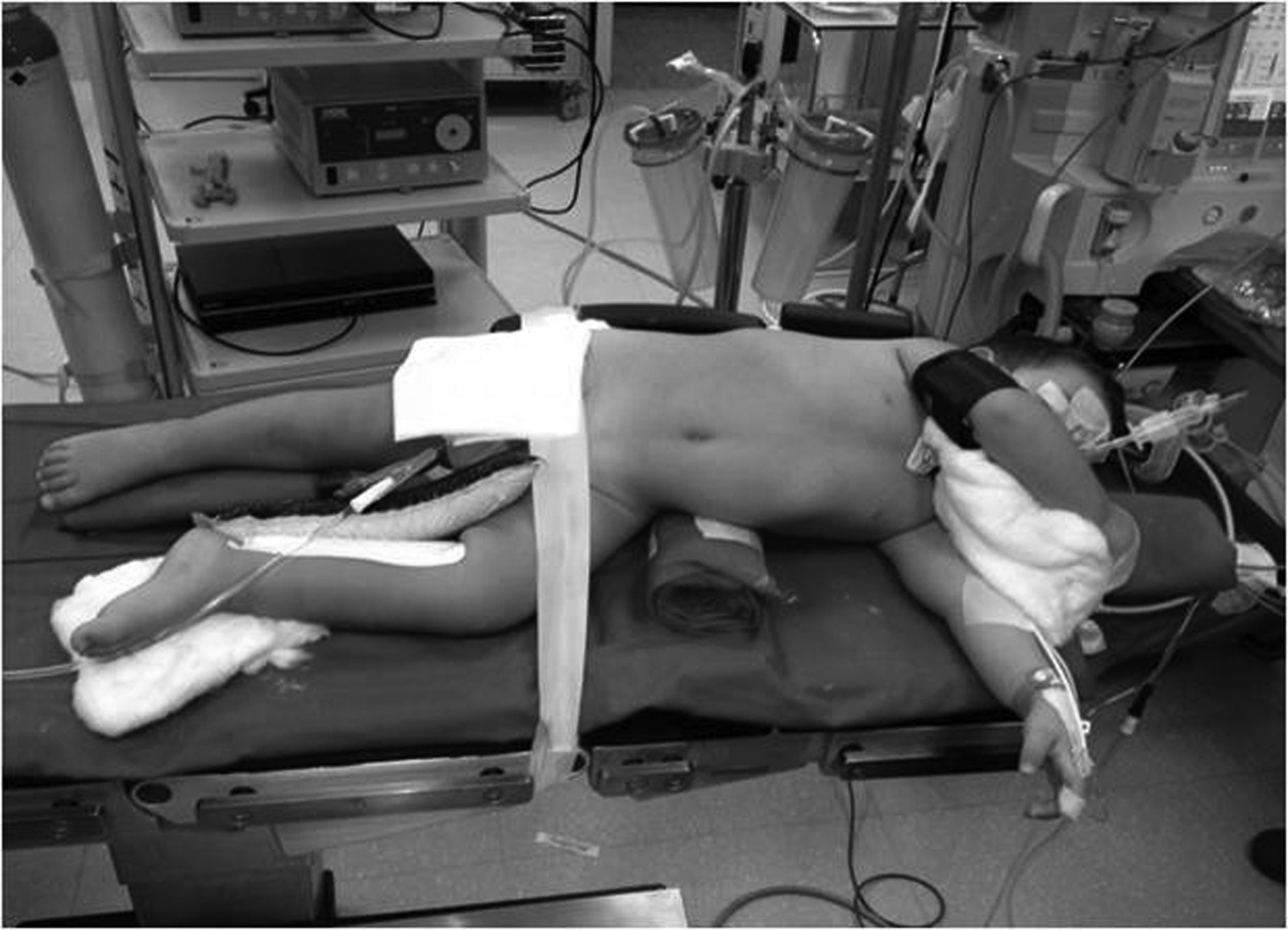
The positioning of the patient on the operative table.
As for the technical difference between upper- and lower-pole PN, it seems that lower-pole PN is technically easier because it is not necessary to decross the ureter from the main hilar vessels as happens in the case of upper-pole nephrectomy.
Probably at the beginning of a surgeon's experience it is better to use always four trocars to have an adequate vision of the operative field. As already reported, a 30° optic has to be used because it allows a better view of the operative field.
As for specimen removal, the nonfunctioning moiety and the dilated ureter can be removed through the optic orifice; it is not necessary to use an endo-bag. As for the need to put in a drainage device, probably it is useful to leave a drainage device for the first 24 hours through one of the trocar orifices. We leave also a bladder Foley catheter for 24 hours after surgery
Our survey shows that RPN still remains a challenging procedure performed only in pediatric centers with extensive experience in this field, and in an analysis of the international literature, our series is one of the largest ones published (Table 1).
According to our opinion, the limit of our survey is that we included only the experience of the last 5 years of surgeons who are already experts.
We reported a longer operative time (255 minutes) compared with that reported in the previous series analyzed and a conversion rate of 6%, mainly due to technical challenges related to a too small operative chamber (Table 1). The complication rate remains high (7/50, or 14%). However, we recorded only Grade II complications according to the Clavien–Dindo grading system, 12 which did not require a new surgical procedure but prolonged significantly the average hospital stay.
We believe that the retroperitoneoscopic approach is better suited to PN in children compared with the open approach. The retroperitoneoscopic magnification provides an excellent exposure of the duplicated anatomy of the kidney and its vascularization, allowing a full dissection in situ without the need to mobilize the remaining part of the kidney. Another advantage is that there is no need for a second inguinal incision to manage the distal part of the ureter as is the case in open surgery. Compared with open surgery, the retroperitoneoscopic approach significantly reduces the postoperative pain due to the minimal muscular trauma of the minimally invasive approach compared with the extensive flank incision required by the open technique. In fact, in our series the average postoperative analgesic requirement was 32 hours. In addition, retroperitoneoscopy allows a very precise dissection of the renal hilar vessels and an excellent differentiation of each hemirenal tissue, separating the nonfunctioning kidney and preserving the maximum function possible for the other half-kidney. Postoperative urinoma or hematoma collections are confined to the retroperitoneum. Drawbacks are the spatial limitations of the narrow retroperitoneal working space, especially in smaller children (younger than 12 months), and the difficulty of removing the entire ureter near the bladder dome. For these reasons, we recommend the retroperitoneal approach in patients older than 12 months who need an upper- or lower-pole heminephrectomy with partial ureterectomy.
On the basis of our results, we can assume that the learning curve for this technique is long, and probably it is still ongoing even for the already experienced surgeons involved in our survey.
We believe that advanced retroperitoneoscopic skills, appropriate instrumentation, and new devices are all critical points for the success of this surgery in infants and children.
Footnotes
Disclosure Statement
No competing financial interests exist.