
Abstract
Abstract
Wandering spleen is an extremely rare clinical condition characterized by abnormal anatomical position of the spleen. Up to now, its etiology remains unknown, and the best surgical procedure is still an area to explore in the future. In this article, we present the case of a 24-year-old woman with wandering spleen combined with congenital heart disease and pigeon chest. Based on the clinical features and our experiences of radiofrequency ablation for preservation of the spleen in traumatic spleen rupture, we successfully performed a modified splenectomy and splenopexy method. Since the operation, the organ has remained in place with good perfusion and function.
Introduction
W
Case Report
The patient was a 24-year-old female. She was a twin and had congenital heart disease and thoracic deformity, but her twin sister was perfectly normal. She had accepted operations to fix a ventricular septal defect 3 years prior and eutocia 2 years prior. The patient had tolerable but persistent mild, dull abdominal pain for 10 days. Diagnosis of wandering spleen in the pelvic cavity was confirmed by computed tomography (Fig. 1). During the operation we found that the spleen was larger than normal, but without ischemic necrosis. The spleen pedicle was much longer than normal and contrarotated about three times. The perisplenic ligament was absent. Cauda pancreatis was accompanied by splenic pedicle torsion (Fig. 2).
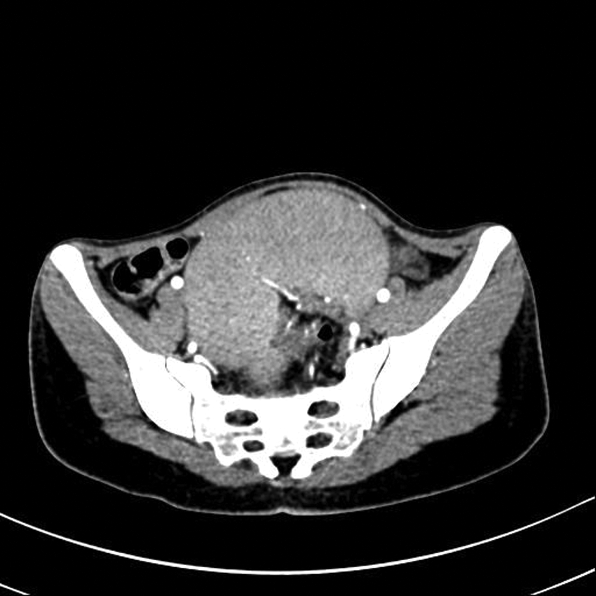
Diagnosis of wandering spleen in the pelvic cavity was confirmed by computed tomography.
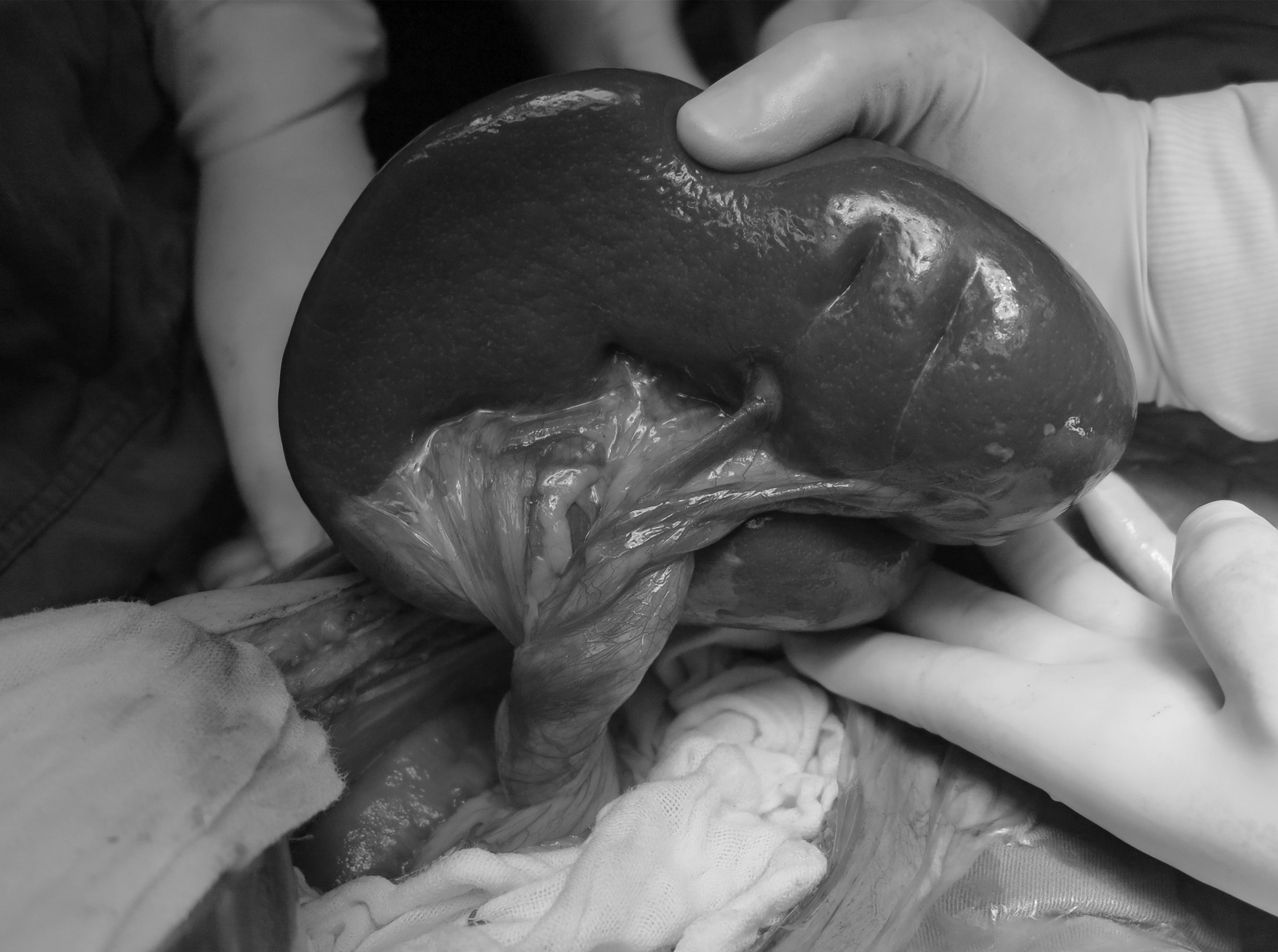
The spleen was larger than normal but without ischemic necrosis. The spleen pedicle was much longer than normal and contrarotated about three times. The perisplenic ligament was absent. Cauda pancreatis was accompanied by splenic pedicle torsion.
Operative procedure
The rotated pedicle was reset, and the vessels of the splenic lower pole were isolated and ligated. After the ligation, the ischemic spleen zone showed a mulberry-like color. The transition zone was ablated with radiofrequency ablation. The resection of the ischemic spleen was then performed directly along the lateral coagulation necrosis zone using a blade (Figs. 3 and 4). The residual spleen was approximately 10×8×4 cm. The spleen and splenic pedicle were then restored to the normal anatomical position, and the coagulation zone and splenic pedicle were directly fixed to the diaphragm, left abdominal wall, mesocolon, and greater gastric curvature using 4-0 polypropylene (Prolene®; Ethicon, Cincinnati, OH) sutures. The operation lasted 100 minutes, and the blood loss was approximately 50 mL.
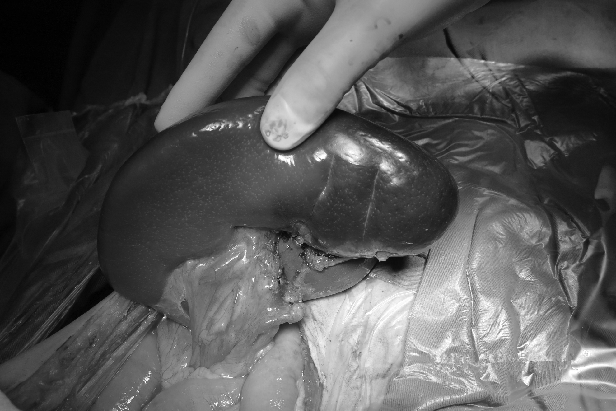
The ischemic spleen zone showed a mulberry-like color after isolation and ligation of the vessels of the lower splenic pole.
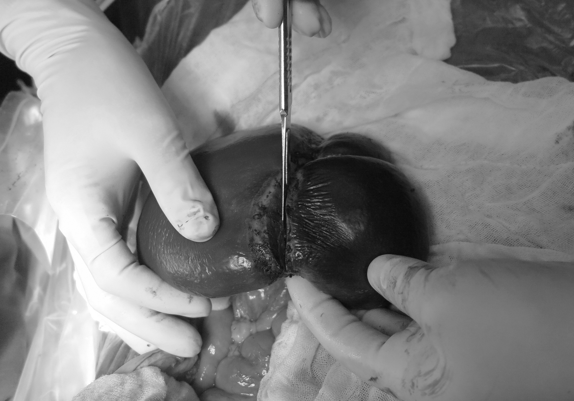
The transition zone was ablated with radiofrequency ablation. The resection of the ischemic splenic was then performed directly along the lateral coagulation necrosis zone using a blade.
Results
The patient did not exhibit postoperative bleeding from the fractured surface of the spleen or fever. The platelets were restored to normal levels by Day 3 postsurgery. The patient was reviewed 3 days after surgery; the spleen position had not changed, with a size of approximately 10×9 cm, and the blood supply was satisfactory. The patient was allowed out of bed after 1 week, was discharged 11 days after the surgery, and was able to resume normal activities after 1 month. She was reviewed using enhanced computed tomography 11 months after the surgery; the spleen size was 10×8 cm, and the spleen remained in the original fixed position with a good blood supply. No abnormalities were found in routine examinations.
Discussion
The first successful treatment of wandering spleen using splenopexy was reported by Sykoff in 1895, as noted by Hall. 4 In recent years, splenopexy has gradually become the most popular surgical treatment for wandering spleen.5,6 Conventional partial splenectomy has the shortcomings of surgical difficulties and more blood loss, which hinder the clinical application and promotion of spleen preservation surgery.
In recent years, with the development of radiofrequency ablation technology and its wide implementation in liver resection, the powerful hemostatic ability of this technology and its potential application for partial splenectomy have become apparent.7,8 The introduction of radiofrequency ablation to spleen preservation surgery has been shown to achieve good outcomes.9,10 In 2010, our department started to implement radiofrequency-assisted partial splenectomy to treat traumatic spleen rupture with a high success rate and low postoperative complications, demonstrating that this type of surgery is convenient and effective. Therefore, we implemented radiofrequency-assisted partial splenectomy plus spleen reduction and fixation in this case.
There are a variety of spleen fixation methods, such as placing the spleen in the posterior peritoneum, as well as fixing the spleen directly to the greater curvature, diaphragm, abdominal wall, and transverse colon. The operation to place the spleen in the posterior peritoneum is relatively complicated, and the spleen is not placed in the splenic fossa, which does not accord with the normal anatomical physiology. Although absorbable mesh fixation is convenient, it is still a foreign body that may induce long-term rejection reactions and chronic inflammation in the abdominal cavity. The easiest approach without introducing foreign material involves direct suture fixation using a needle. However, the spleen has a rich blood supply, which increases the risk of bleeding during the stitching process and spleen tearing. The methods described are not ideal for spleen fixation.
In this case, we used the coagulation zone produced in the radiofrequncy-assisted splenectomy. The coagulation zone is sufficiently robust to prevent bleeding. In the fixation process, we sutured the coagulation zone and the splenic pedicle to the greater curvature, transverse colon, and diaphragm, instead of suturing the spleen directly, and then covered the spleen with the greater omentum. Due to the possibility of failure of the fixation procedure, the patient was required to stay in bed for 1 week. Thus, the coagulation zone may induce avascular necrosis, which may result in dense adhesion between the spleen and the surrounding tissues and further strengthen the spleen fixation. Radiofrequncy-assisted splenectomy is a very convenient procedure, and the generated coagulation necrosis zone makes it relatively simple to fix the spleen; thus, this approach may become a preferred treatment of wandering spleen.
In addition, our case may provide an important clue to discovery the etiology. This patient was a twin and had congenital heart disease and thoracic deformity. However, the woman did not suffer from a wandering spleen before delivering a baby. This shows that genetic factors and hormone changes in pregnancy may play an important role. But, it is regrettable that we did not study this case from the aspect of basic medicine.
In conclusion, based on the experience in previous work, we tried a novel surgical procedure to treat this rare disease and achieved success. Also, this case is helpful for us to think deeply about and discuss the etiology of wandering spleen.
Footnotes
Disclosure Statement
No competing financial interests exist.