
Abstract
Abstract
Introduction:
Hybrid natural orifice translumenal endoscopic surgery (NOTES®; American Society for Gastrointestinal Endoscopy [Oak Brook, IL] and Society of American Gastrointestinal and Endoscopic Surgeons [Los Angeles, CA]) reduces the invasiveness of conventional laparoscopic surgery and overcomes the limitation of pure NOTES, especially in the absence of angulated instruments.
Patients and Methods:
The patients were 66-, 69-, and 32-year-old women with complaints of recurrent flank pain and urinary tract infection due to an obstructed nonfunctioning kidney. Materials used were standard laparoscopic instruments and a 30° 10-mm high-definition laparoscope. Under general anesthesia, each patient was placed in a lithotomy position with the affected side up at 45°. A Veress needle was initially inserted through the umbilicus and was later replaced with a 10-mm laparoscopic port, with an additional 5-mm port also inserted at the affected lower quadrant site. The patient was then positioned in a steep Trendelenburg position, and a 10-mm port was inserted through the posterior vaginal wall under direct vision from the abdominal cavity that was later used for the laparoscope. Nephrectomy proceeded despite noted severe adhesions, and the kidney was placed in the specimen retrieval bag. The vaginal port site was enlarged to 3 cm for extraction of the specimen. A Penrose drain was placed at the lower quadrant 5-mm trocar site. The vaginal wound was repaired using running 2-0 absorbable sutures.
Results:
Three cases of transvaginal hybrid NOTES nephrectomy were successfully completed with a median operative time of 310 minutes and mean estimated blood loss of 300 mL. Median renal dimensions were as follows: craniocaudal, 10.2 (range, 10.6–9) cm; laterolateral, 6.5 (range, 7–5.3) cm; and anteroposterior, 4.8 (range, 6.5–3.9) cm. The patients resumed regular diet as early as Day 1 postoperatively. The drain was removed prior to discharge. The mean date of discharge was Day 3 postoperatively. There were no noted surgical complications according to the Clavien–Dindo grading system.
Conclusions:
Hybrid NOTES transvaginal nephrectomy is a feasible and reproducible procedure in selected patients regardless of laterality for better cosmesis, reduced postoperative pain, and early recovery.
Introduction
U
In hybrid NOTES, the vagina was used not only as a conduit for specimen extraction but also as a working port, in combination with a maximum of two abdominal trocars. Anything beyond was considered a conventional laparoscopic procedure. As to the view with the camera from below, inserted through the vaginal vault, the caudal view of the kidney and the hilum was odd and unfamiliar. This problem, however, can be overcome with strict adherence to anatomic dissection principles, decreasing the invasiveness of conventional laparoscopic surgery and thus overcoming the limitation of pure NOTES.
Case Series
Our patients included 32-, 66-, and 69-year-old women, each complaining of chronic flank pains and recurrent urinary tract infections. They presented with computed tomography scan results of unilateral hydronephrosis and nonfunctioning kidneys on 99mTc diethylenetriamine pentaacetic acid renal scan. Two of the cases involved the right kidney, and the other involved the left. Patients were then oriented to the risks and benefits of undergoing hybrid NOTES. Consent was obtained to undergo such a procedure.
The operative procedure included the following materials: standard laparoscopic instruments and a 30° 10-mm high-definition laparoscope. Under general anesthesia the patient was placed in a lithotomy position with the affected side up at 45°. A Veress needle was initially inserted through the umbilicus and was later replaced with a 10-mm laparoscopic port, with an additional 5-mm port also inserted at the affected lower quadrant site (Fig. 1). The patient was then positioned in a steep Trendelenburg position, and a 10-mm port was inserted through the posterior vaginal wall under direct vision from the abdominal cavity. This was later used for the laparoscope (Fig. 2). Nephrectomy proceeded despite the finding of severe adhesions, and the kidney was placed in the specimen retrieval bag. The vaginal port site was enlarged to 3 cm for extraction of the specimen (Fig. 3). A Penrose drain was placed at the lower quadrant trocar site. The vaginal wound was repaired using running 2-0 absorbable sutures.

Schematic of the left-side transvaginal hybrid natural orifice translumenal endoscopic surgery nephrectomy.

Hybrid natural orifice translumenal endoscopic surgery with a 10-mm port at the umbilicus and posterior fornix of vagina and a 5-mm port at the right lower quadrant.

Transvaginal extraction of the kidney specimen.
Patients' characteristics and results are given in Table 1. The median operative time was 310 (range, 280–335) minutes. The mean estimated blood loss was 300 mL. Median renal dimensions were as follows: craniocaudal, 10.2 (range, 10.6–9) cm; laterolateral, 6.5 (range, 7–5.3) cm; and anteroposterior, 4.8 (range, 6.5–3.9 cm). The patient resumed a regular diet as early as Day 1 postoperatively. The drain was removed prior to discharge. The mean date of discharge was Day 3 postoperatively. There were no noted surgical complications according to Clavien–Dindo grading system. The operative sites (Fig. 4) were dry and well coaptated upon follow-up after 1 week of discharge.
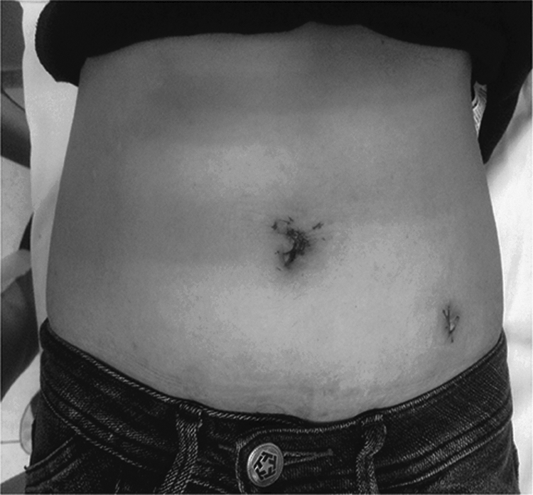
Postoperative photograph of a patient who had left-sided hybrid natural orifice translumenal endoscopic surgery.
IFC, indwelling foley catheter; POD, postoperative day.
Discussion
Minimally invasive surgeons were quite hopeful that further gains could be achieved with NOTES such as (1) lack of skin incisions, (2) reduced postoperative pain, (3) possibility of anesthesia other than general, (4) preferable approach for obese patients and for those with conditions that affect the abdominal wall such as scars, burns, and infections, (5) diminished risk of postoperative hernias, and (6) easier access and other aspects such as early recovery, reduced adhesion development, and shorter postoperative ileus. However, pure NOTES was not a straightforward endeavor and was intended for expert laparoscopist/endoscopists and those with previous animal and human cadaveric model experience. Several limitations were encountered such as (1) difficulty in terms of maneuverability of standard endoscopes and instrumentations within the peritoneal cavity, (2) flexible instruments making it difficult to retract organs such as the kidney, (3) the incongruence of the flexible material to be used in the abdominal cavity, and (4) the limited port access to use good hemostatic devices.1–3
A hybrid NOTES approach may overcome these limitations by increasing safety and at the same time decreasing the invasiveness of conventional laparoscopic surgery. The main advantage of hybrid NOTES was that it allowed the use of conventional laparoscopic devices and overcame the limitation of pure NOTES. Moreover, hybrid NOTES preserved the main advantage of pure NOTES, which was the lack of an abdominal incision for retrieving the diseased organ.1–4
The transvaginal approach seemed to adapt perfectly to the wishes of cosmetic improvement and the absence of scarring that were sought after with the development of NOTES. It also permitted the extraction of surgical samples of considerable size, which made it particularly attractive as an approach route in ablative and even oncological surgery. Several transvaginal NOTES approaches have been used throughout the years using different abdominal trocar sizes (2.5–12 mm) and different vaginal access such as two-channel endoscopes, a 10-mm camera with a deflectable tip, a three-channel trocar (TriPort™; Olympus, Tokyo, Japan), and a 5-mm camera with a deflectable tip and a GelPort® (Applied Medical, Rancho Santa Margarita, CA) wherein surgeons could introduce two 5-mm trocars and one 12-mm trocar as well as a 5-mm deflectable optic. It must, however, be clarified that the strict concept of pure NOTES should not involve transabdominal access ports. Nevertheless, the clinical applications to date were all classifiable as hybrid procedures. Animal experimentation runs in parallel with the clinical implementation of the technique with the aim of achieving the application of pure NOTES to urological surgery.1–8
Recent applications of newer technologies in NOTES included the use of the da Vinci® robotic system (Intuitive Surgical, Sunnyvale, CA) and the magnetic anchoring and guidance system. The evolution of this new technique ran in parallel with its clinical application; basically it was centered on the search for new instruments that permit freer and finer manipulation and for achieving adequate retraction and triangulation in order to guarantee safe and reproducible procedures for the patients.1,9,10
The complications encountered and reported for NOTES included intraabdominal abscess, colon injury, vascular injury, and death. The possible impact on sexual function has been a source of concern since the advent of the transvaginal approach. However, the literature reports low impact on sexual dysfunction after vaginal surgery.1,6,11,12
The transvaginal NOTES nephrectomy specimens include kidneys from live donors, nonfunctioning kidneys, and malignant kidneys. Aside from the different transvaginal approaches in NOTES, there were several reported cases using different orifices, namely, the stomach, bladder, rectum, and ureter.1,6,8–22 The transvaginal access was considered superior to the transgastric or transcolonic accesses because it could be carried out via conventional surgical techniques. Of all the accesses, the vagina was the one that allowed the easiest extraction of surgical samples, especially those of large size as occurred in nephrectomies with low complication rates. Similarly, it allowed the use of rigid instruments and the maintenance of the target organ in direct line with the point of access, improving the questions related to spatial orientation. 1 However, this made it difficult to obtain the correct working angles coming in straight from the vagina, as the instrument passes above the pelvic brim and then has to be twisted posteriorly to work at the upper pole, making it more challenging for dissection. This was due to the difficult angles of view and a “blind spot” just cephalad to the upper pole. Also, the slope of the psoas as it proceeds cephalad directed the upper pole of the kidney in a more posterior plane compared with the lower pole of the kidney.
An additional problem was the considerable distance between the introitus and the upper pole of the kidney, necessitating the use of extralong bariatric type instruments. 20 Another problem was the difference between the anatomy of the left and right kidney due to the presence of the gonadal vein, which can obstruct the view of a rigid instrument.
Moreover, it must be taken into consideration that this technique involved the instrumentation of the peritoneal cavity through the vagina, which could signify a higher peritoneal contamination and consequently an increase in the rate of infections. In spite of all its advantages, the transvaginal access was only available to half of the population to treat due to the question of sex, and within that population any prior pelvic or gynecological surgery may contraindicate this access. 1
In our experience, skills in laparoscopic and robotics surgery paved the way to completing these 3 cases of hybrid NOTES. The uniqueness of our procedure from previously described techniques was that we used a rigid 30° 10-mm high-definition laparoscope together with standard laparoscopic instruments. The view was challenging because it was from the pelvis. The left-side procedure proved to be more difficult due to the gonadal vein obstructing the view, which is usually larger in young females.
Footnotes
Disclosure Statement
No competing financial interests exist.