
Abstract
Abstract
Introduction:
To report the surgical technique, procedure outcomes, and feasibility of robot-assisted simultaneous bilateral radical inguinal with bilateral pelvic lymphadenectomy.
Materials and Methods:
Three consecutive patients of penile and urethral carcinoma with palpable inguinal lymphadenopathy who underwent robot-assisted simultaneous bilateral radical inguinal with bilateral pelvic lymphadenectomy in our institution from May 2013 to October 2015 were included in the study. Surgical technique is described and feasibility of the procedure is assessed.
Results:
Three patients aged 58, 76, and 35 years underwent robot-assisted simultaneous bilateral radical inguinal with bilateral pelvic lymphadenectomy with a mean operative duration of 453.33 minutes (range 420–490 minutes). Average blood loss was 66.66 mL (range 50–80 mL) and mean time to removal of last drain was 44.66 days (range 28–72 days). Mean lymph node yield in left inguinal region, right inguinal region, left pelvic region, and right pelvic region was 18, 14.6, 13.3, and 16.6, respectively. The perioperative period was uneventful. No skin flap–related complications were seen. One patient suffered lymphocele postoperatively, which was managed successfully with needle aspiration. One patient developed lung metastasis in follow-up and none of them had local recurrence.
Conclusion:
Robot-assisted simultaneous bilateral radical inguinal with bilateral pelvic lymphadenectomy is feasible, safe, and may result in decreased morbidity compared to conventional open lymphadenectomy.
Introduction
L
Materials and Methods
We retrospectively analyzed three consecutive patients who underwent robot-assisted simultaneous bilateral radical inguinal with pelvic lymphadenectomy for locally advanced, high-grade squamous cell carcinoma of penis or urethra from May 2013 to October 2015 at our institution. We looked for the feasibility, morbidity, and oncological adequacy of the procedure. Demographic data of these patients are presented in Table 1. These patients were counseled regarding different treatment options with the consent of open conversion if concern arises for oncologic safety. The technique and surgical steps have been described below. The perioperative parameters assessed included the duration of surgery, skin flap or lymph-related complications, time to drain removal, lymph node yield, and histopathological positivity of the lymph nodes.
FNAC, fine needle aspiration cytology; LN, lymph nodes.
Technique
• Patient positioning: After epidural block, the patient was placed in supine position with legs apart (abducted) and padded (Fig. 1) under general anesthesia. The inguinal and groin region was prepared and draped, and Foley's catheterization was performed under sterile conditions. The assistant stood lateral to the right leg for right-sided dissection and between the legs for left-sided dissection.
• Surface marking (right side): Surface marking was done for the extent of lymph node dissection (Fig. 2). About 20 and 15 cm line was drawn vertically from anterior superior iliac spine and pubic tubercle, respectively. Superiorly, a line joining anterior superior iliac spine and pubic tubercle at the inguinal ligament was drawn to mark the limits of radical inguinal lymphadenectomy.
• Ports placement: Port sites were marked one hand breath below the template. Four such ports were placed around 6–8 cm apart (Fig. 2). For camera port, 1.5 cm incision was given and plane was created till the fascia lata. Plane was created by finger dissection or with the back of the Bard-Parker knife handle. The assistant port was placed in the midpoint between the camera port and lateral robotic arm. We used 0° robotic camera for the whole dissection.
• Docking of robot: After positioning the operating table at the lowest possible height, the da Vinci Si robot was docked from the left shoulder (along the side of the patient) (Fig. 3).
• Gas insufflations: The working space was insufflated with CO2 at 12–15 mmHg.
• Retrograde dissection and identification of anatomic limits: First step was to get into the right plane and identify fascia lata, which appears as a whitish sheath of fibrous tissue. We used Bipolar Maryland forceps in the left robotic arm, and Monopolar Hot Shears were used in right robotic arm. The dissection was carried out deep into the fascia lata. After incising fascia lata, dissection was carried out in the femoral triangle between the medial border of the sartorius and the lateral border of the adductor longus. All the lymph nodes bearing fibro fatty tissue were lifted upwards from the underlying vessels after clipping and dividing the small branches of the femoral artery and vein. Saphenous vein along with its branches was also clipped and divided. Dissection proceeded cranially till the white glistening fibers of the inguinal ligament were visible. Thereafter, the lymph node packet was dropped down by separating it from the skin and subcutaneous tissue by dissecting it from distal to the proximal extent of the template. The assistant poked the skin flap with a needle along the boundaries of the template to know the margins of the dissection.
• Retrieval of specimen: The specimen was removed in an improvised impermeable sac and extracted after extending the camera port incision. Suction drain was kept through the most lateral port.
• Dissection on the left side: The robot was maintained at the same position, that is, on patient's left side and similar procedure was repeated on the left side. The assistant stood between the legs.
• Pelvic lymph node dissection: Steep Trendelenburg position was made. Robot was docked between the legs. Standard six ports were placed (12 mm camera port just above umbilicus, three 8 mm robotic ports, and two assistant ports), and bilateral pelvic lymph node dissection was performed. Boundaries of pelvic lymphadenectomy were genitofemoral nerve laterally, bladder wall medially, node of Cloquet distally, and proximally till the bifurcation of common iliac artery.
• Postoperative care: The patients were ambulated on the same day and were allowed orally. Elastic compression stockings were used from the postoperative day (POD) 0 until 3 months after surgery. Low molecular weight heparin was given for 3 PODs.
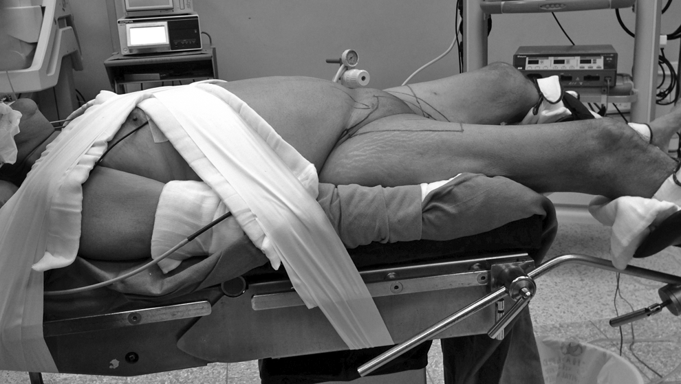
Position of the patient.
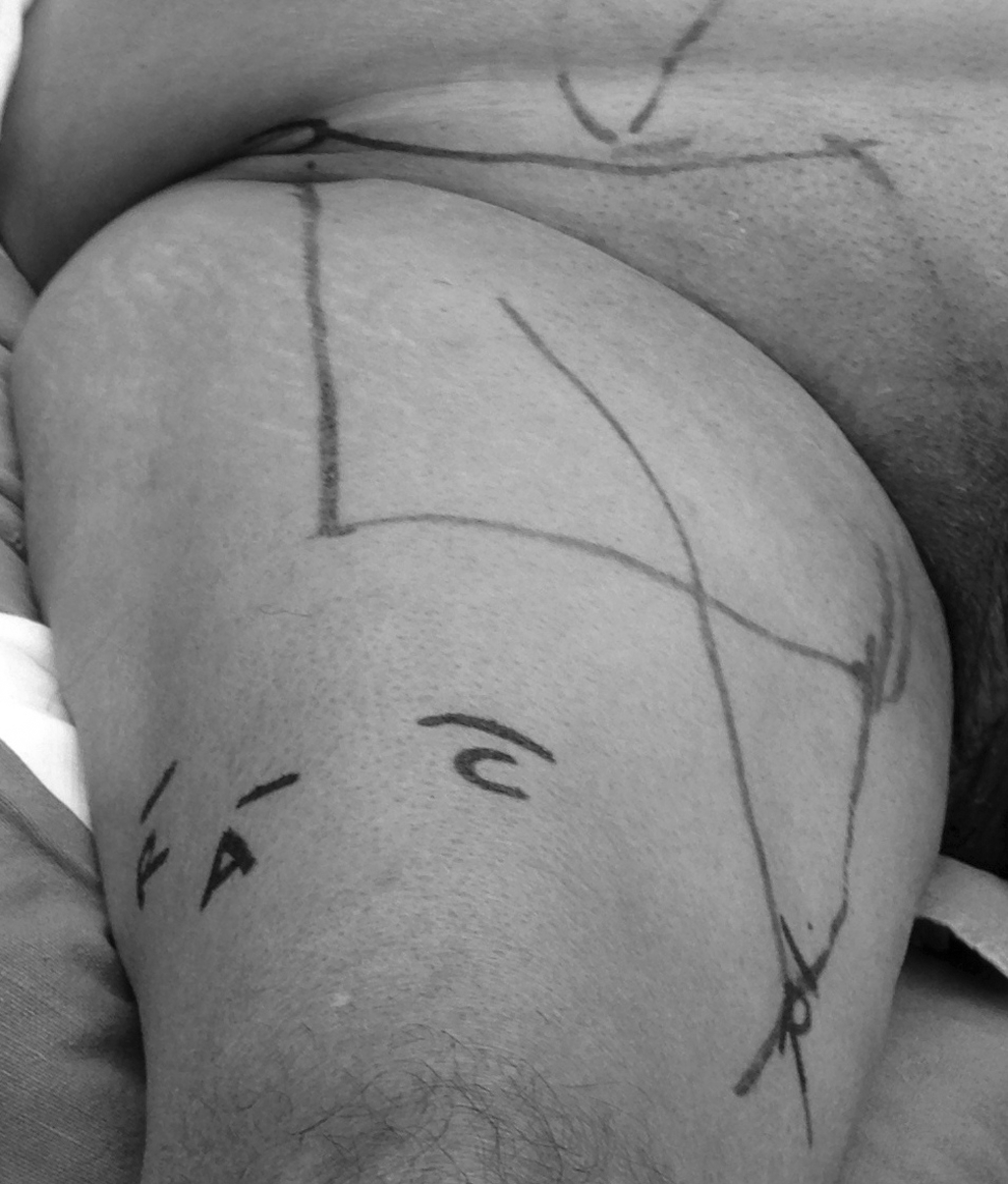
Marking of the template for dissection and placement of ports. A denotes assistant port, R denotes port for robotic arms, and C denotes camera port.

Docking of the robot.
Results
The intraoperative and immediate postoperative period was uneventful in all patients. Average operative time was 453.33 minutes, which include robot docking and console time (Table 2). Mean lymph node yield in left inguinal region, right inguinal region, left pelvic region, and right pelvic region was 18, 14.6, 13.3, and 16.6, respectively. No skin flap–related complications were seen. Lymphocele was seen in one patient postoperatively, which was managed successfully with needle aspiration. Intra-abdominal drain kept for pelvic lymphadenectomy was removed on POD 1 in all patients. Patients were discharged on POD 3 with suction drain in situ (inguinal region). We removed suction drain on outpatient basis when output decreased to 50 mL in 24 hours. Average days of last drain removal were 44.6 days. All patients received chemoradiation as adjuvant therapy. No patient had local recurrence, but one patient developed lung metastasis at 7 months of the follow-up period (Table 2).
I, Inguinal; L, left; LN, lymph nodes; P, pelvic; POD, postoperative day; R, right.
Discussion
Video endoscopic inguinal lymphadenectomy (VEIL) was described in the clinical arena around 10 years ago to duplicate the open template while reducing morbidity without compromising the oncological control. 1 This concept was proposed by Bishoff et al. who showed the feasibility by dissecting two cadaveric models in 2003. 6 In the clinical scenario, first case was successfully operated at ABC Medical School, Sao Paulo, Brazil in 2003. 7 Although VEIL surgery is technically demanding, more than 20 centers have reported their initial experience with similar technical success and improved morbidity rates. A recent study, which compared endoscopic groin dissection with traditional open surgery, reported high complication rate of open surgery (55.8%) compared to endoscopic dissection (7.1%). 8 In another study, Sotelo et al. reported no wound-related complication after 14 endoscopic inguinal lymphadenectomies in eight patients with clinical stage T2 squamous cell carcinoma of penis. 9 Similarly, no skin-related complication was demonstrated in 10 patients who underwent elective VEIL procedure for high-grade penile cancer with nonpalpable lymph nodes. 3 Many more authors had suggested that complications with VEIL were fewer compared to an open surgical procedure and this technique has a potential to reduce postoperative morbidity. In addition, patients who underwent open procedures reported significantly longer hospital stay and time to return to usual activities compared to those who underwent endoscopic procedures. 10 Even when patients were asked about how they felt about both surgeries, they elected the endoscopic approach as the less morbid of the two procedures. 11 So, patients' subjective preference also confirms that VEIL is an attractive minimally invasive technique.
Initial protocols of VEIL did not include patients with palpable inguinal lymph nodes with a fear that the lymph node fixity to femoral vessels would prevent its safe resection. 12 However, Carlos et al. demonstrated that VEIL was a safe complimentary procedure even in patients with palpable inguinal lymph nodes. The preliminary oncological results in these patients were found to be appropriate. 13 Many authors subsequently proved the feasibility of VEIL in N1 patients. 1 By performing a bilateral simultaneous procedure, it was thought that the morbidity (by decreasing operation room and anesthesiology time) would further decrease while maintaining oncological efficacy of conventional VEIL. Pompeo et al. first demonstrated the feasibility and advantage of simultaneous bilateral VEIL. 2 Although the procedure was performed by two different surgeons, there was no clashing between the surgical group. They were able to complete all VEIL procedures without conversion to open, and no complication related to VEIL was recorded. Another group performed bilateral endoscopic inguinofemoral lymphadenectomy using two carbon dioxide insufflators concurrently. They concluded that this approach required careful positioning of the patient, good surgical team, instrumentation, as well as special anesthetic consideration necessary to avoid severe hypercarbia. They found that bilateral simultaneous endoscopic inguinal lymphadenectomy was not only technically feasible but also was an efficient surgical approach. 14 In view of this established safety, we also endoscopically performed inguinal lymphadenectomy in patients with palpable inguinal lymph nodes and did it simultaneously on both sides.
Robot assistance in video endoscopic inguinal lymphadenectomy (RVEIL) can offer a distinct advantage to work in a confined space due to better ergonomics, superior view, and instrumentation. However, the applicability of the robot is newer in this field and only few series have been presented so far (Table 3).5,15–18 Josephson et al. first reported the applicability of robot in inguinal lymphadenectomy in a patient with carcinoma of penis. 15 Similarly, authors from India published their experience of bilateral staged RVEIL in two patients. 16 Sotelo et al. performed robotic bilateral inguinal lymphadenectomy simultaneously without robot repositioning and found it to be feasible and safe. 5
L, left; LN, lymph nodes; NA, not available; R, right; RVEIL, robot assistance in video endoscopic inguinal lymphadenectomy; Sup., superficial.
The key efficacy outcomes for any lymphadenectomy procedure, that is, adequate clearance of lymph nodes are well established for VEIL. Even the oncological adequacy of RVEIL had been prospectively evaluated. 18 Patients with T1-3N0 penile cancer were enrolled prospectively in a study and all of them initially underwent RVEIL. Verification of adequacy of dissection was performed by an independent surgeon through a separate open incision at the conclusion of RVEIL procedure. The author concluded that RVEIL could adequately stage disease in the superficial inguinal region among patients with penile cancer at risk for inguinal metastasis. 18 Similarly, in our series, the number of lymph node dissected out from inguinal region was equivalent to those reported in most of the open conventional or endoscopic series.
Pelvic lymph node dissection is traditionally performed as a staged procedure in patients with two or more positive inguinal lymph nodes, extracapsular nodal extension, or presence of poorly differentiated metastasis. 19 Around 12–20 nodes can be found in this area. 19 In the present series, patients had palpable inguinal lymph nodes on physical examination, which were mobile and less than 4 cm in size with the histopathological staging of penile/urethral carcinoma as T2 or more. Preoperative fine needle aspiration cytology from inguinal lymph nodes was positive. We had also sent the inguinal lymph nodes for frozen section of the contralateral side in the patient (case 2) with unilateral palpable inguinal lymph node. That is why, we performed robotic pelvic (bilateral) lymphadenectomy in the same setting as recommended. 20
Conclusion
Early results suggest that this approach is feasible, safe, and affords an appropriate oncological dissection in selected patients. It has also helped to reduce the hospital admissions or stay and in turn the cost of the Robot. Small sample size and a relatively short follow-up with the lack of long-term oncological outcome constitute the major limitations of the present study. Furthermore, operating time is long, which reflects the beginning of our learning curve.
Footnotes
Disclosure Statement
No competing financial interests exist.