
Abstract
Abstract
Background:
This study focuses on the successful application of three-dimensional (3D) laparoscopic surgeries in the treatment of congenital anomalies and acquired diseases in the young pediatric population. The purpose of this scientific work consists in highlighting the spectrum, indications, applicability, and effectiveness of 3D endosurgery in children.
Methods:
Our experience is based on 110 endosurgical procedures performed in neonates and infants in the 3D format between January 2014 and May 2015. Depending on the type of operations, all patients were divided into the following groups: (1) inguinal herniorrhaphy (IH)—63 patients; (2) Nissen fundoplication (NF)—22 patients; (3) pyeloureteral anastomosis (PUA)—15 patients; (4) nephrectomy (NE)—5 patients; and (5) ovarian cystectomy (OC)—5 patients. The patients of the first three groups were compared with babies who underwent standard laparoscopic surgery, performed in the two-dimensional (2D) format during the same time period. The groups were organized according to patient demographics, operative report, and postoperative parameters.
Results:
The patients were similar in terms of demographics and other preoperative parameters. There were significant differences in mean operative time between 3D and 2D procedures in the groups of patients with hydronephrosis and gastroesophageal reflux, which used manipulation with internal sutures (NF—37.95 minutes versus 48.42 minutes, P = .014; PUA—61.31 minutes versus 78.75 minutes, P = .019), but not in group after IH (15.88 minutes versus 15.57 minutes, P = .681). Postoperative parameters such as length of hospital stay and the number of complications were equivalent between groups.
Conclusion:
In this study, we demonstrated the success of 3D laparoscopy in small babies with inguinal hernia, gastroesophageal reflux, hydronephrosis, ovarian cyst, and multicystic kidney. Laparoscopy in 3D format lessens the duration of complex procedures, which utilize the use of the suture technique into the abdominal cavity. The perception of depth and the presence of tactile feedback make 3D laparoscopic surgery more acceptable when compared to traditional laparoscopy.
Introduction
L
However, conventional laparoscopy still has its limitations. The main weakness is its two-dimensional (2D) image. Another disadvantage of conventional laparoscopy is the lack of depth perception. This weakness has been eliminated with the advent of robot-assisted systems, which were the first to use the three-dimensional (3D) effect. 4
The high cost of equipment and the lack of tactile feedback are limiting factors of robotic technology. The modern 3D system offers most of the advantages of robot-assisted surgery during the performance of traditional thoracoscopic and laparoscopic surgeries with low cost and with the possibility to use conventional laparoscopic equipment. 5
However, 3D video systems are still not widespread in surgery, especially in the pediatric patient population. This trend is mainly due to the fact that the majority of pediatric surgeons are satisfied by a 2D picture of the image that exists during the performance of standard laparoscopy. Another explanation for this phenomenon is the lack of up-to-date equipment, which is necessary for performance of 3D laparoscopic procedures in children.
Pediatric surgeons are faced with the fact that available 3D telescopes have a diameter of 10 mm, which, due to existing opinion, are big for children. In this study, we summarized our own experience of performing laparoscopic surgical procedures in a 3D format in children and opened the veil on a yet unexplored area of application of this technology by demonstrating its benefits for the correction of certain congenital anomalies of the abdominal cavity in the early age group.
Materials and Methods
Our experience of performing endosurgical procedures in a 3D format in neonates and infants is based on the study of the results of 110 procedures completed in the Department of Newborn Surgery at the Irkutsk Children Hospital from January 2014 to May 2015. All patients were grouped according to the type of surgery (Table 1) and were organized in the following cohorts of patients: inguinal herniorrhaphy (IH)—63; Nissen fundoplication (NF)—22; pyeloureteral anastomosis (PUA)—15; nephrectomy (NE)—5; and ovarian cystectomy (OC)—5. All 3D operations were performed by a single surgeon with established background in standard pediatric minimally invasive surgery (MIS) procedures.
2D, two-dimensional; 3D, three-dimensional; IH, inguinal herniorrhaphy; n, number of cases; NF, Nissen fundoplication;
PUA, pyeloureteral anastomosis; SEM, standard error of mean; M, mean value; P, level of confident significance.
It should be noted that the basic steps of operations were exactly the same in the comparison groups. We used the following modern versions of the well-known surgical procedures:
• subcutaneous endoscopically-assisted ligation (SEAL) technique for the treatment of inguinal hernia; • NF with minimal dissection of the esophagus; • Hynes–Anderson pyeloplasty with use of simple interrupted sutures; • standard nephroureterectomy with separate clipping of blood vessels and ureter by the Hem-o-Lock clips; • hybrid method of resection of ovarian cyst after removal.
The modern view of performance of laparoscopic surgeries in 3D system for children is consistent with the choosing of optimal equipment and with implementation of practical skills that are necessary to perform this approach. Our preferences are based on own experience and related to the type of endoscopic equipment for performance of 3D laparoscopic procedures in children, which provides the maximum comfort to an endosurgeon:
• use of endoscopic video heads 3D TIPCAM (KARL STORZ GmbH & Co. KG) with two distal CCD image sensors, viewing direction 30°, a length of 31 cm, which allow the creation of an optimal 3D visualization of internal objects; • use of 3D monitors with diagonal of 32" (KARL STORZ GmbH & Co. KG) located on the optical axis of view of a surgeon and a minimum distance of 1.5 m from the eyes; • use of passive 3D glasses (KARL STORZ GmbH & Co. KG), working on the principle of circular polarization; • use of personal computer and 3D video converter for recording and storing 3D movies.
The need to delicately introduce 10 mm optical systems into the abdominal cavity was the most challenging. However, our own long experience of performance of open or single-port laparoscopic transumbilical procedures has convinced us that the use of the umbilicus as an “invisible” access for the installation of large devices is not accompanied by the additional risk of wound infection, formation of postoperative hernias, and cosmetic defects.
To overcome some of the inconveniences, we performed eversion of umbilicus to outward and cut the skin and fascia of umbilical region (Fig. 1). Further insertion of the optical system in the abdominal cavity did not cause difficulties. The presence of physiological umbilical hernia in youngest patients helped to free installation of the telescope. Sealing of the umbilical incision was carried out by applying of 2–3 aponeurotic sutures.
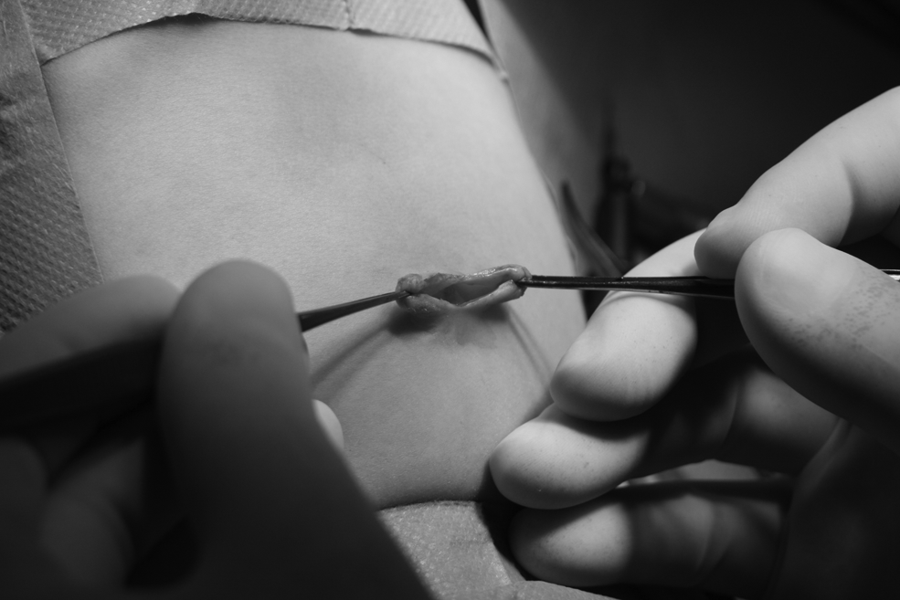
Umbilical eversion for insertion of 10 mm three-dimensional optical system.
The patients of the first three groups in which the number of participants in a cohort was greater than 10 were compared with children who were operated upon during the same period of time by the use of a standard 2D laparoscopy (IH—72 patients; NF—26 patients; PUA—12 patients). Thus, the statistical comparison was performed in patients who underwent IH, NF, and PUA (Table 1). We analyzed the preoperative parameters of small children (weight and age), and intra- and postoperative data of patients (duration of the operation, number of the doses of postoperative analgesia, length of hospital stay). The Mann–Whitney U test was used for calculation of average values. The level of confident significance was set as P < .05.
Results
Before surgery, the comparison patient groups were homogeneous for a number of demographic indicators (Table 1). Comparative analysis showed no significant differences in body weight and age in patients before 2D and 3D laparoscopy (P > .05).
The study of intra- and postoperative parameters showed the differences in some comparison patient groups (Table 2). The study demonstrated the presence of changes in the duration of procedures in patients after NF and PUA (NF—37.95 minutes versus 48.42 minutes, P = .014; PUA—61.31 minutes versus 78.75 minutes, P = .019). However, we found no difference in the duration of surgical procedures in the group where patients underwent IH (15.88 minutes versus 15.57 minutes, P = .681). In our opinion, the revealed phenomenon was caused by a surgeon's better perception of the placing process of endosurgical sutures.
2D, two-dimensional; 3D, three-dimensional; DA, doses of analgesia; DO, duration of operation; IH, inguinal herniorrhaphy; LHS, length of hospital stay; n, number of cases; NF, Nissen fundoplication; PUA, pyeloureteral anastomosis; SEM, standard error of mean; M, mean value; P, level of confident significance.
Postoperative parameters of small children in comparison groups (Table 2) did not differ on the number of doses for analgesia (IH—1.22 versus 1.24, P = .864; NF—3.41 versus 3.27, P = .441; PUA—3.46 versus 3.58, P = .852) and length of stay in a surgical hospital (IH—9.27 hours versus 8.0 hours, P = .120; NF—6.5 days versus 6.92 days, P = .454; PUA—3.33 days versus 3.58 days, P = .299). These data confirm the idea that the minimally invasive procedures performed through the 1 cm umbilical incision, which was required for installation of the 3D optical system, are accompanied by pain and length of postoperative rehabilitation of patients that are comparable with patients who underwent installation of 3 and 5 mm laparoports for 2D laparoscopy.
All endosurgical interventions were performed without conversion to conventional 2D laparoscopy or laparotomy. Early postoperative periods after any format of laparoscopic procedures were uneventful in all patients. We did not record the cases of umbilical wound infection, which could be prone to this complication, because of its depth and difficulties of care. Subsequent examinations of the patients showed no late postoperative complications and recurrence of disease in all children.
The remote monitoring of patients for at least 6 months after surgery revealed the reliability of the basic element of 3D laparoscopy in children—ultrasmall umbilical access, which was confirmed by the absence of postoperative umbilical hernia. High aesthetics of only one incision that is hidden in the depth of the umbilical ring, demonstrated excellent cosmetic results of 3D laparoscopy. As a result, no visible scars on the patients' bodies after the installation of 10 mm devices.
The first laparoscopic surgeries in 3D have been associated with the difficulties of adjusting to a 3D picture image, wearing glasses, and adaptation of previous experience of traditional laparoscopy to the new and improved technical conditions of work in the abdominal cavity. However, after accumulation of experience, subjective feeling left behind, and development of new endoscopic skills led to a preference for the use of 3D laparoscopy for correction of a number of anomalies of abdominal cavity.
Accomplishment of the learning curve was associated with improved outcomes such as decreased operation time. Because the accomplishment of 3D operations in group was discordant, we performed subgroup analyses. Our data show that following the 5th patient from IH group, the operative time decreased by more than 16 minutes. The operative time after NF continually decreased, with the most dramatic reduction achieved following the 10th patient till 40 minutes. We did not design a learning curve at patients with hydronephrosis because of the small number of participants. Thus, overcoming the learning curve is manifested by decreased operative time in two subgroups enough patients and also confirmed our assumption of the fast period of mastering of 3D technologies.
We did not note any additional inconvenience during endosurgical procedures such as eye strain, dizziness, blurred vision, double vision, and nausea, which may be associated with the appearance of new visual experiences. The sense of depth and tactile feedback improved the perception of internal organ anatomy and ensured the progress in the implementation of complex endosurgical skills: suturing and knotting.
Discussion
Traditional laparoscopy is based on 2D images, displayed on a conventional flat screen that requires a surgeon to use additional visual skills to judge the position of an instrument in the abdomen and the depth of its impact. This limitation is a significant problem due to the increased needs in special precision of manipulations performing by endoscopic surgeons.
Progress in imaging technologies for laparoscopy allows the removal of existing obstacles. In the early 1990s, there was a boom of 3D video systems in laparoscopic surgery. At least, several manufacturers offered a device for 3D visualization in medicine after the first appearance of 3D video for laparoscopy, which was first developed in the Nuclear Research Center in Karlsruhe. 6 At that time, the 3D format was unable to establish itself in clinical practice due to technical and ergonomic shortcomings of 3D monitors that had a standard resolution. Currently, 3D displays have been improved, and now they provide the ability to use high-definition television.
Principles of 3D visualization in endosurgery
Depth perception is the visual ability of a human to judge the distance of objects and spatial relationships of objects that are located at different distances. How a 3D world is projected onto a 2D retina and how this view provides information about the depth will be clear after further discussion on this topic.
Stereoscopic vision is most important for the perception of depth. It occurs due to the distance between eye pupils, which enables each eye to have a slightly different view of the same scene. Only brain can then merge the two images into a single 3D image in the so-called process of binocular vision. Laparoscopic surgeons who perform their work using a conventional 2D image, in fact, work with one eye closed. This is one of the reasons that 2D laparoscopy leads to emotional stress and headaches.
A 3D system has two chambers and two optical lens systems that transmit two mutually offset images onto a 3D monitor. When a surgeon wears 3D glasses, the two images merge into one, and there is a sense of depth. It also provides a more precise location of the imaging organs in the abdominal cavity, allowing more successful manipulation and suturing in real time due to tactile feedback.
It should be briefly mentioned that wearing 3D glasses is only the first step to getting the maximum benefit from the extra dimension (Fig. 2). The key to understanding visual ergonomics, which exists when performing laparoscopy in 3D, is the capture of a small image of the abdominal cavity and displaying it magnified on a 3D monitor in the operating room. The distance from the eyes to a screen is from 0.5 to 5 m. This is 12 times greater than the distance between the front lens of a laparoscope and the object of the procedure.

External view on OP team while performing three-dimensional laparoscopic operation.
The width and height of the objects, which are equivalent to the size of the surgical field captured by a laparoscope, are transferred to a 3D monitor. The transfer of image depth, and therefore the establishment of a realistic view, is determined by features of the 3D camera that captures and connects the left and right half of the image. There is the concept of “comfort zone” of binocular vision for 3D images 7 —the objects that are too close and those that are located too far away are the diverging lines of sight and they form on the retina the fields of “rivalry” (B. Mendiburu calls them as “painful areas”), which should be avoided. The “comfort zones” in the 3D device adjusted so that the images that are close to the point of our interest and the images that are far away from this point match each other while watching.
3D visualization in robot-assisted surgery
The advantages of robotic surgery are due to largely 3D visualization. The technical superiority of these systems is also in increased degrees of freedom to work with tools and elimination of tremor. These properties of robot-assisted surgery can improve the quality of manipulation during laparoscopic procedures. The robot offers the potential benefit in promotion of a number of minimally invasive procedures, especially in complex narrow fields of surgery, such as laparoscopic prostatectomy or reconstruction of heart valves.
Some authors believe that the robot technique can be a useful method to reduce the learning curve in some sections of endosurgery. 8 However, other researchers have found that the learning curves for the series of operations with using of conventional laparoscopy, for example, in gynecology, were significantly lower than those reported for surgeries, in which robotic technology was used.4,9
It is the established opinion in gynecology that robotic surgery should not be used as a substitute of traditional laparoscopy, especially in cases where laparoscopy is a daily standard. 10 A survey of women revealed that they preferred the traditional laparoscopic approach to the robotic approach when undergoing endosurgical intervention. 11
A major disadvantage of robotic surgery is the lack of tactile feedback, and the inability to change the configuration of an operating table after the robot arm is attached. 12 Another major constraint is the cost of the da Vinci device, which is from $ 1.0 million to $ 2.3 million in the United States, depending on configuration. Related annual cost to maintain the complex is about $ 180 thousand. 13 For comparison, the total cost of 3D laparoscopic system is $ 250 thousand and the annual cost of maintenance is $ 25 thousand.
Thus, robotic surgery is an innovative and cutting-edge 3D endosurgical technology. However, it should be understood that use of robotics in cases where traditional laparoscopic approaches can achieve the same clinical results and accompanied by lower costs is doubtful. The question to be answered in the near future, “What helped to improve the quality of surgery: the use of robotic technique or the 3D image on the screen?”
Comparison of 2D and 3D laparoscopy
Today, there are a limited number of studies about the comparison between the procedures performed with using of 3D video systems and surgeries performed with using of 2D devices. Three hundred forty articles have been analyzed in the recent systemic scientific review, where 3D laparoscopy was discussed. 14 This report included 31 randomized clinical trials, only 3 of them were clinical and the others were conducted on simulation models. The duration of surgery and number of errors were used to evaluate the outcomes of these procedures. Approximately 71% of the studies showed a reduction in the duration of surgeries and 63%—significant decrease in errors when 3D laparoscopy was performed.
The question about the superiority of 3D laparoscopic systems over 2D systems is still unresolved, although the potential benefits of 3D visualization are well known by now. These benefits include improved performance due to appearance of spatial depth measurement and tactile feedback, which are missing in 2D laparoscopic and robotic surgeries. The quality of 3D laparoscopy was assessed during the performance of endosurgical procedures, as well as on the basis of simulation and experimental models.
A limited number of clinical studies were presented with comparison between 3D and 2D laparoscopies. Initially, scientific examinations that were aimed at investigations of advantages and disadvantages of 3D systems revealed conflicting results. Some of them reported that 3D visualization significantly improved the performance of a surgeon,15,16 others claimed equivalence of the results of operations and could not demonstrate any superiority of 3D visualization to resolve a variety of laparoscopic tasks.17,18
However, it should be noted that these findings were made when working with the early generation of 3D systems.5,19 Subsequent clinical comparisons have shown that surgeries performed by the new generation of 3D systems needed less time than traditional. This statement was supported by the study conducted by Wagner, 3 in which he showed that labor productivity of a surgeon can be increased by 60%–70% with use of a 3D imaging system. A recent scientific review showed that 3D technology significantly reduces the duration of laparoscopic cholecystectomies. 20 In our study, the duration of 3D operation for complex procedures (NF and PUA) was significantly shorter than traditional 2D surgeries (NF—37.95 minutes versus 48.42 minutes, P = .014; PUA—61.31 minutes versus 78.75 minutes, P = .019), but not in subgroup after relative simple procedure of IH (15.88 minutes versus 15.57 minutes, P = .681). Therefore, this new technique may be of additional success in complex pediatric endosurgical procedures.
Some studies showing the superiority of a 3D system were performed with the usage of simulation and experimental surgical models.21,22 The comparison of 2D and 3D visualization on the simulation models has limitations due to a lack of standardized skills assessment system for laparoscopic surgery. Several years ago, the Society of American Gastrointestinal Endoscopic Surgeons (SAGES) developed a comprehensive program, “Fundamentals of Laparoscopic Surgery” (FLS), taking into consideration the didactic and manual skills for evaluation of basic laparoscopic skills.
The effectiveness of this program has been confirmed in numerous studies evaluating the qualifications and training of surgeons in laparoscopy.23,24 Recent studies have shown that 3D imaging accelerates the solving of basic tasks of the FLS program, mainly for the more difficult assignments3,25–28 and for acquisition of skills by endosurgical beginners.25,28 Despite the benefits provided, there are some reports about adverse effects of 3D visualization such as dizziness and eye fatigue of a surgeon. 29
3D laparoscopy in pediatric surgery
Despite growing popularity, 3D laparoscopy is not widely used in pediatric surgery, especially in neonates and infants. Most surgeons categorically deny the possibility of performing laparoscopic surgery in 3D format in children and explain their decision by saying that the optical system for 3D laparoscopy is “massive” for a child's body.
However, the introduction of a 3D telescope through an elastic and pliable umbilicus, used as a “keyhole” for performing a number of transumbilical open and laparoscopic surgeries, allows to completely eliminate a scar from the abdominal wall. The additional benefit of slightly increased umbilical incision is that it can be easily transformed into advanced transumbilical incision, for example, for performing “hybrid” procedures that combine the principles of open and laparoscopic surgeries.
Scientific publications about 3D laparoscopy in children are rare. There are few reports about use of 3D laparoscopy in children: in 22 pediatric patients in Tübingen (Germany) 30 and in 48 pediatric patients in Russia. 31
It is reasonable that reporting about any of new technique, it is adopted to demonstrated the learning curves of this innovation. After acceptance of new operations begins the period of the studying characterized by worse results which decrease with experience. In pediatric surgery, case volume and frequency of diseases are low and forming of curves demands a long time to reach a plateau. In our study, two groups of patients (NE, OC) have been excluded from an assessment. It is the first limitation of our research. Another limitation—diverse groups of patients. Discrete laparoscopic methods do not allow to construct effective curves. In this study, we have presented the cohorts of patients (3D and 2D), including various types of simple (IH) and difficult (NF, PUA) operations. Only in two subgroups we could construct learning curves—63 patients after IH and 22 patients after NF. We suggest that only one measure may be universal—operative time and this is applicable to any minimally invasive procedure in any patient population. However, the IH procedure is easy and therefore the time of the development was short—after five operations we have reached a plateau. It means that we expect to overcome the learning curve after five surgeries for inguinal hernia. NF is a more difficult operation. For this reason, a plateau has been reached on the 10th patient. This indicates that only 10 surgeries are needed for a trained minimally invasive surgeon to complete the learning curve of NF in 3D format. Reasonings on production of curves in a series of patients after PUA are premature and we need the extra time of accumulated experience to construct schedules for this operation in 3D format.
On the basis of our own scientific results, we are inclined to conclude that 3D laparoscopy is feasible in children and it is an excellent alternative to traditional laparoscopic surgery in treatment of many diseases, including those in small children. In our opinion, some advice that has appeared in the course of accumulation of our personal experience is important for the initial mastery of skills in 3D laparoscopic surgery:
• all medical monitors have a lower brightness in 3D mode than in 2D mode. Thus, an operating room should be dimmed to increase your visibility of 3D images; • if visibility becomes poor, a surgeon should try to increase the power of the light source; • in case of image deterioration, from time to time a surgeon should separately close left and right eyes, to check contamination of one of the optical channels and, if necessary, to clean it; • in 3D mode when the object is too close to the frontal lens, the picture may be blurry. Correction of 3D visualization is necessary to slightly remove a laparoscope to outward or temporarily use the 2D mode.
Thus, the use of 3D laparoscopy in the pediatric population begins its story. The prejudice related to the fact that the existing optical system is too large for children is unreasonable, given the potential of child umbilicus to hide a scar after surgery. In addition, 3D technology in an incredible way changed the depth perception of child's internal organs and preserved the tactile feedback, which is absent in robotic devices, and improved the skills of talented surgeons.
The main question of whether 3D endosurgery in feasible in children in general, as well as a safe technology compared to traditional laparoscopy, was confirmed by the results of our study. Restoring depth perception allows more precise and faster dissection, suturing, and knotting even in narrow operating spaces such as subdiaphragmatic and retroperitoneal spaces. No conclusions should be drawn with regard to the superiority of 3D over 2D vision in pediatric endosurgery because of the nonrandomized setting and the small number of patients. Supported by the results, we conclude that 3D vision can be safely changed to conventional 2D pediatric endosurgical procedures without adjusting current surgical protocols.
Conclusion
In the last few years, 3D movies made an incredible amount of money. To make the movie “Avatar,” J. Cameron took 10 years. This is a courageous and impressive film that inspired many, and not just artists. 3D laparoscopy is also a courageous and impressive work of engineering, which improved the quality of modern surgery. Since its emergence, surgeons were delighted by its method and each surgical discipline is trying to find an appropriate role for it.
In our study, we demonstrate the ability to perform 3D laparoscopic surgery in neonates and infants with inguinal hernia, gastroesophageal reflux, hydronephrosis, nonfunctioning kidney, and ovarian cyst. 3D laparoscopy allows to decrease the duration of complex operations, where endosurgical stitching was used.
In our view, 3D laparoscopy is one of the most promising directions of development of pediatric endosurgery. The quality of 3D image improves the accuracy of manipulation and eye coordination, accompanied by a decrease in both capital and annual equipment costs.
We hope that the development of 3D optical technology will allow to overcome some of the disadvantages of modern 3D devices for laparoscopy and will raise the levels of skill of a surgeon. It should be noted the importance of conducting randomized controlled trials to test the effectiveness of 3D laparoscopy in children to determine its role in pediatric minimally invasive surgery.
Footnotes
Disclosure Statement
No competing financial interests exist.