
Abstract
Abstract
Purpose:
To show the benefit of three-dimensional (3D) reconstructions of preoperative imaging for surgical performance.
Methods:
A laparoscopic training environment with 15 hidden lymph nodes was designed. Three of them were marked with radiographic contrast agent and were only distinguishable from unmarked nodes via CT imaging. Thirty-six surgeons were divided into two groups. To group 1 the unprocessed CT data were shown. Group 2 was additionally shown a 3D reconstruction of the anatomy. Time of studying the imaging was recorded. All surgeons had to find the three target lymph nodes laparoscopically. Time to fulfill this task and errors was measured. Afterward, the 3D reconstruction was also shown to group 1. Then, all participants completed a questionnaire. Furthermore, 3D reconstructions were used in 15 clinical cases of partial nephrectomy or lymphadenectomy, and surgeons' opinion was evaluated with an additional questionnaire. The imaging and 3D reconstructions were available on a mobile device.
Results:
The time of studying the imaging to gain confidence was significantly shorter with the 3D reconstruction. Laparoscopic intervention time was shortened and errors were reduced significantly within group 2. The clinical application of 3D reconstructions in difficult cases was believed to be helpful.
Conclusions:
3D reconstructions of preoperative imaging lead to better surgical performance in a difficult laparoscopic training environment. Surgeons gain a 3D impression of patients' individual anatomy easier, faster, and more reliable. Providing 3D reconstructions previous to surgery should be routinely implemented for patients with complex anatomical situations.
Introduction
L
In this study, we evaluate the benefit of 3D reconstructions of preoperative imaging to surgical outcome. The transfer of these 3D reconstructions, containing relevant anatomical structures, on mobile devices allows surgeons to study the specific anatomic conditions directly in the operation theater. In this article, we present a controlled experimental study to evaluate the benefit on surgical success of these applications.
Material and Methods
The study consists of an ex vivo and an in vivo phase. For ex vivo evaluation, we used a custom-designed laparoscopic training environment within a pelvitrainer containing silicone-based organs and vessels (Fig. 1). Fifteen nodular structures (1–2 cm) simulating lymph nodes were randomly placed in between the organ models. Three of these nodular structures were marked with radiographic contrast agent and were thus only distinguishable by CT imaging. These three lymph nodes were defined as targets to be identified during laparoscopy. The whole scenery was covered by an intestine model consisting of tube-shaped balloons, 2–3 cm in diameter, and filled with water (Fig. 1).
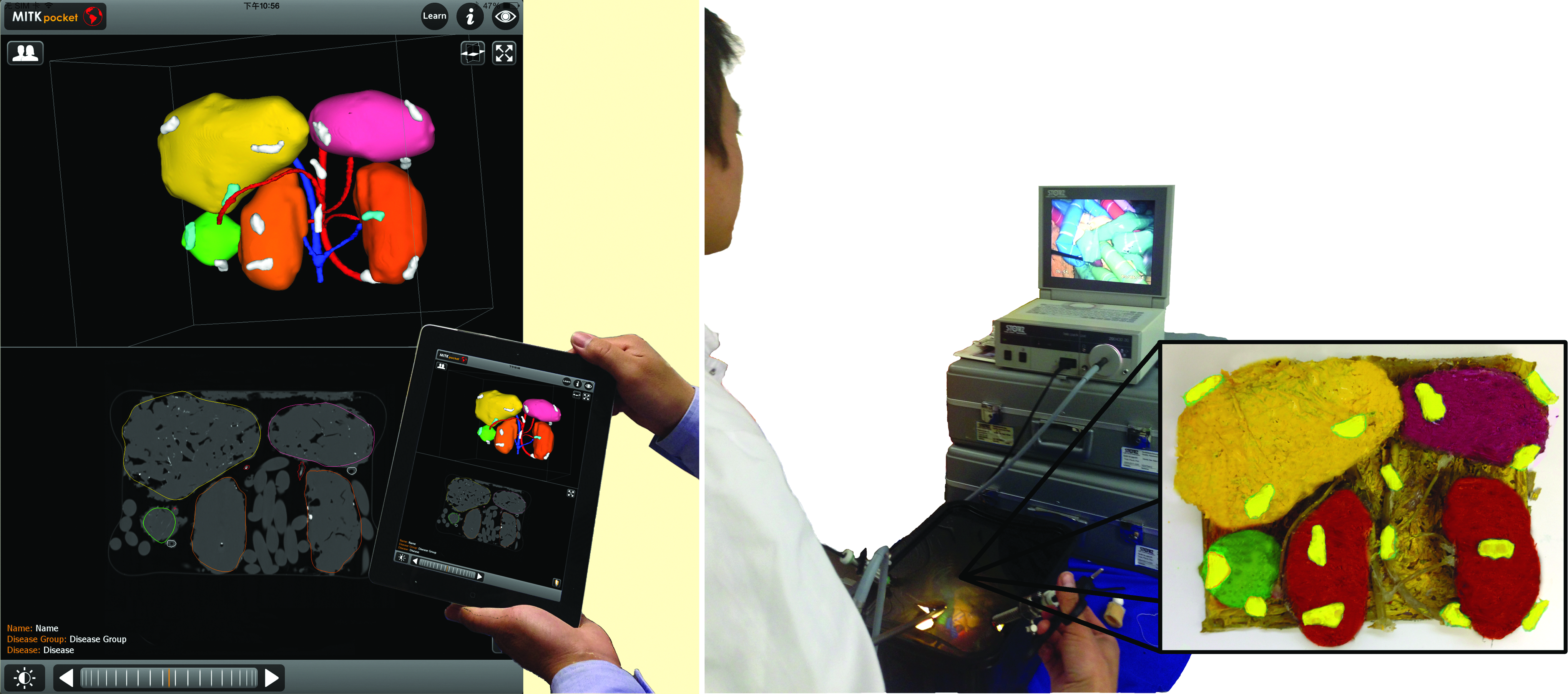
Left: Study setup—screenshot of mRay showing the 3D reconstruction (top) of the CT data in a coronal view (bottom). Vessels (blue/red), kidneys (orange), liver (yellow), spleen (pink), tumor (green), and lymph nodes (white = without contrast agent, turquoise = with contrast agent-enhanced targets) are segmented. The intestine model is not segmented and not displayed in the 3D view. Right: After studying the imaging, the surgeon (here author Z.L. is visible) is searching for the targets laparoscopically. Inside the pelvitrainer, the silicone model (schematic colored illustration inset right) is placed and covered with an intestine model.
A high-resolution CT scan of the pelvitrainer was performed and imported in the software MITK (Medical Imaging Interaction Toolkit, www.mitk.org; German Cancer Research Center, Heidelberg, Germany). 5 By semiautomatic segmentation, a detailed virtual 3D model of the custom anatomy was elaborated. The processed database was conducted to the software mRay (www.mbits.info; mbits GmbH, Heidelberg, Germany) available for Android or Apple iOS mobile devices. The mobile device in this study was an iPad third generation (Apple, Inc., Cupertino, CA). With mRay, users can easily switch from any plane view to multiple plane view or to 3D view. While the used mobile device is only able to show a 2D perspective of the 3D reconstruction, the 3D impression arises from motion (e.g., rotating the 3D reconstruction). The interactive input of the surgeon's finger on the touch screen is intuitive and allows its use also for unexperienced users. Data were stored encrypted, and access to the imaging data bank was available via WiFi. A total of 36 surgeons, either trained in urology (n = 14) or general surgery (n = 22), were randomized into two groups. The first group (group 1) included 19 surgeons who were only allowed to study the unprocessed CT data. The second group (group 2) included 17 surgeons who additionally were provided with the virtual 3D reconstruction. The groups were equal concerning the age and level of laparoscopic surgery experience.
Each surgeon was provided with a manual, explaining the procedures of the whole task, as well as a short standardized demonstration of the hardware and software.
First, the surgeons studied the imaging with or without 3D reconstruction according to its group. The time needed to be confident with the anatomy was recorded. Immediately afterward, laparoscopy was started to fulfill the task of identifying the three radiographic marked lymph nodes. When the surgeon claimed that he had found one out of the three target nodes, instant verification (correct or false) was given. The time until all target lymph nodes were found or the surgeon quit was recorded. The number of erroneous chosen targets was counted. Afterward, the 3D reconstruction was also shown to group 1.
Finally, both groups had to answer a specific questionnaire using a ranking scale from 1 to 5, while 1 meaning “definitely not” and 5 “absolutely.” The questions for both groups were as follows. (1) Was the visualization method of your group helpful to achieve a good impression of the anatomy? (2) Was the imaging according to the visualization method of your group easy to understand and able to speed up the surgery? Three additional statements had to be rated by group 1 as follows. (3) If I had the 3D visualization before, surgery would have been much better/faster than it was. (4) The 3D visualization would have been helpful to achieve a better impression of the anatomy. (5) The anatomy would have been easier to understand and would have speeded up the surgery with the mobile device.
Statistical analyses were performed with the Statistical Package for Social Science 18.0 (IBM Corporation, Armonk, NY). P < .05 is considered to be statistically significant adopting the paired two-sided Student's t-test.
For the in vivo phase, we evaluated the visualization method in a clinical setting. Fifteen consecutive urological cases scheduled for lymph node dissection or partial nephrectomy of endophytic tumors were chosen for a demonstration of the technique. The only requirement for patient selection was that intraoperative localization of the targets was assumed to be difficult. Patients' imaging received detailed 3D reconstruction of relevant structures. The surgeons were not the same who participated in the ex vivo study. They were asked to study the imaging right before the surgery in the midst of the operation room on the mobile device. After intervention, a short questionnaire was completed, using the same ranking scale named above. The questions were as follows. (1) How intuitive is the interaction with the software? (2) How helpful was the method for the intervention? (3) Do you think the impression of the anatomy is better after studying the 3D reconstruction on the mobile device? (4) Do you think you have found the target(s) easier/faster/better after studying the 3D reconstruction? (5) Will you use the mobile device in future?
Results
As shown in Table 1, there was no difference in median age or experience level between both groups. The time until surgeons were confident with the anatomy was significantly faster in group 2 (310 seconds versus 114.1 seconds; P < .001). Also, total laparoscopic intervention time was shortened significantly with the 3D reconstruction (373.8 seconds versus 229.1 seconds; P < .001). Five surgeons, all in group 1, quit before they found all targets. Significant less errors were made by group 2 identifying the targets (2.3 targets versus 1.3 targets; P = .0047). The results of the questionnaire are shown in Table 1. The two questions asked to both groups were scored with significantly higher scores by group 2 using the 3D reconstruction as follows: (1) it helped to achieve a better impression of the anatomy (P = .0012), and (2) the imaging was easier to understand and able to speed up the surgery (P = .002).
Significant difference, brating from 1 (definitely not) to 5 (absolutely), data represent means.
The time of creating the 3D reconstructions in the 15 cases of the in vivo study, done by experienced staff, was 4–15 minutes (6.8 ± 3.2 minutes; mean ± standard deviation). The results of the questionnaire, asked after intraoperative demonstration of the 3D reconstruction, are illustrated in Table 2. To all surgeons the software was intuitive to interact (Rating 4 and 5; Question 1). Eighty percent of the surgeons believed that the method helped to perform the intervention (Rating 4 and 5; Question 2), whereas 20% were undecided (Rating 3; Question 2) or disagreed (Rating 1 and 2; Question 2). 93.3% thought that their impression of the anatomy was better with 3D reconstructions (Rating 4 and 5; Question 3); no surgeon disagreed, but one was undetermined (Rating 3; Question 3). Finding targets easier/faster/better with 3D reconstructions was believed by 80% of surgeons (Rating 4 and 5; Question 4). Further use was demanded by 86.7% of the surgeons (Rating 4 and 5; Question 5); no one declined, while 13.3% were undecided (Rating 3; Question 5).
Rating from 1 (definitely not) to 5 (absolutely).
Discussion
Modern medical imaging allows for subtle diagnostics and reveals high-resolution and detailed images of the anatomy. However, the transfer of the image data to the surgical field is a mental task to be mastered by the surgeon. Its success depends on inconsistent, uncontrollable, and unreliable factors such as the surgeon's knowledge, skill, or stress level. However, a reliable transfer of patient's anatomy from radiology to surgery might be crucial, as it allows safe and fast intraoperative targeting of lesions and risk structures. This may therefore reduce operation time, complication risk, and trauma. To bypass the source of human errors while transferring image data to intraoperative anatomy, computer-assisted surgery systems aim to provide surgeons with additional visual information of pre- or intraoperative imaging. However, especially in soft tissue surgery, due to organ deformation and shifting landmarks, these systems have not reached generic use, yet.6,7
In the meantime, other methods are used to improve surgeons' 3D impression of complex anatomical structures. One example, volume visualization of CT imaging, is applied since more than one decade. Defined intervals of gray values are visualized using specific color and opacity. Especially, contrast-enhanced vessels or bones can be visualized clearly and relieve the spatial relation better than transversal 2D CT images. 8 However, tissues of lower contrast changes are still lacking of clear discrimination by means of volume visualization. Therefore, risk structures in soft tissue surgery often have to be defined separately and manually. The creating of 3D reconstructions that only contain structures of interest allows for tailored imaging. Mitterberger et al. compared volume visualization and manually created 3D reconstructions in its use previous to adrenal-sparing surgery. 9 They concluded that 3D reconstructions provide more accurate information.
As a consequence, individual 3D printing models were fabricated. Using these models, different groups describe benefits for the hands-on experience and surgical planning, which lead to improved surgical dissection. 10 The 3D printing models presented by Igami et al. were used for surgical planning in two cases of patients with residual small liver metastases after chemotherapy. 11 They stated this method as a good alternative to ultrasonography, where small lesions were not always visible. They assumed that it improved the feasibility in these cases. However, the use of 3D printing models is limited by the cost, the speed of production, and necessary postproduction steps. Moreover, no accurate method of measuring outcomes compared with the current practice was proposed by the authors using 3D printing models.10,12,13
By creating an ex vivo environment mimicking a difficult anatomic scenario for laparoscopic surgery, we generated the conditions for a controlled study. Surgical success was measured by finding three previously radiographic marked lymph nodes comparing the effect of preoperatively studying unprocessed CT data or CT data combined with its 3D reconstruction.
Our findings show a clear benefit of the use of 3D reconstructions previous to surgery, concerning time consumption as well as surgical success. The time needed to be confident with the spatial relationship of the structures was reduced significantly, if 3D reconstruction was available. Furthermore, solving the given task of finding all lesions was also faster and associated with significant fewer mistakes. In this study, surgeons of group 1 took almost three times longer for studying the imaging. They needed longer to find all targets and were more often wrong. This implies that a visualized 3D reconstruction leads to a better memorization of the imaging by the surgeons. The data show that in difficult anatomic scenarios, there is a need for 3D reconstruction before laparoscopic surgery, as it improves the surgical success. Whereas studying unprocessed imaging data seems to be less sufficient.
Similar to our findings, Komai et al. also could find significant improvement in surgical performance using 3D reconstructions of preoperative imaging during difficult surgical procedures. 14 Furthermore, our ex vivo results are in concordance to the impression of the surgeons in the in vivo study, who stated in the postinterventional questionnaire to profit from visualized 3D reconstructions instead of having the CT image alone.
The acceptance of new devices or technology pushed into surgical workflow depends on many factors. The system has to relieve the surgeon relevantly and the benefits have to be clear. It has to be intuitive to use, safe, and handy. The system used in this study seems to meet these requirements. After using it on a trial base in clinical routine, no negative feedback was given and future use was demanded.
Conclusion
3D reconstruction of preoperative imaging is effective in a difficult laparoscopic environment. It allows for an easier, faster, and more reliable way of gaining a 3D impression of patients' individual anatomy. This leads to a more accurate and therefore faster intervention. The adoption of the system to real surgery may also offer these advantages. Furthermore, the effect may be strengthened by the integration of mobile devices into clinical routine due to their ability for intuitive and unbounded application. Further application and evaluation within clinical routine have to be awaited.
Footnotes
Disclosure Statement
No competing financial interests exist.