




Abstract
Abstract
Background:
Open surgery is a preferred treatment for primary obstructive megaureter (POM) in cases where the conservative treatment fails, with reported success rates of 90%–96%.
Objective:
To describe our initial experience in the treatment of POM by laparoscopic-assisted extracorporeal ureteral tapering repair (EUTR) and laparoscopic ureteral extravesical reimplantation (LUER) by following Lich Gregoir technique as an alternative to open surgery.
Design, Setting, and Participants:
A total of 7 patients with POM underwent laparoscopic-assisted extracorporeal ureteral tapering repair and ureteral extravesical reimplantation by following Lich Gregoir technique between 2011 and 2014. Postoperative follow-up included the following: Renal and bladder ultrasound, voiding cystourethrogram (VCUG), and mercaptoacetyltriglycine (MAG3) renogram were done at 6 months.
Outcome Measurements and Statistical Analysis:
Statistical analysis was performed by using the SPSS software package (version 15.0; SPSS, Chicago, IL), and P < .05 was considered statistically significant. Paired tests and Wilcoxon test were performed to compare pre- and post-measures.
Results:
LUER and EUTR were completed successfully in all patients without conversion. A postoperative MAG3 renogram showed nonobstructive pattern in all patients. Statistical analysis revealed significant differences before and after surgery in the average time of elimination on the MAG3 renogram (T½ 59.10 minutes versus 13.57 minutes, P < .0001). After medium-term follow-up, the overall POM resolution was 100%. One case of vesicoureteral reflux (VUR) was found during VCUG control. A total of 7 patients were asymptomatic without recurrence of POM.
Conclusion:
Laparoscopic-assisted extracorporeal ureteral tapering repair and ureteral extravesical reimplantation by following Lich Gregoir technique for POM constitutes a safe and good option when the first line of treatment fails, with a success rate similar to the open procedure. Nevertheless, larger randomized prospective trials and long-term follow-up are required to validate this technique.
Introduction
P
The guidelines of British Association Paediatric Urologists (BAPU) propose that an initial differential renal function (DRF) below 40%, or a drop in DRF of 5% on serial scans, and an increasing dilatation on serial ultrasound (US) scans are considered suggestive of obstruction. 2
Regarding morphologic appearance of POM, the Pfister-Hendren classification established three types of megaureters: Type I involved the distal ureter without associated hydronephrosis; type II extended to both ureter and pelvis; and type III was associated with severe hydroureteronephrosis and ureteric tortuosity.3,4
Around 80% of perinatally detected megaureters are spontaneously resolved.5–7 Conservative treatment appears to be the current option for an initial treatment.2,8 Nevertheless, when increasing hydronephrosis on a US scan, a decrease of renal function, urinary tract infection (UTI), and stone formation are observed, and surgical correction should be recommended.8–10
The surgical objectives of ureteroneocystostomy are the same as for nondilated ureters. Distal ureteral tailoring is often necessary to achieve an adequate length-to-diameter ratio that is required for successful nonrefluxing reimplantation. The tailoring of megaureters has been reimplanted with standard cross-trigonal techniques.11–13 Historically, ureteral reimplantation and tapering by extravesical or transvesical open surgery has been the treatment of choice.14,15
There are multiple possibilities for minimal invasive treatment, including endoscopic, laparoscopic, and robotic approaches. The concept of minimal invasive treatment was introduced by Angulo et al. in 1998, with the use of balloon dilation of the ureterovesical junction (UVJ) by cystoscopy. 11 It has gained space in the treatment of POM, with goods results and advantages of minimal invasiveness.10,12–15 However, multicenter studies and prospective trials with long-term results should be encouraged to provide more definitive evidence on its benefits. 10
Laparoscopic or robotic repair for POM can be performed transvesically or extravesically. Nevertheless, purely laparoscopic reconstructive surgery can be technically challenging, even for the most experienced laparoscopic surgeons.
The first report of laparoscopic repair for POM was described by Kutikov et al. in 2006. 16 After that, short series and cases reports have been described by transvesical and extravesical approaches, which have shown a promising technique that makes laparoscopic or robotic ureteral reimplantation simpler to perform, with a success rate similar to the open procedure.16–20
The objective of this study is to describe our initial experience in the treatment of POM by laparoscopic-assisted extracorporeal ureteral tapering repair and ureteral extravesical reimplantation by following Lich Gregoir technique as an alternative to open surgery. When the first line of treatment fails, and to describe the evolution, we evaluated the results and benefits of this technique.
Patients and Methods
We retrospectively reviewed the charts of 14 patients, with prenatal diagnosis of POM and deterioration of renal function on isotope renography. They were treated by minimal invasive techniques between January 2011 and December 2014.
An attempt of balloon dilatation by cystoscopy was performed as a first line of treatment in 14 patients. It was successfully carried out only in 7 patients.
The other 7 patients who were included in this study were 5 men and 2 women, and their mean age was 28 months (10–47 months). Five of them were treated on the left side, and 2 of them were treated on the right side (Table 1). The mean distal ureter diameter was 19.4 mm (15–25 mm). The surgical indications according to BAPU included an initial DRF < 40%, presence of severe hydroureteronephrosis, ureteric tortuosity, and failure of conservative management (breakthrough febrile UTIs, stone formation, pain, worsening dilatation, and deteriorating DRF on serial scans).
DRF, differential renal function; DUD, distal ureter diameter; HUN, hydronephrosis; MAG3, mercaptoacetyltriglycine; P-H, Pfister-Hendren classification; VUR, vesicoureteral reflux.
All patients underwent laparoscopic-assisted extracorporeal ureteral tapering repair and ureteral extravesical reimplantation by following Lich Gregoir technique. Detrusor myotomy was done in vertical fashion, and ureteral tapering repair was performed according to Hendren technique (Table 1).
Surgical Technique
Under general anesthesia, the patient was placed in supine position with the legs apart; a urine catheter was inserted at the beginning of the surgery. Three ports were used in all cases: 5 mm-30° for the telescope and two 3 mm trocars. The surgeon was positioned at the head of the patient, the assistant to the left, the nurse to the right, and the monitor at the lower end of the table. The telescope was inserted through a transumbilical incision, and the other two 3 mm trocars were placed at the left and right lower abdomen under direct vision (Fig. 1).
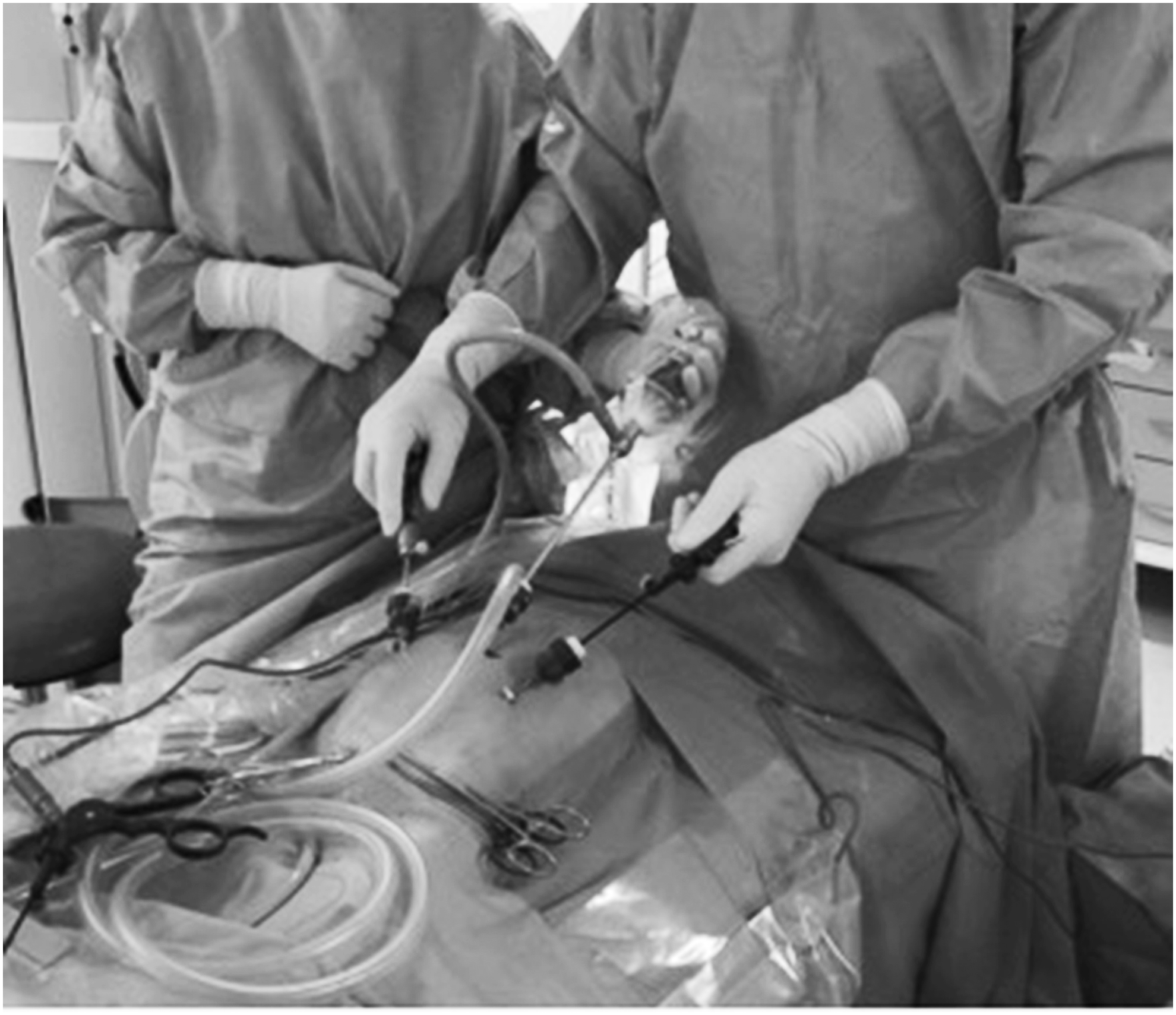
Trocars and surgeons' position.
Two stay sutures were inserted through the abdominal wall and placed on each side of the posterior bladder to pull the anterior wall of the bladder up and to expose the UVJ (Fig. 2). The retroperitoneum was incised just to identify the distal ureter that was isolated and dissected toward the UVJ. The ureter was mobilized to achieve sufficient freedom for a tension-free reimplantation. Once the stenotic part of the ureter was completely dissected, the bladder was filled with CO2. Using the monopolar scissors, the peritoneum was incised to expose the muscular wall of the bladder and to create an optimal tunnel with a length that was about four times the size of the ureter (Paquin law). The detrusor muscle fibers were cautiously divided with scissors vertically to create a submucosal tunnel, until the mucosa was exposed (Fig. 3).

The stay sutures are placed through the abdominal wall by percutaneous stitches (arrow) level to the bladder to expose the ureterovesical junction.

Detrusormyotomy. Exposition of bladder mucosa.
The distal ureter was transected at the level of stenosis and was tapered in all cases. The ureteral tapering technique used was extracorporeal Hendren technique, with exteriorization of the ureter through the ipsilateral port (Fig. 4), and ureteral tailoring was done by using continuous absorbable sutures. A stent was placed intraoperatively by laparoscopy. Vesicoureteral anastomosis was carried out after opening the bladder mucosa, by two continuous 6/0 PDS® sutures. Extravesical ureteral reimplantation by following Lich Gregoir technique was done. The ureter was placed in the new tunnel, and the detrusor muscle was reapproximated with absorbable sutures (Fig. 5). Neither peritoneal drain nor bladder catheter was used. The stent was removed at 6 weeks postoperatively.

Extracorporeal ureteral tapering according to Hendren technique.

Vesicoureteral reimplantation.
Postoperative follow-up consisted of a renal and bladder US scan at 1, 3, 6, and 12 months. Voiding cystourethrogram (VCUG) and mercaptoacetyltriglycine (MAG3) renogram were done at the sixth month.
Good results were defined as absence of leakage, anastomotic stenosis, vesicoureteral reflux (VUR), and voiding dysfunction.
Statistical analyses were performed by using the SPSS software package (version 15.0; SPSS, Chicago IL), and P < .05 was considered statistically significant. Paired tests and Wilcoxon test were performed to compare measures before and after surgery.
Results
Laparoscopic-assisted extracorporeal ureteral tapering repair and ureteral extravesical reimplantation by following Lich Gregoir technique was completed successfully in 7 patients without conversion. A vertical detrusor myotomy and extracorporeal ureteral tapering repair by Hendren repair was done in all cases.
The definition of success was the absence of obstruction. There were no major intraoperative complications. The mean surgical time was 154 minutes (130–170 minutes). The postoperative analgesia was managed by acetaminophen 15 mg/kg/6 h and continuous nubain 1 mg/kg/day. The mean hospital stay was 2.8 days (2–3 days). No urinary leakage occurred in the early postoperative period. The mean follow-up period was 28 months (11–47 months). None of the patients experienced postoperative voiding dysfunction. At 3 months postoperatively, 1 patient presented a febrile UTI, and a VUR grade III was diagnosed by VCUG. A redo laparoscopic surgery was done, showing partial disassembling of reimplantation, and then, the tunnel was extended to increase the length of antireflux, by following Lich Gregoir technique with an uneventful course in a long-term follow-up.
A postoperative MAG3 renogram showed nonobstructive pattern in all patients (Table 1). Statistical analysis revealed significant differences both before and after surgery in the average time of elimination on the MAG3 renogram (T½ 59.10 minutes versus 13.57 minutes, P < .0001). According to obstruction, the overall success rate of POM resolution was 100%. After medium-term follow-up, all patients were asymptomatic without recurrence of POM or VUR.
Discussion
The management of POM has evolved over the past 20 years. In 1989, Peters et al. reported 47 infants < 8 months-old with POM. Forty-two patients (89%) of these with moderate-to-severe obstruction underwent open surgical correction. The mean age was 1.8 months-old in the prenatally diagnosis group and 3.8 months-old in those presenting after birth. All infants showed functional and structural urographic improvement with a mean follow-up of 2.3 years. 21 Keating et al. suggested that the decision to surgical correction was based on absolute renal function; therefore, the expectant treatment avoids surgery in a maximum of 87% of patients. 5 Chertin et al. published that renal function < 30%, grade III and IV hydronephrosis, and ureteric diameter >1.33 cm are statistically significant and independent predictive factors for surgery. 22 The BAPU consensus supported initial conservative management and the surgical indications when initial DRF < 40%, especially when associated with massive hydroureteronephrosis and failure of conservative management (breakthrough febrile UTIs, pain, worsening dilatation, or deteriorating DRF). 2
The standard open surgery involves excision of the aperistaltic and/or narrow ureteral segment, reduction of caliber of the distal dilated ureter, and ureteral reimplantation into the bladder in an antireflux fashion, with reported success rates of 90%–96%. 23 However, complications and morbidity may occur, especially during the first year of life. 10 The major short-term complication is urinary leakage, and long-term complications include VUR or persistent obstruction. 23
In our experience, after >150 procedures performed by Laparoscopic Extravesical Ureteral Reimplantation by following Lich Gregoir technique for the correction of VUR, we never allow a urethral catheter to be included in bilateral cases. Nevertheless, in POM, where vesicoureteral anastomosis is done, and a stent is recomended, it is enough to avoid the urinary leakage.24–26
During the past decade, laparoscopy was successfully used for obliterative and reconstructive management of different urological procedures, with high success rates and advantages of minimum invasiveness. Laparoscopic procedures requiring sutures are technically demanding, even for experienced laparoscopic surgeons. Different techniques of ureteral reimplantation demonstrating feasibility by pneumovesicoscopy, laparoscopy, and robotic-assisted technique are encouraging and have been reported to be beneficial in terms of results, decreased postoperative pain, allowing shorter hospital stay and a quicker return to normal activity.17,19,20,24–26,27–30,31 In 2006, Ansari et al. presented 3 cases of laparoscopic extravesical transperitoneal ureteral reimplantation by Lich Gregoir technique, with extracorporeal tailoring of the ureter by using Hendren technique. After a 1 year follow-up, no patients presented VUR and the renal function was preserved in all cases. 19 In 2013, Bondarenko reported 10 cases of POM who underwent laparoscopic extravesical ureteral reimplantation and intracorporeal plication of the ureter by using Starr technique. The mean follow-up was 13.6 months, and the improvement of the dilatation of the pelvicalyceal system and the ureters occurred in all patients. 20
Laparoscopic and robotic-assisted techniques by the transvesical approach have been developed as an alternative to open surgery, with promising outcomes. Nevertheless, larger series and prospective randomized trials in pediatric patients with long-term results are needed.17,27–30,32
The laparoscopic transperitoneal extravesical approach provides a large operative space for reimplantation and does not limit the manipulation of the bladder and ureter.20,24–26
The potential complication in open extravesical surgery has been associated with voiding dysfunction in bilateral cases, which can be up to 10%. 31 However, Palmer published data on 60 patients from 0.9 to 10.5 years-old (mean age 5.1 years-old), who underwent extravesical ureteral reimplantation by open surgery, including 36 bilateral procedures. None of the patients presented voiding dysfunction. 33 In our series, these complications have not been found.
The most popular techniques used for tailoring megaureters are either plication reported by Kalicinski and Starr,34,35 or tapering reported by Hendren. 36 Kalicinski et al. described a folding technique whereby the lateral avascular part of the ureter was excluded from the lumen by a longitudinal running suture, and then folded posteriorly before reimplantation. 34 Starr modified this procedure, introducing many interrupted sutures that folded the ureteral wall inwards. 35 Hendren advised excisional tapering, whereby a longitudinal strip of the redundant ureter is excised, and the ureter is sutured in two layers. 36
In the laparoscopic approach, to reduce ureteral caliber, some authors have adapted the classic tailoring techniques, with results comparable to open surgery.17,18
Landa-Juarez et al. decribed a new method without ureteral taloring for laparoscopic ureterovesical repair for megaureter treatment, which consisted of incising the stenotic portion of the ureter longitudinally and maintaining its posterior wall attached to the bladder; the anterior wall of the ureter was anastomosed transversally to the bladder mucosa to liberate the obstruction. 32
In our series, Hendren technique was used in 7 patients with extracorporeal tapering and it is technically easier than the intracorporeal technique; nevertheless, it is recommended to not perform ureteral stenting to avoid postoperative leakage. 36
Balloon dilatation by cystoscopy can be used as the first line of treatment for the correction of POM. Nevertheless, when endoscopic management of POM is performed, the stenotic UVJ is bypassed with a guidewire, but sometimes cannulation of the urethral meatus is very difficult. Farrugia et al. reported 16 infants with 19 RU managed by stent insertion at a median age of 24 weeks. Six stents were inserted endoscopically and 13 (68.4%) were inserted by the open procedure, after a failed cystoscopic attempt. 37 Castagnetti et al. needed open insertion of the stent in 5 of a total of 10 neonates and infants after failure of an attempt of endoscopic insertion, in patients with POM treated only with the stent. 38 In our initial series of 14 patients, a balloon dilatation by cystoscopy was performed in all cases, as a first line of treatment, with success only in 7 patients (50%). In the other 7 patients, it was impossible to pass the guidewire through the UVJ; therefore, LUER and ureteral tapering repair was performed. Thus, all our 7 patients benefited from the minimal invasiveness.
VUR is one of the most frequent postoperative complications. Peters et al. published data on 47 infants <8 months-old with POM. Forty-two patients of these underwent open surgical correction. In a mean follow-up of 2.3 years, VUR was seen in 8 patients (19%), with spontaneous resolution observed in 3 of them. The 5 remaining patients were followed and this resulted in redo reimplantation in 3 patients. 21 The presence of VUR after endoscopic balloon dilatation is controversial, because a VCUG is not systematically performed. Some authors just only perform VCUG in cases of persistent dilatation or if a UTI occurs. 13 García-Aparicio et al. reported 13 patients, 10 men and 3 women, with a mean age of 7 months-old (4–24) treated by endoscopic balloon dilation. Postoperative VUR was found in 2 patients (15.3%) in a systematically postoperative VCUG at 6 months. One patient with high-grade VUR was treated with open ureteral reimplantation, and the other was treated conservatively. The authors concluded that the presence of VUR after this procedure is greater than after open ureteral reimplantation. García-Aparicio et al. reported that 5 patients needed redo balloon dilatation when the stent was removed because the cystoscope was not able to pass the UVJ; therefore, in 8 patients (61%), the endoscopic balloon dilatation was successful after the first attempt. 15 In 2015, the same author presented a total of 22 RU who underwent balloon dilation to treat POM in 15 boys and 5 girls with a mean age of 14 months-old (3–103 months). After endoscopic treatment, 6 RU (27.7%) developed VUR. Four of the RU were managed surgically and in the other two VUR disappeared in a second VCUG. 39 In our series, 1 patient (14.2%) presented a febrile UTI at 3 months postoperatively and a unilateral VUR grade III was diagnosed by VCUG. In this case, a redo laparoscopic extravesical ureteral reimplantation by following Lich Gregoir technique was done with an uneventful course in a long-term follow-up. For this patient, the parents were informed about endoscopic and laparoscopic options, and the decision was taken in conjunction with the surgical team. In postoperative MAG3 renograms, the results showed statistical differences before and after surgery (T½ 59.10 minutes versus 13.57 minutes, P < .0001) and no obstructive pattern occurred.
In a comparison with historical series in open surgery by the extravesical approach for megaureters, in 1994, McLorie et al. reported that 23 children underwent extravesical megaureter repair. Three of them had bilateral megaureters. There were 14 obstructing and 12 refluxing ureters. Only 8 of the 26 ureters were tapering.
For obstructive megaureters, a stent was placed, and drains were placed whenever the ureter was dismembered or tapered but not otherwise. A urethral catheter was left indwelling for 4 or 5 days. Regarding complications in this series, 4 children had postoperative UTIs, 2 had stent-related complications, including a dislodged stent and an encrusted stent. Transient voiding difficulty was observed in 3 children and was managed by clean intermittent catheterization (CIC). One of them had a unilateral reimplant for high-grade reflux, and CIC was employed for 1 week after catheter removal. The other 2 underwent bilateral reimplantations for reflux and continued CIC for 1 and 4 months, respectively. Spontaneous voiding was achieved in all 3 children. 40
In our series, there were no major intraoperative complications. The mean hospital stay was 2.8 days. No urinary leakage occurred in the early postoperative period. None of the patients experienced postoperative voiding dysfunction. Only 1 patient presented a VUR, and a redo laparoscopic surgery was done with an uneventful course in a long-term follow-up. The overall POM resolution of obstruction was 100%. After medium-term follow-up, all patients were asymptomatic without recurrence of POM or VUR.
In conclusion, this study represents our initial experience of laparoscopic approach in POM, which shows that laparoscopic-assisted extracorporeal ureteral tapering repair and ureteral extravesical reimplantation by following Lich Gregoir technique is a feasible and effective procedure, with a success rate comparable to open surgery. We consider it a safe and good option when the first line of treatment fails; nevertheless, larger randomized prospective trials and long-term follow-up are required to validate this technique.
Footnotes
Disclosure Statement
No conflict of interest or financial disclosure has been declared by M.L., R.G., G.R., F.V., and M.A. Ethical standard: This study was done with anonymized patient data, and therefore, ethics committee approval was not necessary.