
Abstract
Abstract
Introduction:
Gastropleural fistula (GPF) is a complex pathology that can present as a result of surgery, trauma, peptic ulcer disease, malignancies, radiation, or chemotherapy. Management typically includes endoscopic or surgical intervention along with intraabdominal or intrathoracic drainage of pre-existing infection. Traditionally, surgical approaches have been through exploratory laparotomy or thoracotomy, subjecting already ill patients to additional morbidity.
Case Report:
We describe and demonstrate a laparoscopic minimally invasive approach to the management of a GPF with a wedge resection of the stomach, along with a review of the current literature regarding GPF treatment.
Conclusion:
GPF repair can be performed through laparoscopy and may lead to improved patient outcomes and faster recovery.
Introduction
A
Diagnosis of GPF can be made with oral-contrasted studies such as fluoroscopic upper gastrointestinal (UGI) imaging or computed tomography (CT) scans. Upper endoscopy can also be performed for diagnostic and therapeutic purposes. Treatment algorithms vary depending on patient presentation and the underlying etiology of the GPF. Most patients, before any surgical interventions, undergo placement of tube thoracostomies for drainage and control of pleural effusions. For smaller fistulae, endoscopic interventions have had recent success6,9,20; however, large defects typically require surgical repair or resection. Multiple published cases have described approaches through laparotomy or thoracotomy, but the invasive nature of the operations along with the patients' chronic illnesses and acute conditions makes GPF repair a high-risk surgery.2–3,5,8,14–17 Case reports of laparoscopic repairs are described in the literature, with relatively good patient outcomes.7,13,18,19 We present a patient with a GPF who underwent laparoscopic resection, with accompanying videos showing our technique. 21
Case Report
Patient presentation
The patient is a 58-year-old female with a remote history of a Nissen fundoplication, for medication refractory gastroesophageal reflux disease, who was admitted to the intensive care unit at an outside institution for pneumonia. She then developed a parapneumonic effusion, which subsequently evolved into a left-sided empyema. She was taken to the operating room and underwent a video-assisted thoracoscopic surgical (VATS) decortication with surgical findings consistent with an empyema. Chest tubes were placed at the end of the surgery. The patient initially improved, but on postoperative day 3, her condition deteriorated. A chest X-ray suggested a left-sided consolidation with empyema (Fig. 1a), a CT scan was obtained with oral contrast showing a GPF with free flow of oral contrast into the left thoracic cavity (Fig. 1b–d).

The patient was then transferred to our institution for further management. Endoscopic closure was attempted, but observation of the defect was difficult as the stomach was unable to be distended. Given the severe sepsis and the presence of an ongoing leak into her left thoracic cavity from her GPF, she was taken urgently to the operating room for a laparoscopic exploration.
Surgical technique
The patient was placed supine in a split leg position. Entry should be performed with great care as there may be an intraabdominal inflammatory component. In this case, a 12-mL optical trocar port was placed in the supraumbilical position just left of midline for optimal observation of the hiatus and into the mediastinum. Two 5-mL working ports were placed in a left and right subcostal position, and a third 5-mL port was placed on the left far laterally for retraction. An additional subxiphoid incision was used for the placement of a liver retractor.
In patients with previous foregut surgery, such as a fundoplication or gastric resection, the anatomy may be distorted, and care must be taken not to injure the vascular supply to the stomach, create additional gastrotomies, or extend the length of the pre-existing fistula. Using a combination of electrosurgery and sharp dissection, the hiatus was exposed and the fundoplication was undone. Once proper anatomy was restored, the fistula was fully assessed and noted to be greater than 5 cm in length and located on the fundus of the stomach. It was decided to resect this area to provide a healthy tissue bed for wound healing. Stay sutures were placed to ensure a full thickness resection of the fistula and the defect was removed using a laparoscopic linear stapler and the staple line was oversewn. Intraoperative endoscopy was used as a bougie to guide resection and to confirm patency.
Hospital course
Postoperatively, the patient was kept nothing by mouth until an UGI series was obtained on postoperative day 2, demonstrating a patent gastroesophageal junction without the presence of a leak (Fig. 2). The patient was then started on a clear liquid diet and advanced to a full liquid diet on postoperative day 4. On postoperative day 7, all chest tubes were removed and the patient was discharged. The patient was seen in clinic for follow-up 1 week after discharge and her diet was advanced to solid food. At her final follow-up 2 months after discharge, she was recovering well without any further complications.
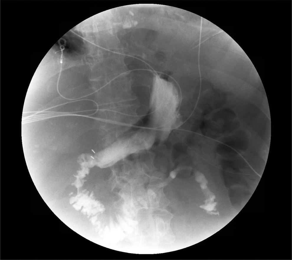
Upper gastrointestinal series with oral contrast on post-operative day 2 showing patency of the gastroesophageal junction without the presence of a leak.
Discussion
A MEDLINE search from 2000 to 2015 of all published cases of GPF with known treatment approaches was performed and shown in Table 1. A majority of these articles utilize either a laparotomy or thoracotomy approach.2,3,5,8,14–17 Other groups employed endoscopic treatments, which appeared to be effective if the GPF is a small defect.6,9,20 Patients treated with only tube thoracostomies were placed on palliative care and expired relatively soon after diagnosis.4,11 Other published case reports of laparoscopic management of GPFs appeared to be technically successful, but the patients' clinical courses were ultimately determined by the original disease process.7,13,18,19
VATS, video-assisted thoracoscopic surgical.
To our knowledge, this is the first reported case of a GPF developing within a Nissen fundoplication after a thoracoscopic decortication. The progressive course of pneumonia, empyema, VATS decortication, and finally the formation of the GPF suggests that the fistula developed iatrogenically rather than through a primary gastric process. The patient presented here had a disease process that was curable and the minimally invasive approach we employed aided in expediting the recovery process.
Conclusion
For surgeons experienced in laparoscopic management of complex gastrointestinal disease processes, a laparoscopic approach for patients with GPF can be safely performed. As compared with traditional thoracotomy or laparotomy, laparoscopy affords patients a faster recovery process and less morbidity in an already highly debilitating disease process.
Footnotes
Disclosure Statement
No competing financial interests exist.