
Abstract
Abstract
Background:
Gastrointestinal (GI) bezoars are the most common foreign bodies causing obstruction in the GI tract. They are frequently seen following upper GI tract surgery and surgical intervention is required often. The aim of this study is to describe the surgical management of GI bezoars.
Materials and Methods:
A retrospective cohort study, including all patients diagnosed with bezoars between May 2008 and May 2017, was conducted. Patient charts were reviewed, and demographics, clinical, surgical, and postoperative data were collected and analyzed.
Results:
Forty-five patients were included, with a mean age of 62.04 years (Range 18–91). Thirty patients underwent previous surgery (66.6%), most commonly surgical interventions for peptic ulcer disease (22 patients, 73.3%). Obstruction was most common in the ileum (27 patients, 60%). Thirty-nine patients (86.7%) required surgical intervention. Laparoscopy was attempted in 20 patients (51.2%), but conversion to open procedure was required in 11 patients (55%). Postoperative complication rate was 41%. No preoperative factors were found to be correlated with postoperative complications. Postoperative complications were associated with a longer length of stay (P = .006) and a higher readmission rate (P = .04). Patients treated with laparoscopy tended to have a lower BMI (P = .04), less previous surgeries (P = .04), and a bezoar located more proximally (P = .03), however, laparoscopy showed no benefit in complications rate, readmissions, and length of stay.
Conclusions:
GI bezoars require surgical intervention at high rates. Postoperative complications are common. Completion of an upper GI endoscopy is important and should be performed at an early stage of management.
Introduction
G
Clinical manifestation is commonly that of a small bowel obstruction. Symptoms include abdominal pain, abdominal bloating, nausea, and vomiting. High clinical suspicion for bezoars should be raised with any history of upper GI tract surgery and/or recent ingestion of persimmons, known to be a risk factor for bezoar formation. 3 Special attention should be made to psychiatric disorders and clinical signs as alopecia or halitosis. Confirmation of the diagnosis is achieved with a computed tomography (CT) scan. 4
Management goals for GI bezoars include removal of the foreign object and preventing recurrence. Therapeutic strategies are varied, and they depend mainly on the type and location of the bezoar. Gastric phytobezoars could often be treated without surgical intervention, using chemical dissolution or endoscopy. Gastric trichobezoars, on the contrary, take time to form and are resistant to enzymatic or chemical dissolution and are commonly treated surgically. Phytobezoars can be treated without surgical intervention, depending on their anatomical location. When located in the stomach, surgical intervention can usually be avoided. 5 However, they present more commonly in various parts of the small bowel, mainly at the ileocecal valve, and prompt surgical intervention is required. 2
In this study, we aimed to analyze our collected experience with the surgical management of GI bezoars over the last 10 years, to help physicians, dealing with the management of this uncommon entity based on our experience.
Materials and Methods
A retrospective-cohort search of all patients admitted to our tertiary medical center for GI bezoars between May 2008 and May 2017 was conducted. We identified 45 patients who were entered into the database. Medical charts were reviewed for demographic data, presurgical data, operative data and findings, and postoperative course and complications. All patients were diagnosed with a CT scan. Endoscopic reports for patients who underwent additional endoscopic evaluation for bezoar remnants were also identified and registered. Admission data, including length of stay and readmissions, were also recorded and analyzed. Postoperative complications were analyzed based on the Clavien–Dindo score, used to classify surgical complications based on their severity and the level of intervention they require.
Statistical analysis was done using Fisher's exact test and the χ2 test to evaluate differences between qualitative variables. A t-test was applied to compare quantitative variables. A P-value of <.05 was considered significant. The statistical analysis was performed by using SAS/STAT for Windows version 9.4 (Statistical Analysis System Corp., NC). The study was approved by the local Institutional Review Board before collection of data.
Results
Overall, 45 patients were admitted for GI bezoars over the 10-year study period. The study included 27 males (60%) and 18 females, with a mean age of 62.04 years (range 18–91 years). Additional demographic data, including BMI, Charlson comorbidity index, American Society of Anesthesiology (ASA) score, and smoking habits, are all listed in Table 1.
ASA, American Society of Anesthesiology; BMI, body mass index.
Thirty patients (66.6%) underwent abdominal surgical interventions in the past, most commonly surgical procedures related to the stomach in general and to a peptic ulcer disease specifically (22 patients, 73.3%). Surgeries for peptic ulcer included vagotomy alone (6 patients, 20%), vagotomy and pyloroplasty (8 patients, 26.6%), Bilroth operation type II (6 patients, 20%), and Bilroth operation type I (2 patients, 6.6%). Other less common surgical interventions included hysterectomy (2 patients 6.6%), open appendectomy (2 patients, 6.6%), various bowel resections (3 patients, 10%), and one open abdominal aortic aneurysm (AAA) repair.
The most common type of bezoars were phytobezoars (Fig. 1), most of them with unknown content of indigested food (44 patients, 97.7%). One patient with trichotillomania was found to have a stomach filled with hair (trichobezoar; Fig. 2). Bezoars were most common in the ileum (27 patients, 60%) followed by the jejunum (14 patients, 31.1%) and the stomach (4 patients, 8.9%).

Coronal CT image shows the typical CT appearance of SBO due to a phytobezoar in the distal jejunum. The bezoar appears as an intraluminal mottled-appearing mass that contains air bubbles in its interstices and is located at the site of the obstruction (long arrow). Notice transition point distal to the bezoar (short arrow). CT, computed tomography.
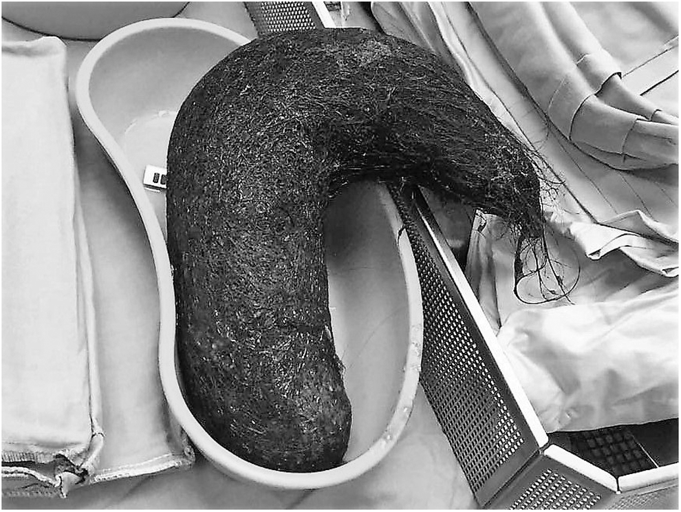
A stomach shaped trichobezoar specimen extracted surgically from a 18-year-old female with trichotillomania.
Thirty-nine patients (86.7%) required surgical intervention. Exploratory laparotomy and “milking” of the bezoar past the ileocecal valve was the most common surgical intervention (18 patients, 46.1%) followed by diagnostic laparoscopy and “milking” (10 patients, 25.6%). Enterotomy was performed in 10 patients as well, most of them using an open surgical technique and 1 patient using laparoscopic technique. Only 1 patient required bowel resection (2.5%). The majority of patients were operated within 24 hours from arrival (26 patients, 66.7%, range 0–1 days) with a third of patients operated 2 or more days following presentation (13 patients, 33.3%, range 2–8 days). The delay was correlated in some cases with a therapeutic attempt to allow passage of the bezoar and in some cases with a delayed diagnosis when a CT scan was performed after the acute obstruction has not resolved with conservative management. Average time from presentation to surgery was 1.61 days (range 0–8).
Laparoscopy was initially attempted in more than half of the patients (20 patients, 51.2%) but conversion was required in 55% of cases (11 patients), mostly due to adhesions and lack of working space due to dilated bowel loops. Comparison between patients who were operated using a laparoscopic technique and an open technique showed that patients who completed a laparoscopic procedure tended to have a lower BMI (P = .047), had less previous surgical procedures in the past (P = .049), and were found to have bezoars located more proximally (Stomach/Jejunum, P = .038). Overall operating time varied and averaged at 74.2 minutes (range 33–162 minutes), and it was comparable between the laparoscopic (mean 75.89 minutes, standard deviation [SD] 27.14) and the open (mean 73.70 minutes, SD 26.46) approaches. No differences were seen in postoperative complication rate, readmission rate, and length of stay. The analysis between the laparoscopic and the open surgery groups is detailed in Table 2. No significant intraoperative complications or blood loss were seen in any of the cases.
ASA, American Society of Anesthesiology; SD, standard deviation.
Postoperative complication rate was fairly high at 41% (16 patients). The most common postoperative complication was ileus, seen in 8 patients (20.5%), followed by wound infection (5 patients, 12.8%). Three patients suffered from both ileus and wound infection. Analysis according to the Clavien–Dindo score yielded 12 (30.7%) minor postoperative complications and 4 major complications (10.2%). All major complications required an additional surgical intervention and 1 patient eventually died from postoperative complications, after suffering from bowel evisceration and fatal arrhythmia following the second surgical intervention.
Endoscopic evaluation of the upper GI tract following or during the surgical procedure was performed in 35 patients (89.7%). Remnant bezoars were found in a large portion of the patients (14 patients, 40%). One patient required a second upper GI endoscopy due to inability to retrieve the entire remnant indigested material during the first attempt.
Analysis of factors contributing to postoperative complications yielded no definitive results in all demographic characteristics analyzed. No risk factors for postoperative complications were identified regarding surgical data, such as operating time, surgical technique, and whether an endoscopy was performed or not. Length of stay and readmission rate were both significantly higher in patients who suffered from postoperative complications. The analysis is summarized in Table 3.
ASA, American Society of Anesthesiology; SD, standard deviation.
Discussion
GI bezoars have been described since ancient times and were considered for centuries as an antidote for poisonous substances. 6 They were used as therapeutic agents until the 16th century, when the Famous French surgeon Ambroise Pare conducted an experiment demonstrating bezoars have no healing powers. 7 Nowadays, the term “bezoar” refers to an indigested content that can cause a significant bowel obstruction that requires early recognition and prompt intervention.
Although there are many reports of GI bezoars, those were initially case reports and small case series, published throughout the early decades on the 20th century. 8 Bezoars are still considered a relatively rare cause of bowel obstruction, presenting as the obstructive cause in less than 1% of patients presenting with bowel obstruction. 9 In addition, gastric bezoars were also found in less than 0.5% of esophagogastroduodenoscopies in several large series.10–12 Certain types of indigested food products were noticeable as prone for the formation of GI bezoars. Celery, pumpkins, grape skins, prunes, raisins, and, in particular, persimmons are among the most common causatives of phytobezoars. 13 Unsurprisingly, that rates of GI bezoars are higher in regions where these types of food products are more frequently consumed, like the middle east14,15 and southeast Asia. 16 These types of food products consumed along with a history of abdominal surgery, specifically upper GI tract surgical procedures, should raise a clinical suspicion of GI bezoars when examining a patient presenting with obstructive symptoms and abdominal pain. The relationship to upper GI tract surgery, specifically surgical procedures related to ulcer peptic disease have been showed by several authors in the past.3,17 These surgical procedures, that usually included vagotomy and/or pyloroplasty can cause a reduction in gastric motor activity, rendering the digestive motor complexes ineffective at the level of the stomach.18,19 In addition, the procedures can reduce the evacuation of solid indigestibles by reducing the secretion of chlorhydropeptic substances, causing a slower digestion of these solids. 17
Nowadays, GI bezoars are fairly easily diagnosed using either radiological studies, mainly CT scans, 20 and/or upper GI endoscopy, which can also be therapeutic in gastric bezoars.
Surgery was the only possible treatment until reports of endoscopic recovery of bezoars started to appear in the early 1970's. 21 Researches started noticing the increased incidence of GI bezoars after gastric-related surgical procedures that were common for the treatment of peptic ulcer disease and upper GI bleeding, before the widespread use of proton pump inhibitors. 3 A study by Robles et al. 22 showed high rates of previous gastric procedures, up to 70%, in patients presented with GI bezoars, of which the majority underwent vagotomy and pyloroplasty for peptic ulcer disease. A more recent study by Koulas et al., 23 which included 23 patients treated for bezoars in two centers in Greece, reported that 57% of patients had previous gastric surgery. A similar study from Turkey, including 34 patients, reported a similar rate of 55.88% of previous gastric procedures. 24 In both studies, postoperative complication rate was around 30%. Most reports of laparoscopic treatment for GI bezoars are case reports,25,26 and there are no large scale series describing the experience with laparoscopic technique. A study by Yau et al. 27 compared 10 patients treated with laparoscopy to 14 patients treated with open procedure and concluded that laparoscopy was associated with a shorter operating time, less postoperative complications, and a shorter length of stay. However, our findings did not show significant advantage to laparoscopy in these parameters. In addition, conversion rate was relatively high at 55%. However, our analysis showed that patients successfully treated with laparoscopic technique were thinner, with less prior surgical interventions and with a proximally located bezoar. These parameters might help clinicians decide whether laparoscopic surgery is the better option for patients with GI obstruction from bezoars.
Another important point raised in our study is the need for immediate endoscopic evaluation when the diagnosis of a GI bezoar is made. Our study found that a significant rate (40%) of patients may have remnants of the bezoar in the proximal GI that can be retrieved with an upper endoscopy, perhaps preventing an additional bowel obstruction, thus aiding to the resolution of the clinical state caused by the bezoars.
Our study has several limitations. Despite being one of the largest series in the literature, the number of patients in this series is too small to draw firm conclusions, and the retrospective nature of this study may also impact the results and level of recommendations. However, based on our experience, we suggest early suspicion of GI bezoar as an etiology for bowel obstruction in patients with previous gastric surgery and anamnesis that included the consumption of food products containing fruit skins and a significant load of fiber.
Conclusions
Endoscopic evaluation should be a routine part of the workup and treatment either during or following surgery. Surgeons should be aware of the significant postoperative complication rate following surgical intervention for GI bezoars, specifically postoperative ileus, as resolution of the obstruction may be delayed. In the hands of experienced surgeons, laparoscopy is feasible, but conversion rates are high and our series failed to show significantly improved outcomes when compared with open surgery.
Footnotes
Disclosure Statement
No competing financial interests exist.