
Abstract
Abstract
Background:
Minimally invasive surgery (MIS) has gained increasing importance in neonatal surgery but the effects on neonatal physiology remain unclear. We aimed to characterize the impact of capnoperitoneum on physiologic parameters in a small animal model for neonatal MIS.
Material and Methods:
Twenty-four 10-day-old Sprague Dawley rats underwent inhalative anesthesia (1% isoflurane in 100% O2 250 mL/minutes) and were allowed to breathe spontaneously. CO2 was insufflated into the abdominal cavity for 1 hour via a 24G cannula. Anesthetized litter mates without insufflation served as sham controls, those without any treatment as external controls. Continuous monitoring included O2-saturation, heart and respiration rate, pulse and breath distension. After euthanasia, blood gas analysis was performed.
Results:
All animals survived the experiment. Capnoperitoneum was best tolerated at a pressure of 2 mmHg and a flow of 0.5 L/minutes. A significant decrease in heart rate was observed within the first 30 minutes of insufflation comparing the CO2 and sham group (P < .05). In both, the CO2 and sham group, postmortem pH-levels were lower and pCO2 levels were higher compared to external controls (P < .05). Additionally, levels of pCO2 were higher but pH levels remained unchanged in the CO2 compared to sham group (P < .05).
Conclusion:
We established a small animal model for neonatal laparoscopy. A pressure of 2 mmHg and flow of 0.5 L/minutes induced physiologic alterations but was well tolerated by the animals. These settings can be used in future studies on the impact of the capnoperitoneum in neonatal MIS.
Introduction
T
In addition, MIS procedures are usually performed under general anesthesia, which may alter hemodynamics and acid-base metabolism. 12 Moreover, neurotoxic effects have been shown for all types of anesthesia in animal studies, even though long-term effects on human neurodevelopment could recently not be found.13–15
Taken together, the impact of the capnoperitoneum in MIS on the developing organism is still poorly understood. The aim of the study was to establish a small animal model for neonatal MIS and to characterize the impact of capnoperitoneum on physiologic parameters.
Materials and Methods
Animals and study design
Sprague Dawley rats were bred and kept at the Medizinisch-Experimentelles Zentrum of the University of Leipzig. Animals were housed in a temperature-controlled environment with an artificial 12:12 hours light-dark cycle and had free access to food and water. All experimental procedures were performed in accordance with the European Council Directive of 2010 (2010/63/EU) and were approved by the Local Ethics Committee for Animal Experimentation (TVV 05/17).
Ten-day-old (P10) Sprague-Dawley rats were selected for the study as their developmental stage corresponds to that of full-term newborns.16,17 Twenty-four animals were randomized to one of the three study groups and underwent either inhalative anesthesia and capnoperitoneum (CO2 group, n = 8), anesthesia only (sham group, n = 8), or none of these (external controls, n = 8) (Fig. 1).

Study design. Twenty-four animals of both sexes were included in the study and randomly assessed to one of the three study groups. Inhalative anesthesia and capnoperitoneum (CO2 group, n = 8), anesthesia only (sham group, n = 8) or none of these (external controls n = 8) were applied.
Experimental procedures
Twenty-four male and female rats (P10) were separated from the dam and kept warm under a heat lamp. Anesthesia was established in an induction chamber applying 5% isoflurane (Baxter, Deerfield, IL) in 100% oxygen (O2) for 5 minutes. Subsequently, isoflurane concentration was reduced to 1% in 100% 250 mL/minutes O2 and only adjusted during the intervention if necessary. For peri- and postoperative analgesia, metamizol (100 mg/kg bodyweight; Ratiopharm, Ulm, Germany) was dissolved in 0.9% warmed NaCl and administered subcutaneously to the scape of the neck in a final volume of 50 μL. The depth of anesthesia was assessed by the pedal withdrawal reflex. 18
Animals were placed on a heating plate (37°C) equipped with a ventilation chamber to maintain anesthesia and oxygen supply. A specific sensor was applied to the upper thigh of the rat and connected to a small animal pulse oximeter (MouseOxPlus, Harvard Apparatus, Holliston, MA). 19 After disinfection, a 24-gauge modified “Veress needle” (BD Insyte, Heidelberg, Germany) was inserted into the abdominal cavity and connected to an insufflator allowing control of pressure (1–15 mmHg) and flow (0.1–15 L/minutes) (Endoflator 40 SCB; Karl Storz, Tuttlingen Germany). Pressure levels between 1 and 5 mmHg and flow levels between 0.5 and 1 L/minutes were tested to reveal the optimal setting for our experiment. Finally, capnoperitoneum was maintained for 60 minutes at an insufflation pressure of 2 mmHg and a flow of 0.5 L/minutes.
Throughout the entire experiment animals were continuously monitored using the MouseOxPlus. Five physiologic parameters were assessed: O2 saturation (SpO2 in %), heart rate (bpm), respiration rate (brpm), pulse distention (μm), and breath distension (μm). Pulse and breath distension were calculated using the change in distention of the peripheral arterial blood vessels. Pulse distension, which is a direct measurement of changes in the local blood volume, allows an indirect measurement of the peripheral arterial blood pressure. Breath distension, which is based on the pressure changes of the thoracic cavity by in- and expiration, determines the breathing effort of the animal.19–21
At the end of the experiment, animals of all three groups were euthanized by an overdose of pentobarbital (600 mg/kg bodyweight; Release, Wirtschaftsgenossenschaft deutscher Tierärzte eG, Garbsen, Germany). A single blood sample was obtained for blood gas analysis by thoracotomy using a capillary (95 μL) from the left cardiac ventricle. Partial pressure of oxygen (pO2) and carbon dioxide (pCO2) in addition to pH levels were measured immediately using an ABL 800 Basic (Radiometer, Kopenhagen, Denmark) blood gas apparatus. Moreover, brain, liver, heart, kidneys, and lungs were harvested and fixed for future analysis.
The main outcome measure was survival of the experiment for 1 hour. Secondary outcome measures included O2-saturation, heart and respiration rate, pulse and breath distension, and a final blood gas analysis (pH, pCO2, and pO2).
Statistical analysis
Statistical analysis was performed by SPSS (Statistical Package for the Social Science V24). Graphs were prepared using Origin 8.0. After testing for normal distribution by Shapiro–Wilk test, parameters of continuous monitoring and blood gas parameters were compared by Kruskal-Wallis test and post hoc Mann-Whitney test. Significance level was set as P < .05. Data are presented as mean ± standard deviation if not indicated differently.
Results
Experimental setup and survival
Twenty-four Sprague-Dawley rats (n = 8 per group at P10) weighing 26.3 ± 3.2 g, were included in the study. Inhalative anesthesia was applied with 1% isoflurane in 250 mL/minutes O2. Capnoperitoneum was maintained with a pressure of 2 mmHg and flow of 0.5 L/minutes. None of the treated animals died during the experimental period. Capnoperitoneum at 2 mmHg and flow of 0.5 L/minutes induced a markedly distended abdomen in rat pups. Higher pressures (>2 mmHg) and an increased flow (>0.5 L/minutes) led to a relevant desaturation (SpO2 < 87.5%) and decreased heart rate (<200 bpm) within 5 minutes and were therefore abandoned.
Physiologic parameters
After induction of anesthesia (5 minutes period), the mean heart rate did not differ within both treatment groups (CO2 group: 303 ± 44 bpm, sham group: 282 ± 35 bpm; P > .05). However, mean heart rate significantly decreased 10 minutes after establishment of the capnoperitoneum in the CO2 group (CO2 group: 260 ± 28 bpm, sham group: 291 ± 25 bpm; P < .05) and remained significantly reduced for 20 minutes before it assimilated the level of the sham group (CO2 group: 231 ± 15 bpm, sham group: 268 ± 28 bpm; P > .05) (Fig. 2A). Pulse distension, an indirect measurement of arterial blood pressure, remained unaltered over time and did not differ between the groups (Fig. 2B).

Physiologic pulse parameters differed between groups. Heart rate
Mean respiration rate was 42 ± 14 brpm in the CO2 group and 38 ± 14 brpm in the sham group (P > .05) after induction of anesthesia. Neither mean respiration rate nor breath distension differed significantly between both treatment groups during the entire experiment (Fig. 3A, B).
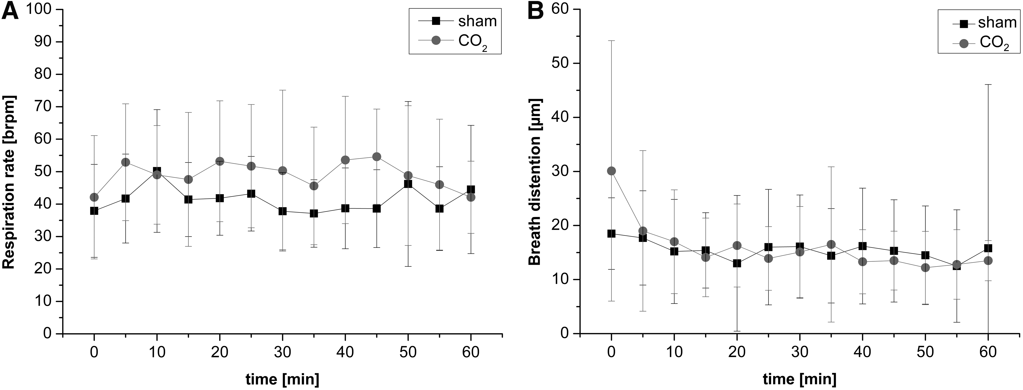
No alterations of physiologic breathing parameters. Continuous monitoring of respiration rate
Mean O2 saturation was 99% ± 0.5% in the CO2 group and 98 ± 2.6% in sham group (P > .05) at the beginning of the experiment and remained constantly stable at ∼100% in both groups throughout the entire experiment (data not shown).
Blood gas analysis
Mean postmortem pH levels were significantly lower in the sham group (pH 7.13 ± 0.06; P < .001) and in the CO2 group (pH 7.09 ± 0.04; P < .001), compared to external controls (pH 7.24 ± 0.07), but did not differ significantly among each other (Fig. 4A).
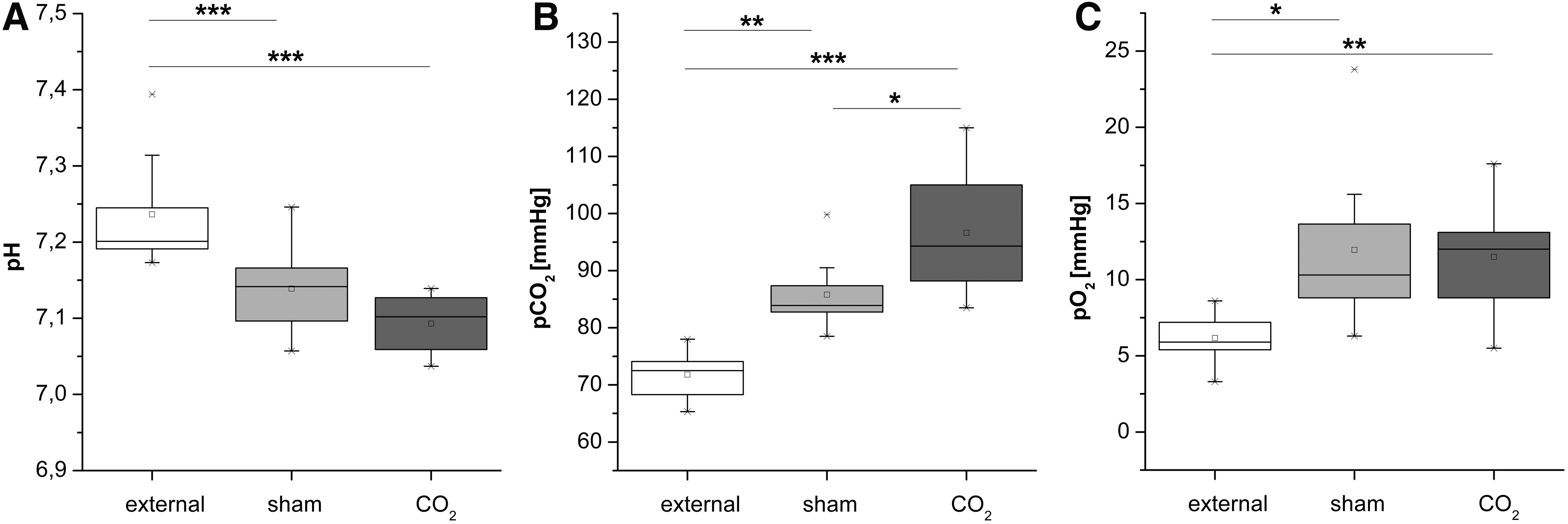
Blood gas parameters pH, pO2, and pCO2 differed between external controls and treatment groups.
Mean postmortem pCO2 levels were significantly increased in the sham group (86 ± 7 mmHg; P < .01) and in the CO2 group (97 ± 11; P < .001) compared to external controls (72 ± 4 mmHg). Levels also differed significantly between both treatment groups (P < .05; Fig. 4B).
Mean postmortem pO2 levels were significantly increased in both treatment groups (sham group: 12 ± 5 mmHg, CO2 group: 11 ± 4 mmHg) compared to external controls (6 ± 2 mmHg; P < .05), but did not differ significantly among each other (Fig. 4C).
Discussion
Systemic effects of MIS and its feasibility have been widely studied using adult animal models.22–24 At the same time, minimally invasive techniques have gained increasing importance in pediatric surgery. In this study, we established a small animal model mimicking neonatal MIS and characterized the peri- and postoperative impact of the capnoperitoneum on physiologic parameters.
Physiologic parameters
Between 10 and 30 minutes of treatment, mean heart rate significantly decreased in the CO2 group when compared to the sham group. In adult rats, bradycardia has only been observed for insufflation pressures greater than 5 mmHg, suggesting a more susceptible circulatory system in neonatal rats.23–25 In contrast to earlier studies, the blood pressure, as assessed by continuous measurement of the pulse distension, remained unaltered during our experiments. 4 This was most likely due to the low insufflation pressure of 2 mmHg we applied, as hemodynamic effects of the capnoperitoneum strongly depend on the intraabdominal CO2 pressure and corresponding CO2 blood levels.23–25 Comparable to adult rats, oxygen saturation, respiration rates, and breath distension were normal in both treatment groups during the entire experiment.22,23 In line with others, we observed a respiratory acidosis in the sham and CO2 group when compared to external controls.24,26
Impact of capnoperitoneum versus inhalative anesthesia
It has been postulated that high pressure levels within the abdomen as induced by a capnoperitoneum may restrict diaphragmatic excursion. Decreased spontaneous breathing in turn can result in respiratory acidosis. 27 This could not be confirmed by our results. We did not observe an additional decline in pH levels comparing sham and CO2 group, even though pCO2 levels were higher in the latter. Likewise, no gasping or other signs of dyspnea were observed in both treatment groups, assuming an effective breathing without an impact of the applied capnoperitoneum. 19 Thus, the observed respiratory acidosis in both the sham and CO2 group might be an effect of inhalative anesthesia with isoflurane that decreases respiration rates but not of the capnoperitoneum alone.12,28,29 In contrast, others observed an increase in respiratory acidosis by a capnoperitoneum between 6 and 12 mmHg.24,26 However, alteration in pH levels caused by the capnoperitoneum is a pressure-dependent effect. 24 Thus, a mild insufflation pressure of 2 mmHg did not exacerbate the anesthesia-induced respiratory acidosis, which is one of the main issues during laparoscopy.24,30
Feasibility of the model
In this study we established a small animal model for neonatal MIS, which is feasible, reproducible, and well tolerated by the animals. Each animal survived the whole experimental period. Ten-day-old rat pups were chosen because they represent the developmental stage of human newborns with regard to cortical maturation.16,17 According to recent recommendations for pediatric/neonatal MIS, mild levels of CO2 insufflation were chosen to establish an appropriate capnoperitoneum while keeping side effects to a minimum.3,22
Hemodynamic effects of capnoperitoneum in adult rats have been shown to strongly depend on the level of intra-abdominal pressure, corresponding pCO2 levels, applied anesthetics, and respiratory management during the procedure.23–25 In our rat pups, a capnoperitoneum of 2 mmHg induced a significantly distended abdomen comparable to that necessary for neonatal laparoscopy. This corresponds to the experimental setup of others. 23 Moreover, the use of low pressure insufflation allowed spontaneous breathing instead of endotracheal ventilation, which can eliminate high pCO2 levels by hyperventilation but is also tedious and time consuming. 25
Limitations
We are aware of several limitations of this study. As this work was designed as a pilot study, we investigated a small number of animals only. Under spontaneous breathing, pCO2 could not be regulated by ventilation during the experiment. Moreover, the applied CO2 pressure was limited by our ventilation management although an appropriate capnoperitoneum could be established. We analyzed blood gas parameters postmortem instead of intraoperatively when animals were still alive. However, each animal was euthanatized and analyzed according to a standardized protocol, ensuring a comparability of the samples. Finally, the correlation of animal models to human data is, as a matter of principle, difficult.
Conclusion
We established a small animal model for laparoscopy in newborns. Applying a pressure of 2 mmHg and a flow of 0.5 L/minutes induced physiologic alterations but was well tolerated by the animals. All animals survived the experiment. These settings can be used in future studies on the impact of the capnoperitoneum in an appropriate animal model mimicking neonatal MIS.
Footnotes
Acknowledgment
This work was supported by the International Pediatric Endosurgery Group (IPEG) through awarding the IPEG Research Grant 2017 to Dr. S. Mayer.
Disclosure Statement
No competing financial interests exist.