
Abstract
Abstract
Introduction:
In pediatric patients, thoracoscopic wedge-resection of pulmonary nodules is an established therapy. However, intraoperative localization of small lesions is still challenging. Purpose of this study was to evaluate the efficacy of preoperative computed tomography (CT)-guided wire-marking of small lung nodules.
Materials and Methods:
Between 2012 and 2017 a total of six cases receiving thoracoscopic resection of CT-marked lung nodules were analyzed. The nodules were preoperatively tagged by a wire, which was attached to the thoracic wall by sterile dressing. Characteristics of interest were stability of wire, complete resection, and prevention of open thoracotomy.
Results:
Six procedures were performed on five patients, including four men and one woman. Median age at intervention was 16 years (range 11–19 years). All patients had a history of primary malignancies, including osteosarcoma (n = 4) and synovial sarcoma (n = 1). A total of 10 nodules were visualized in CT of which 9 were marked by wire. The median expected size of nodules was 6 mm (range 2–23 mm). Two patients had bilateral, two left-sided and two right-sided lung lesions. There was no wire slippage. In five procedures wedge resection was possible; one case needed a near total lobe resection. In one case a mini-thoracotomy at port insertion site was performed to extract the specimen. There was no conversion to thoracotomy. Histopathology showed R0 resection in all patients. Malignancy was found in all specimens. In one patient postoperative hemorrhagic anemia necessitated transfusion.
Conclusions:
Preoperative wire-localization of small lung nodules is a safe and effective tool to enable thoracoscopic resection in children and to avoid thoracotomic interventions.
Introduction
Thoracoscopic resection in pediatric patients is justified by minor traumatization of the growing ribcage, less cytokine production, less pain, and better pulmonary function within the first days after surgery compared to open thoracotomy. 1 The efficiency and safety of thoracoscopic resection in pulmonary nodules has been shown in both adult and pediatric patients.2–7 The number of nodules found incidentally with unclear dignity has risen dramatically over the last years, due to higher resolution of computed tomography (CT) and increased availability. This leads to the Society for Pediatric Radiology (SPR) Thoracic Imaging Committee warning against unnecessary CTs being performed, to avoid misinterpretation. 8 Interpreting these CT scans proves more difficult in the pediatric population than in adults. Where adults show specific signs of nodules being benign, such as calcification patterns and stability over time, these criteria have not been established in pediatric patients.9–11
Some authors showed retrospectively that nodule size and presence of calcification, and peripheral location correlated with malignancy in patients with osteosarcoma and Ewing sarcoma.12,13 Several other authors found lung nodules in children with extrapulmonary malignancies showing a variety of patterns on CT, with none of the nodule features reliably differentiating benignity from malignancy. 14 Contrary to reports in adults, both tiny nodules <5 mm and well-circumscribed lesions have a higher risk of malignancy in the pediatric population.10,11 Malignancy can therefore not be separated from benign disease by CT established criteria in children.10,11
Small lung nodules or those not located near the pleural surface are often difficult to access thoracoscopically because of common visibility and reduced tactile sensation. Nodules at risk of not being found upon thoracoscopy are typically situated distant to the pleura and small. 15 Preoperative marking of small indeterminate pulmonary nodules should therefore be considered when the distance to the nearest pleural surface is >5 mm and in cases of lung nodules smaller than 10 mm. 15 This necessitates additional options for intraoperative detection of small pulmonary nodules, including wire-markers, intraoperative ultrasound, or application of radiotracers.5,16–24
There are few cases of primary malignancies presenting as small pulmonary nodules in the pediatric population. More urgent, however, is the diagnosis in patients with known malignant diseases in their medical histories and new nodules detected during follow-up. Metastatic tumors account for ∼80% of all lung tumors in children and more than 95% of malignant tumors of the lung in this population. The most frequent primary tumors are osteosarcoma and Wilms tumor, with pulmonary metastases of osteosarcomas being typically asymptomatic and detected by imaging studies. 25 Positron emission tomography positive osteosarcomas are at the highest risk of developing recurrent pulmonary metastases. 26 New pulmonary lesions in known cancer patients should therefore always undergo excisional biopsy to confirm metastatic disease. 26
This retrospective analysis evaluates the experience in our institution with preoperatively CT-assisted wire-marked small pulmonary nodules and the consecutive thoracoscopic resection.
Patients and Methods
Inclusion criteria
We analyzed the data of all pediatric patients undergoing thoracoscopic resection of previously wire-marked pulmonary nodules (n = 5). The patients were treated between January 2012 and December 2017. Preoperative localization was performed on patients with nodules ≤10 mm, a distance to the nearest pleural surface of over 5 mm or when visibility during thoracoscopic inspection was uncertain. 15 Patients were excluded from wire-marking when there was critical proximity to vital structures, such as heart or major blood vessels.
The data were reviewed for demographics of the pediatric population, technique and specifics of both localization and thoracoscopic resection, clinical and histological characteristics of the pulmonary nodules, and follow-up.
Preoperative CT-marking
For preoperative localization of the nodules, the patients were put under general anesthesia in the surgical theater and transported to the department of interventional radiology. In cases of adolescent patients, with an expected uncomplicated management, induction of anesthesia was performed at the radiological department.
After low dose thorax CT imaging was acquired, the needle path was planned with dedicated software for CT-interventions (Siemens, Forchheim, Germany) in consensus with the pediatric surgeons. A coaxial needle system was placed under sterile conditions with repeated reimaging throughout the process. After confirmation of correct position a lung marker system (Somatex lung coil, Art. No. 272012; Teltow, Germany) was placed adjacent to the lesion (Fig. 1). A sterile dressing was used to secure the wire to the chest wall, as well as to cover it.
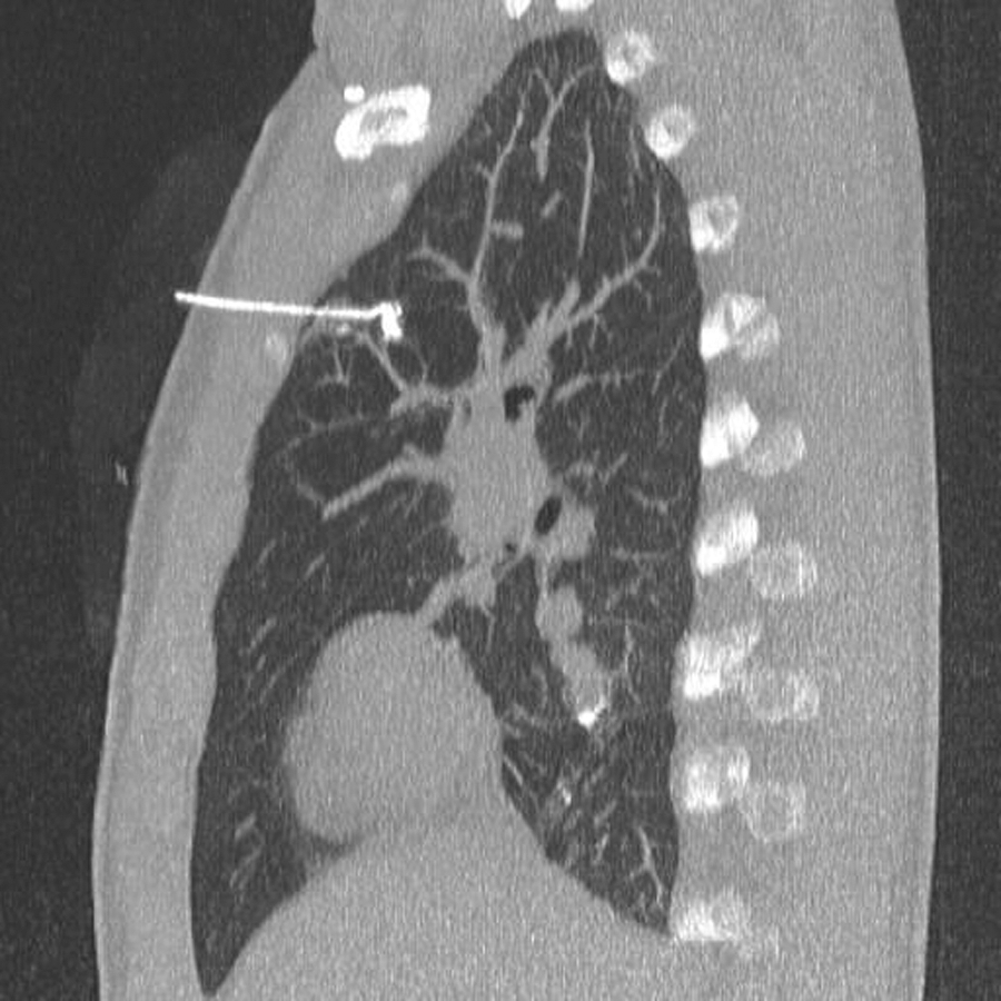
CT-guided wire-marking of the nodule. CT, computed tomography.
During the whole process of transportation and placement of the wire the patients were kept under general anesthesia and relaxation to prevent coughing and secondary dislocation of the wire.
Technical details of operation
The patients were placed in lateral decubitus position for a standard thoracoscopic intervention. Three port sites (1 × 12 mm, 2 × 5 mm) were established in a triangular setup. A primary survey of the lung and positioning of the marker was performed and the external fixation of the wire loosened before establishing one-lung ventilation. Gentle traction was applied to the wire to facilitate tenting of the lesion (Fig. 2). If this was not possible due to an instable fixation, the lung was elevated by forceps. Then a wedge-resection was performed using a linear endoscopic stapling device (Ethicon ETS 35; Ethicon Deutschland, Norderstedt, Germany). The specimen was placed in an endobag and removed from the thoracic cavity through the largest port or directly through the chest wall at the port site. If necessary this entry was widened in the style of a mini-thoracotomy.

Thoracoscopic view of lung tenting up.
A pleural drain was placed in all patients, which was removed by the second to third postoperative day.
Results
Six procedures were performed on five patients (four men and one woman) and a total of nine wire-marked lesions were excised (Table 1). The median age of patients was 16 years (range: 11–19 years). The primary cancer in four of the patients was an osteosarcoma of the lower extremities and a synovial sarcoma of the right thigh in the fifth patient. Lung metastases upon primary diagnosis were found in three patients, of which two had been treated primarily by thoracotomy. Apart from one case, all other lesions treated were metachronous appearances and not part of the initial treatment. The date of intervention was set at a median of 17 months after initial cancer diagnosis (range: 3–48 months). Time between diagnosis of the lesion to intervention ranged from 5 to 112 days with a median of 19.5 days and an average duration of 41 days. Excluding the primarily treated patient, as the delay was due to the neoadjuvant chemotherapy, the average duration sinks to 27.2 days.
Patient Demographics
M, male; F, female; L, left; R, right; B, bilateral.
In total, there were 10 nodules found in all patients of which 9 were marked preoperatively. The lesions were distributed evenly to both sides and bilateral affliction (right = 2, left = 2, bilateral = 2), there was a solitary nodule in three patients, two nodules in two patients and three nodules in one patient, who only had two of these wire-marked. In this patient bilateral marking was avoided due to adequate size and position to pleura of the contralateral nodule. Four of the lesions were set in the upper lobes and five in the lower lobes. All of the marked lesions were set deep to the pleural surface, making a preoperative marking necessary, with six of the lesions being smaller than 10 mm on CT scan. CT-graphic size did correlate well with the nodule size found upon histopathologic workup with a median of 1.25 mm difference. None of the nodules were in critical proximity to vital structures, they all allowed wire-marking.
Localization via CT-guided needle and coil placement was possible in all patients, with lesions being as small as 2 mm. The coil was described by the treating radiologist as being placed behind the lesion in all cases. In one patient the wire-marked resection was performed twice with a tumor-free interval of 6 months in between. Upon initial placement of the wires, no adverse events were reported. All wires remained stable during transfer to the operating room. There was one minor parenchymal hemorrhage. On reevaluating the scans we found a minimal pneumothorax of 5 mm without apparent influence on respiration in one patient and missing archived scans postmarking in one patient. No further intervention was necessary due to these events.
Thoracoscopy was performed on all patients, with a mini-thoracotomy being necessary in one patient (case 5) to retrieve the lung-tissue. Operating times ranged from 33 to 106 minutes with a median of 82 minutes (Table 2). In the three cases of solitary nodules, operating times averaged 58 minutes, as opposed to 105 minutes in the cases with multiple lesions. Upon surgery all wires were seated in the tissue, there was no reported wire slippage during surgical manipulation. In case 5, where both nodules were marked in the right upper lobe, significant adhesions and insufficient isolation of the marked areas necessitated a subtotal lobectomy and mini-thoracotomy to remove the specimen. Intraoperatively no increased blood loss or necessity of transfusion was documented, but wound surface was significantly larger than in wedge resections. The same patient developed anemia 3 days postoperatively and received 2 U of fresh frozen plasma, and 2 U of concentrated red cells. The anemia was most likely caused by diffuse bleeding over the pleural drainage.
Surgical Data
CT, computed tomography.
The median hospital stay between intervention and discharge was 5 days, ranging from 3 to 9 days.
Pathologic workup proved metastatic lesions in all resected nodules (Table 3), with the secondary lesion not being found in the resected upper lobe of case number 5. All other marked nodules were found in the resected tissue, with all resection borders being tumor-free. The sizes of resected nodules ranged from 2 to 26 mm with a median of 6 mm. Comparing CT-graphic sizes to histopathologic workup a median difference of 1.25 mm or 25% in size was found.
Nodule Characteristics
LLL, left lower lobe; LUL, left upper lobe; RLL, right lower lobe; RUL, right upper lobe.
All patients with osteosarcoma received regular follow-up postoperatively according to EURAMOSS/EUCOS protocol, including CT-studies to control outcome.
Case 1 and 3 are two interventions in the same patient, with case 3 being the primary intervention. After the first resection the patient did not show new metastatic disease in the CT-scans performed 1 and 4 months postoperatively. In the 6 months post-op scan a lesion was found in a different location. After resection of the nodule the patient showed massive progress of tumor growth with new metastases and involvement of hilar lymph nodes on the 1 month post-op scan. Within weeks the patient needed bronchial stenting, developed malignant pleural effusion, and died 7 months after surgery.
Case 2 had taken a 3 months post-op CT-scan, which showed multiple new pulmonary nodules. The patient was then operated on in a different hospital, where he underwent open metastasectomy. Within 6 months he again developed metastatic disease and had open metastasectomy redone. Since then the patient has been tumor-free for 6 months.
In case 4 no new nodules were seen for 18 months. The patient then developed two pulmonary lesions in a new location with pleural connection, which were resected thoracoscopically. After three more months the patient showed massive tumor progress. Open metastasectomy, pleurectomy, and partial resection of the pericardium, diaphragm, and the fourth rib were performed. Three months postoperatively a new pulmonary lesion was seen that disappeared under chemotherapy. At 5 months postoperatively the patient currently shows unclear pleural consolidation.
For case 5 no additional CT-scans of the lungs were performed due to new bone metastases of the skull being found after thoracoscopic resection. The patient died within 9 months.
Case 6 showed new metastatic disease 4 months postoperatively and underwent open metastasectomy. The patient then went on to develop renal metastases and died of acute bleeding due to a brain metastasis 12 months after our intervention.
Discussion
Surgery is the method of choice in suspected pulmonary metastases in cancer patients. All patients had a known primary tumor with a high risk of developing pulmonary metastases and all resected lesions were proven as malignant by histopathology. As surgical resection of lung metastases shows a proven survival benefit in patients with osteosarcoma, compared to solitary chemotherapy, surgery will remain our primary goal.11,25
However, the difficulty of localization of pulmonary nodules remains. Preoperative marking of small indeterminate pulmonary nodules should be considered when the distance to the nearest pleural surface is >5 mm and in cases of lung nodules smaller than 10 mm. 15 On account of negative experiences with the inability to localize a lesion upon thoracoscopy, we set a generous indication for CT-marking in case of assumption of altered anatomy, caused by prior infections, surgery, adhesions, or pulmonary regions with bad visibility during thoracoscopy. For an experienced interventional radiologist correct identification and marking appears to be possible even in tiny lesions.
In most cases upon thoracoscopy, apart from the wire, nodules are invisible, proving the necessity of preoperative marking. Additionally, the wire also assumes active function during surgery as it allows manipulation. 3 Also, the wire being a fixed marker does not require an extra access to the patient and frees one hand of the surgeon when compared to alternative methods of localization, such as sonography or radiotracing. The possibility of dual localization with added methylene-blue reduces the risk of failure due to a single method, even though we did not experience wire dislodgement or false marking in our study population. 27
The risks of CT-guided marking of the lesions are low. Asymptomatic pneumothorax, as found in one of our patients, and parenchymatous bleeding appear to stand in no correlation to treatment and postoperative recovery. Although high accuracy is reported, we do not mark nodules close to vital structures such as heart, major blood vessels, and hilum, as critical complications such as tension pneumopericardium have been reported. 28 Risks associated with wedge-resection in peripheral nodes also appear to be low. In cases where more parenchymatous dissection is necessary, leading to larger wound surfaces, the risk of bleeding is increased. Therefore, we would advise to always primarily attempt tissue sparing wedge-resection. Also, there should always be diligent preoperative evaluation for the risk of adhesions.
Surgical operating times are short in thoracoscopic resections, which patients benefit from. Total anesthetic time including transfers and CT-marking, however, is longer. Still with an average postoperative stay of 5 days, our patients showed faster recovery than we usually see after thoracotomy. This further adds to the reduction of traumatization in patients with pulmonary metastasis and allows repetitive interventions if necessary. 25
Overall, the importance of well-coordinated organization needs to be heavily emphasized. There is not only a high skills level needed in the surgeon, anesthesiologist, and radiologist, but also communication and a well-structured planning of the whole procedure are essential to achieve successful treatment.
Concerning recurrence of disease, our numbers are too small for statements on the techniques effect on survival. Most of our patients, being secondarily metastasized osteosarcoma patients, already have a bad prognosis and a high likelihood of recurrent disease.25,29 Current studies addressing this issue in the adult population most commonly agree that palpation of the lung is necessary to resect all detectable disease. However, there is no evidence for a survival difference between thoracotomy and thoracoscopic resection.30,31 Greenwood and West therefore advocate the less traumatic approach. 31 We also support this approach in our pediatric patients. However, to definitively see whether this reduction in traumatic therapy and therefore improvement in quality of life outweighs the effect on survival, larger studies are needed.
In conclusion, preoperative localization via CT-guided coil-wire placement and thoracoscopic resection of small pulmonary nodules is a safe and effective procedure. There is little traumatization and fast recovery in the children, especially under the aspect of the low overall outcome in metastasized solid tumors in children.25,29 Nevertheless, close cooperation of radiologists, anesthesiologists, and surgeons is needed to provide successful identification and resection of small pulmonary nodules in children.
Footnotes
Acknowledgment
The present work was performed in partial fulfillment of the requirements for obtaining the degree “Dr. med.”
Disclosure Statement
No competing financial interests exist.