




Abstract
Abstract
Objective:
To compare the clinical effectiveness of two approaches of minimally invasive esophagectomy (MIE) in esophagectomy patients.
Materials and Methods:
We retrospectively screened the perioperative data in a total of 49 patients who underwent MIE. Among them, 30 patients underwent thoracoscopy combined with laparoscopy (Group A), while the rest received mediastinoscopy combined with laparoscopy (Group B). Clinical effectiveness and postoperative complications were comparatively analyzed.
Results:
The patients with mediastinoscopy showed a shorter average surgical time, less blood loss during surgery, diminished drainage volume in the first 3 days after surgery, and reduced hospitalization time, compared with the patients with thoracoscopic approach (P < .05). However, the mediastinoscopic route seems to render a higher incidence of postoperative hoarseness than thoracoscopy combined with laparoscopy (P < .05). In mediastinal lymph node dissection, the number of right recurrent laryngeal nerve lymph nodes (RLN LNs) in mediastinoscopic route was significantly less than that of thoracoscopy (P < .05).
Conclusion:
Mediastinoscopy was less invasive, with shorter surgical time, faster recovery, and better patient comfort, although it had certain limitations in the dissection of right RLN LNs. It is beneficial to patients with poor pulmonary function and no obvious mediastinal lymphadenectasis.
Introduction
E
Several routes have been practically used to perform MIE, that is, thoracoscopic, laparoscopic, mediastinoscopic, hybrid, total, and robotic-assisted MIE. Compared with traditional esophagectomy, mediastinectomy has the advantages of not only avoiding thoracotomy and reducing bleeding but also removing mediastinal lymph nodes completely. 8 Presently, two alternative routes of MIE, that is, pneumatic mediastinoscopy combined with laparoscopic resection and thoracoscopy combined with laparoscopic resection are clinically used according to the route of entrance. The preferential MIE access largely depends on the experience and the operative skills of surgeons who may generally favor and stick to one of certain surgical methods. So far, systematically comparison of MIE with different access route in the context of surgical period and postoperative complications remains still incomplete.
The purpose of present study was to retrospectively compare the clinical effects of video-assisted mediastinoscopic resection and video-assisted thoracoscopic resection in 49 esophageal cancer patients and to reveal whether and to what extend these two surgical approaches impact the clinical effectiveness and postoperative complications.
Materials and Methods
Clinical data
A retrospective analysis was made on a total of 49 esophageal cancer patients who received surgical resection by MIE from October 2016 to August 2017 in Changzheng Hospital Affiliated to the Second Military Medical University. Of the 49 patients, 30 underwent thoracoscopy combined with laparoscopy (Group A), while the rest 19 received mediastinoscopy combined with laparoscopy (Group B). All the patients had the anastomotic site at the neck and none of the patients received preoperative neoadjuvant therapy. Pathologic tumor stages were grouped according to the American Joint Committee on Cancer (AJCC) 8th edition TNM staging system. Clinical effects, including surgical time, total blood loss during surgery, number of lymph node dissection, amount of drainage 3 days after surgery, visual analog scale (VAS) scores the first and fifth day after surgery, rate of hoarseness after surgery and recovery time, postsurgical complications, and hospitalization days after surgery were documented.
For the comparison of two methods, we included patients with following criteria: (1) tumor length was ≤60 mm, and preoperative examination revealed that lymphadenectasis or distal lymphatic metastasis could be excluded; and (2) the patients had no history of thoracic surgery on the affected side and had no presurgical adjuvant chemotherapy and radiotherapy. Surgical operations of the two groups were all performed by the same team of surgeons.
Anesthesia and surgical methods
A single lumen endotracheal tube was used in general anesthesia. The patients in Group A first took the left lateral decubitus position and four trocars were placed. The first 10-mm trocar for the camera was inserted in the seventh or eighth intercostal space on the midaxillary line and the incision was 1.5 cm; the second and third 5 mm trocars were inserted in the sixth and ninth intercostal space on the posterior axillary line as working trocars for the assistant and the incisions were all 1 cm; the fourth 10 mm trocar was inserted in the fourth intercostal space on the anterior axillary line for the instrument and the incision was 1.5 cm. Insufflation with carbon dioxide with a positive pressure of 8–10 cmAq was initiated to allow for an adequate view of posterior mediastinum. The mediastinal pleura was cut longitudinally to separate the esophagus. The arch of the azygos vein was divided and then cut with endoscopic vein vascular stapler. The entire intrathoracic esophagus was mobilized from the thoracic inlet to the diaphragmatic reflection. The adipose tissue around the esophagus, the surrounding lymph nodes, subcarinal lymph nodes, and para-recurrent laryngeal nerve lymph nodes (RLN LNs) were also resected completely. The esophagus was exposed down to the diaphragmatic hiatus and up to the neck. Upon completion of chest surgery, two negative pressure absorbing balls were placed on the inferior and superior mediastinum, respectively.
The patient was then turned to the supine position. The observation hole should be around 1.5 cm at the left of the umbilicus. A 10 mm trocar was put in, and the camera was inserted. One main working port was made on the lateral margin of the right rectus abdominis and a 10 mm trocar was put in with an incision of 1.5 cm. The surgical hole should be close to the bilateral clavicle midline. Two 5 mm trocars were put in the two ports, which were located at midclavicular line under the left and right costal margin, and a 5 mm trocar was inserted just inferior to the xiphoid as a working trocar for the assistant. Then, a pneumoperitoneum with CO2 at a pressure of 10–12 cmAq was established. Then the operator performed laparoscopic lower esophageal dissection, gastric mobilization, and abdominal lymphadenectomy. After enlarging the diaphragmatic hiatus, the lower esophagus was divided through the diaphragmatic hiatus till the level of inferior pulmonary vein. After abdominal operation, the incision inferior to the xiphoid was further extended to form a standard gastric tube. Gastric tube was completed along the greater curvature, and the esophagogastric junction was then dissected. Then, the gastric tube was pulled to the neck, with esophagogastric anastomosis by using 21 mm circular anastomat (Ethicon Endo-Surgery, Guaynabo, PR). In the end, one negative pressure absorbing ball was placed on the splenic recess, and the other was placed in the upper mediastinum through the neck incision.
In Group B, a 5 cm incision was made on the medial margin of the left sternocleidomastoid muscle. After separating the esophagus from the neck segment, a home-made 70 mm neck seal protector was put in place (Fig. 1). A 10 mm trocar was placed at the thumb position on the protective glove as the observation hole and the ring finger and small finger positions were placed in two 5 mm trocars as the operating holes. Then, insufflation with carbon dioxide at a pressure of 8–10 cmAq was initiated. The loose tissue around the esophagus, lymphatic, and anathreptic blood vessels were safely coagulated with 5 mm laparoscopic curved shears (LCS; Ethicon Endo-Surgery). Then mediastinoscopy was gradually moved to the distal esophagus (Fig. 2A). The thoracic lymph nodes were exposed and dissected (Fig. 2B). After thoracic esophageal dissociation was completed, abdominal surgery was performed in the same way as Group A. The negative pressure ball was placed in the neck incision. After surgery, patient-controlled pump analgesia was used in all the patients.
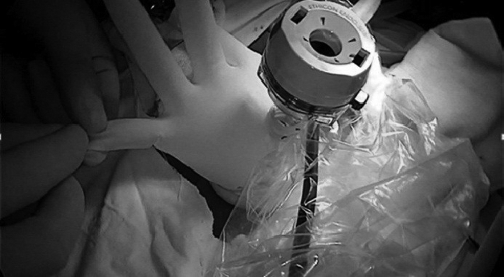
Home-made 70 mm neck seal protector. A 10 mm trocar was placed at the thumb position on the protective glove as the observation hole, and the ring finger and small finger positions were placed in two 5 mm trocars as the operating holes.

Views through the mediastinoscope looking from the neck down into the abdomen.
Evaluation of medical data
The entire period of surgery, total blood loss during surgery, number of lymph node dissection, amount of drainage 3 days after surgery, VAS scores the first and fifth day after surgery, rate of hoarseness after surgery and recovery time, postsurgical complications, and hospitalization days after surgery were compared between the two groups.
Statistics
The patient's data were collected and analyzed by using the SPSS19.0 statistical software. The data are expressed as mean ± standard deviation, and the comparative analysis between the two groups were performed by using unpaired t-test. The categorical variables were expressed as frequencies and analyzed by using χ2 test. The difference was considered as statistically significant when P < .05.
Results
All the surgeries were successful in both groups, and there was no case of death during perioperative stage. There were no statistical differences in age, gender, tumor site, TNM stage, or pathological type when comparisons were made between the two groups (P > .05). The patients' information is summarized in the Table 1. As tableted in Tables 2 and 3, surgical time, blood loss during surgery, amount of drainage 3 days after surgery, hospitalization days after surgery, and VAS scores of Group B were all superior to those of Group A (P < .05). With the exception that the incidence of such complication as postsurgical hoarseness in Group B was significantly higher than that in Group A (P < .05), there was no statistical significance in the rate of other complications (P > .05).
Patient Demographics and Tumor Sites of the Two Groups
Comparison of Perioperative Data of Two Groups of Patients
VAS, visual analog scale.
Comparison of Postsurgical Complications in the Patients of the Two Groups
In mediastinal lymph node dissection, the number of right RLN LNs in Group B was significantly less than that of Group A (P < .05). There were no statistical differences in the number of dissected lymph nodes around esophagus, subcarinal lymph nodes, left RLN LNs and the number of positive lymph nodes, when comparisons were made between the two groups. The statistical analysis is summarized in Table 4.
The Comparison of Lymph Node Dissection Between the Two Groups
RLN LNs, recurrent laryngeal nerve lymph nodes.
Discussion
With the updating and upgrading of endoscopic systems and surgical instruments over the last decade and repeated clinical practice by surgeons, thoracoscopy combined with laparoscopy is a surgical skill mastered by more and more thoracic surgeons. However, due to poor pulmonary function of certain patients, they can by no means tolerate open thoracic surgery. In such a case, the minimally invasive surgical method of mediastinoscopy combined with laparoscopy is the preferred option as it could shorten surgery time. In addition, two-lung ventilations in mediastinoscopy are beneficial to patients with poor lung function, whereas one-lung ventilation is necessary in thoracoscopic surgery. Therefore, the video-assisted mediastinoscopy can be safely implemented in patients with compromised pulmonary function.9,10 As pneumatic mediastinoscopy is applied with a pressure of 8–10 cmAq, a broader and better surgical field vision in mediastinal anatomic structure could be obtained. 11 Through repeated practice, we have found that video-assisted mediastinoscopy has the following potential advantages: (1) keeping integrity of the pleural membrane through cervical approach and reducing stimulation to the lung and heart9,10; (2) easier anesthesia compared with that in the conventional thoracoscopy and prevention of adverse effects of single-lung ventilation on the patient during surgery, and as a result, requirements on cardiopulmonary functions of the patient are reduced; (3) avoidance of posture change and further sterilization, resulting in shorter surgical time and less anesthetic effects; (4) significant decrease in VAS scores after surgery with higher sense of comfort 12 ; and (5) less trauma, less blood loss during surgery, lower volume of drainage after surgery, and fast recovery. 13
At the same time, we have also found that there are disadvantages or limitations in video-assisted mediastinoscopy: (1) limited manipulating space and field vision, so skilled surgeons are required 12 ; (2) compared with thoracoscopy, mediastinoscopy may encounter difficulty in the dissection of mediastinal lymph nodes, especially for the right RLN LNs that cannot be dissected by routine methods, so a right neck incision is necessary for further dissection. Therefore, it is particularly important for the surgeons to evaluate the clinical stages and choose proper cases before surgery in the light of patient symptoms and radiographic scanning.14,15 Mediastinoscopy is suitable for patients without obvious enlargement of mediastinal lymph nodes; and (3) higher incidence of hoarseness which mostly occurred 1–2 days after surgery and all the cases recovered 6 months after surgery, while in the patients with video-assisted thoracoscopic surgery, there were fewer cases of hoarseness, but often with delayed recovery. Video-assisted mediastinoscopy can be an ideal approach for the exposure of bilateral recurrent laryngeal nerve, 16 although it is improbable to disrupt RLN through manipulation itself and surgical routes.
Evidences have showed that cervical lymph node dissection, right recurrent nerve lymph nodes dissection, reconstruction routes, and sites of anastomosis were identified as independent risk factors for recurrent laryngeal nerve paralysis. 17 With the exclusion of other routine factors for the lesion of RLN, we consider that higher incidence of hoarseness might be attributed to the following factors: (1) Close surgery to esophagus and compression or stretching of adjacent tissues during surgery could lead to lesion and edema of nerve tissues. (2) The dissection of RLN LNs may overexpose RLN, affecting local blood supply to nerves and resulting in a high incidence of hoarseness 1–2 days after surgery. When compensatory blood circulation is established, symptoms would be gradually improved.
Conclusion
These two methods of radical esophagectomy are technically safe and effective. Pneumatic mediastinoscopy combined with laparoscopic resection has the features of less trauma, shorter surgical time, faster recovery and higher sense of comfort, although it has limitations in the dissection of right RLN LNs. Therefore, mediastinoscopy is beneficial to patients with poor pulmonary function and no obvious mediastinal lymphadenectasis.
Footnotes
Acknowledgments
This work was supported by the Shanghai Education Development Foundation and Shanghai Municipal Education Commission [16SG32], and the Nature Science Foundation of Shanghai [16ZR1436900].
Disclosure Statement
No competing financial interests exist.