
Abstract
Abstract
Introduction:
Cholecysto-choledocolithiasis is a rare entity in children and its management is still challenging and controversial. The laparoendoscopic rendezvous (LERV) procedure, consisting of laparoscopic cholecystectomy and simultaneous endoscopic retrograde cholangiopancreatography for the management of symptomatic choledocholithiasis is well described in adult patients. However, in the literature, few reports about its application in the pediatric population have been recorded.
Aim of the Study:
The aim of the present study is to report our first successful cases of symptomatic cholecysto-choledocholithiasis LERV treatment.
Methods:
Two girls suffering of hemolytic disease presented to our third referral center with acute abdominal pain due to cholecysto-choledocholithiasis. Preoperative, perioperative, and postoperative data were retrospectively reviewed.
Results:
Surgery was performed without complications. The girls were dismissed once re-alimentation and re-canalization were achieved and had no other episodes of biliary cholic. Reported advantages of LERV include: a shorter in-hospital stay, a reduction in the number of procedures and anesthesia, and a reduced overall risk of complications.
Conclusions:
The promising result with our 2 cases suggest that, when performed in highly specialized centers, LERV is a safe procedure, which leads to considerable benefits, despite logistic and organizational difficulties.
Introduction
Cholecysto-choledocolithiasis is an uncommon event in pediatric population and its management is still debated. Although laparoscopic cholecystectomy (LC) is commonly performed nowadays, the treatment of choledocholithiasis is still controversial. The “Laparoendoscopic Rendezvous” (LERV) procedure is a combined approach in which LC occurs simultaneously with endoscopic retrograde cholangiopancreatography (ERCP) and endoscopic Oddi's sphincterotomy. This approach minimizes the risks of early and late-onset complications after endoscopic procedures on Vater's papilla for the management of symptomatic choledocholithiasis. ERCP, as well as LC, is considered a demanding technique in pediatric patients. These procedures are considered safely conducted only in highly specialized centers.1,2 However, primarily for epidemiological and technical reasons, their performance is still far less common than in the adult population and even rarer is their association in a one-stage procedure.
This article reports the first short series of pediatric patients successfully treated with LERV in a single pediatric institution.
Materials and Methods
Both procedures were performed at the Pediatric Surgery Department of Padova University Hospital (AOU, Azienda Ospedaliera Universitaria di Padova) by a fully trained surgeon in LC, and an endoscopist with expertise in pediatric ERCP. Both patients underwent general anesthesia, performed by a pediatric anesthesiologist. Data on indications, preoperative assessment and final diagnosis, liver and pancreatic function tests before and after procedure, imaging, analgesic and antibiotic treatment, and postoperative follow-up have been collected.
Results
Two females, of 11- and 10-years of age respectively, were admitted in our Center for acute vomiting and progressive upper quadrant abdominal pain. In the first case, the patient had already a diagnosis of sickle-cell disease. Blood tests showed: cholestasis (total bilirubin 21 mg/dL, conjugated bilirubin 15.4 mg/dL, Gamma-glutamyl transferase (gGT) 376 U/L, Aspartate Aminotransferase (AST) 216 U/L, Alanine Aminotransferase (ALT) 813 U/L) and elevation of pancreatic enzymes (amylase 988 U/L, lipase 3420 U/L). In the suspect of cholecysto-choledocholithiasis complicated by acute pancreatitis, ultrasound (US) and magnetic resonance cholangiopancreatography (MRCP) were performed. The latter exam revealed several gallstones in the gallbladder and in the biliary tree together with a dilated common bile duct (CBD) (8 mm). No Wirsung dilatation was present. The patient of the second case already had a diagnosis of hemolytic anemia and was already in treatment with ursodeoxycholic acid (UDCA) for previous episodes of abdominal pain due to biliary gallstones. Despite medical treatment, she was admitted in our emergency room for a further episode of biliary colic associated with jaundice and hyperchromic urine. Blood tests revealed isolated cholestasis markers (total bilirubin 15.9 mg/dL, conjugated bilirubin 9.8 mg/dL, gGT 143 U/L, AST 386 U/L, ALT 771 U/L), in the absence of pancreatitis. US and MRCP showed a large steatotic liver, a hydropic gallbladder filled with several calculi and a dilation of the extrahepatic biliary tree (common hepatic duct 11 mm, CBD 9 mm), associated with the presence of a small calculus (2 mm) in the intrapancreatic tract of the CBD. No Wirsung dilatation was reported.
In both patients cholecysto-choledocholithiasis was consequently diagnosed. Indication for the LERV procedure was given. The procedure was performed as described in literature. 3 Patients were set in supine “French” position. Three 5 mm trocars were inserted with Hasson technique and the cystic duct was isolated. A percutaneous 5 Fr catheter was then inserted in the cystic duct under direct laparoscopic vision (Fig. 1). A guidewire was advanced through the catheter into the duodenum and the catheter was clipped to the cystic duct. The first jejunal loop was identified and clamped to avoid bowel insufflation during the endoscopic procedure. Duodenoscopy was then performed with an Olympus JF-140 R duodenoscope®. The guidewire was advanced into the duodenum and it was pulled out of the patient's mouth. The sphincterotome was then introduced along the guidewire, the papilla was directly cannulated, and sphincterotomy was then performed. Once the sphincterotomy was completed, a Dormia basket was introduced into the papilla and CBD along the same guidewire, to clean the biliary tree. This procedure was repeated twice in both patients, although only in one of them a direct outflow of calculi and biliary sludge from the papilla was observed. The guidewire was removed through the mouth, and the endoscopic procedure was ended. LC was finally completed after clipping of the cystic duct. After extraction of the gallbladder from the umbilical port, ERCP was repeated again: contrast infusion in the papilla showed CBD's clearance (Fig. 2).

Cannulation of the cystic duct.
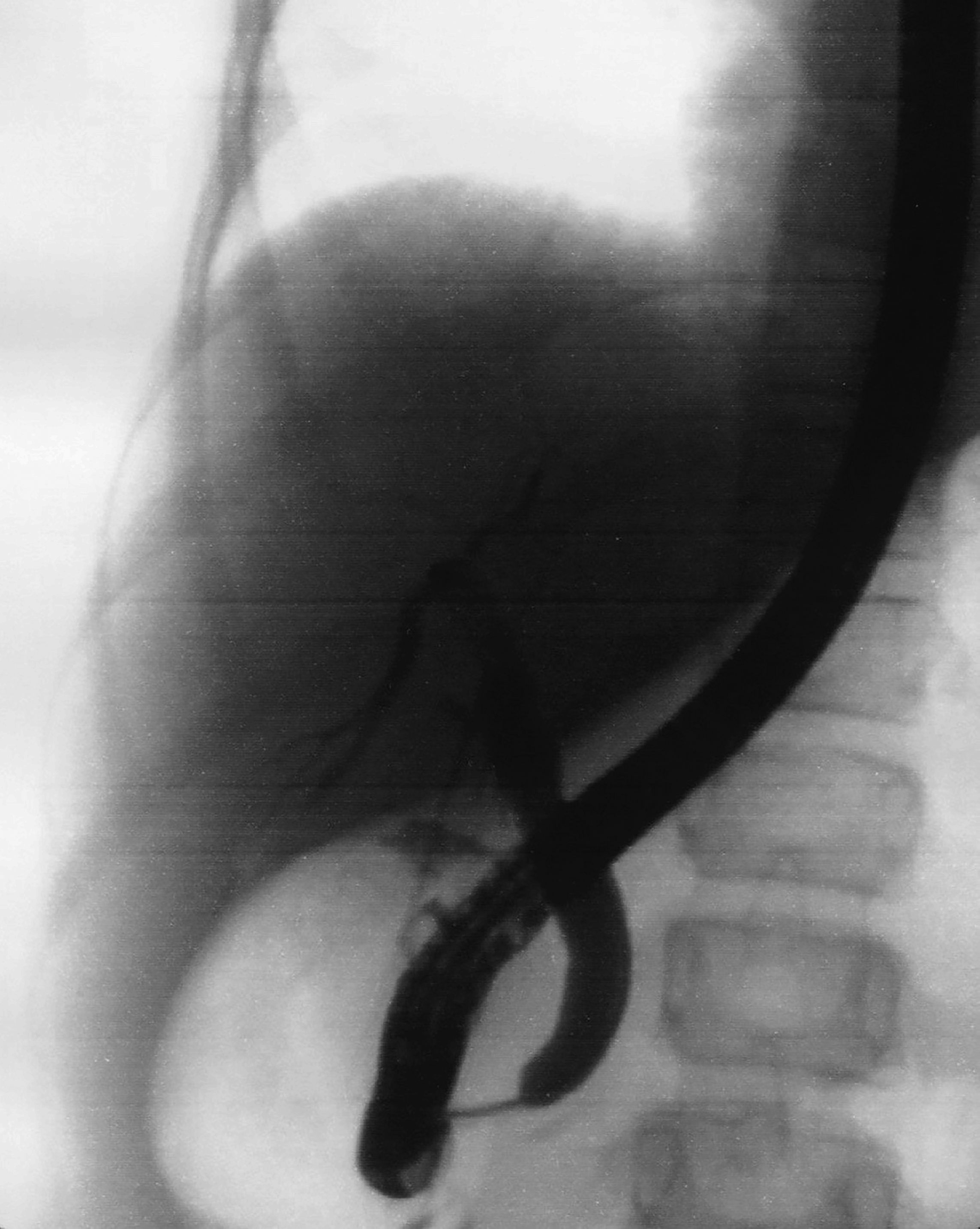
Intraoperative cholangiogram.
Mean surgery time was 147.5 minutes and no intraoperative complications were observed. In postoperative management, fasting was maintained for less than 24 hours in both patients and oral painkillers were needed for an average of 6.5 days. The length of stay after surgical procedure was of 6 days in the case in which pancreatitis coexisted. In this case, intravenous antimicrobial therapy with Cyclosporine was given during hospitalization and oral therapy with Amoxicillin/Clavulanic acid was continued for 2 weeks after discharge. On the contrary, in the second case, intravenous therapy was given only perioperatively in three administrations and the length of stay was of 5 days only. The girls were dismissed once realimentation and recanalization were achieved. UDCA treatment was continued for 3 months after discharge. Two weeks after the procedure, blood exams were performed as follow-up tests and showed normalization of cholestasis and pancreatitis indexes in both cases. The 2 girls had no further episodes of biliary colic.
Discussion
The advantages of the LERV technique for the treatment of cholecysto-choledocholithiasis are well known and described in adult patients. A high success rate, a shorter in-hospital stay, the requirement of a single anesthesia, and a lower incidence of post-ERCP pancreatitis are the main benefits. 4 A recent meta-analysis regarding management of gallstone disease with ductal calculi conducted in an adult population, compared four different surgical approaches: pre-LC ERCP, LERV procedure, post-LC ERCP, and LC with laparoscopic common bile duct exploration (LCBDE); LERV procedure was found to be the most successful and safest technique. In particular, when compared with LC-LCBDE, it avoids the risk of biliary leak and/or the need of a biliary drainage. 5 Thus, LERV procedure is the treatment of first choice in our Center. Up to date only one case of pediatric rendezvous 6 and a few cases of pediatric LCBDE are reported in literature. In particular, Lau et al. reported a successfully performed LCBDE using a cholangiography or CBD Exploration Kit. 7 Nevertheless, these approaches seem considerable alternatives for ductal calculi treatment. They require high training and expertise, thus their application remains limited because of the small population in which they might be performed.
A standardized technique is still needed, especially considering the recent increase in incidence of these pathologies. In particular, in the United States, the prevalence of cholelithiasis in children younger than 16 years was 0.15% in the sixties, being estimated around 1.9%–4% in 2012.8–10 Cholelithiasis in the infant is related to prematurity, total parenteral nutrition use, abdominal surgery, or sepsis. Hemolytic disease was historically considered the most prevalent comorbidity in the adolescent. The recent incidence increase in teen-pregnancy, oral contraceptive use, and obesity has changed adolescent's epidemiology of gallbladder disease, increasing its frequency and, accordingly, the number of cholecystectomies.11–14 Even if in our 2 cases, the hemolytic disease underlined the gallbladder disease, the rendezvous procedure can be applied to all causes of cholecysto-choledocholithiasis.
ERCP application in children has been limited for many years, because of the lack in development of small fiberoptic duodenoscopes. 15 Despite the disadvantages of both patient and equipment sizes, ERCP success rates in children of a wide range of ages are estimated to be between 89.5% and 100%, and these results are comparable to the adult patients.16,17 The most common complication of ERCP is pancreatitis, however, other nonspecific complications of endoscopy (i.e. hemorrhage, perforation, infections) are possible. 18 The selective cannulation of the CBD is known to be one of the most difficult maneuvers in ERCP and can often end in failure of the procedure or lead to pancreatitis for multiple attempts or accidental pancreatic duct cannulation and contrast injection. In the LERV procedure, the direct view of the laparoscopic camera, ensures an easy, selective, and safe cannulation. This represents the major technical advantage of LERV when compared with the two-step procedure. 19 A meta-analysis reported that there is no significant difference in the clinical outcome of patients undergoing one- versus two-step procedure. Nevertheless the two-stage approach was shown to have significantly higher costs, related to a longer length of hospital stay or two distinct hospitalizations, and to the requirement of two procedures. 20
The main disadvantages of the LERV technique are logistics, demanding the simultaneous availability and presence of surgical and endoscopic teams and their equipment. Moreover, in the particular case of the pediatric population, in addition to a pediatric surgeon with adequate laparoscopic expertise, a trained pediatric endoscopist is needed, contributing to the organizational issue. However, despite the abovementioned difficulties, in both our cases, the surgery time was short and in accordance with the literature. 3 The length of stay was brief, as well as postsurgical fasting time, and it was possible to dismiss our patients in 5–6 days. Moreover, no more hospital accesses or surgical procedures were needed.
Conclusions
The frequency of gallbladder disease in pediatric population is expected to rise in the next years. The LERV procedure appears to be a valid tool for the treatment of cholecysto-choledocholithiasis, as shown in our two cases. In fact, LERV does not only reduce the costs for the institution but it also decreases the risk of complications, such as post-ERCP pancreatitis and infection while exposing the patient to only one general anesthesia and its related implications. The need of an expert and trained team specialized in children health care remains a crucial element, along with proper organizational skills. These two aspects continue to be the main obstacle to a larger application. In conclusion, the LERV procedure seems to be a valid therapeutic option for the treatment of cholecysto-choledocholithiasis, particularly in children, and should be considered as first-line treatment in centers where LC and ERCP are already separately performed. Nevertheless, for its intrinsic surgical, endoscopic, and managerial complexities, it should be performed only in highly specialized centers.
Footnotes
Disclosure Statement
No competing financial interests exist.