
Abstract
Abstract
Background:
This study aimed to investigate the efficacy and safety of retroperitoneal laparoscopic tuberculous nephrectomy (RLTN) and open tuberculous nephrectomy (OTN).
Materials and Methods:
One hundred fifty-eight patients treated with RLTN were enrolled in the RLTN group. One hundred patients treated with OTN were enrolled in the control group. Surgical parameters and postoperative conditions were recorded to assess the effect of the operation, and complications were recorded throughout the follow-up time. The follow-up time was 2–72 months.
Results:
Eight cases in the RLTN group were moved to the OTN group due to intraoperative bleeding. There were significant differences in the diameter of the incision between the 2 groups (P < .01). The resumed time and length of hospitalization in the RLTN group were significantly shorter than those in the OTN group (P < .05). During surgery, 6 cases (3.79%) in the RLTN group presented pus overflow due to a rupture of the empyema space. The same happened to 3 cases (3.00%) in the OTN group. After surgery, there was 1 case of abdominal tuberculosis (TB) in the RLTN group. There was no tuberculous sinus or systemic disseminated TB during the follow-up period.
Conclusions:
Both treatment approaches were safe and effective, but RLTN presented more advantages. Therefore, RLTN can be used as a first-line method for tuberculous nephrectomy.
Introduction
Retroperitoneal laparoscopic nephrectomy has advantages such as clear operative view, moderate injury, reduced bleeding, low number of complications, and fast recovery, and, therefore, it is the first-line treatment of nephrectomy.1–4 Advanced renal tuberculosis (TB) usually has serious perirenal adhesion due to a strong inflammatory response. In the past, because of the difficulty to dissect the renal pedicle, open surgery was preferred in clinical practice. 5 With the recent developments in laparoscopic operative technology, retroperitoneal laparoscopic tuberculous nephrectomy (RLTN) has gradually replaced conventional open operation.6–8 Moreover, only a few studies focused on the RLTN procedure for the treatment of tuberculous kidneys. This study uses a large sample size to assess the clinical efficacy and safety of the two surgical methods. Moreover, the surgical experience of RLTN is also summarized in this study.
Materials and Methods
Patients
This study has been approved by the Ethics Committee of General Hospital of PLA. All subjects gave their consent to participate in this study. Between January 2004 and August 2015, a total of 158 patients treated with RLTN were enrolled in the RLTN group, whereas 100 patients treated with open tuberculous nephrectomy (OTN) were enrolled in the control group. Groups were not divided randomly but according to the laparoscopic technique used at the time of surgery. Before 2010, open surgery was the treatment of choice for nephrectomy; however, after 2010, laparoscopy has become the first choice for the treatment of nephrectomy. Only a few special cases were intraoperatively switched into conventional open surgery. Demographics and baseline clinical characteristics of patients in both groups are shown in Table 1. Among them, 25 patients presented pulmonary TB (9.7%), and 138 cases previously had pulmonary TB (53.5%). TB of the male reproductive tract was observed in 54.8% of patients. Renal TB was identified by preoperative contrast-enhanced computed tomography (CT; or magnetic resonance imaging [MRI]) and laboratory tests. Postoperative pathology results showed renal and ureter TB. Operative parameters and postoperative conditions in both groups were periodically recorded. All patients were available for regular follow-up, and complications were recorded.
Baseline Demographics in Both Groups
ASA, American Society of Anesthesiologists; BMI, body mass index; OTN, open tuberculous nephrectomy; RLTN, retroperitoneal laparoscopic tuberculous nephrectomy.
Enhanced-contrast CT features of renal TB
Multiple cystic low-density shadows without enhancement were seen in the involved kidney. There was calcification on the wall of some holes and the slightly intensified surrounding renal cortex, representing a change known as “hoof of the Gervus nippon Temminck.” The wall of the ureter was thick and stiff and was usually accompanied by ureterectasia (Fig. 1). There were 204 patients with unilateral renal TB and 54 patients with bilateral renal TB.

CT characteristics of end-stage renal tuberculosis. Typical CT characteristics represent as multiple cystiform hypodense shadows as petal-shaped arrangement, classification, enhancement slightly around the renal cortex, accompanied by dilatation of ureter, thickening of the wall.
Anti-TB therapy
After at least 15 days of regular anti-TB therapy, the following regimen was followed: (1) rifapentine capsule (Wuxi Fortune Pharmaceutical Co., Ltd., WuXi, China), 0.45 g daily, twice a week; (2) para-aminosalicylic acid isoniazid (Chongqing Huapont Pharmaceutical Co., Ltd., ChongQing, China), 0.3 g three times a day; (3) ethambutol hydrochloride (Shenyang Hongqi Pharmaceutical Co., Ltd., LiaoNing, China), 0.75 g daily; (4) pyrazinamide (Aiwan City of Guangdong Pharmaceutical Co., Ltd., GuangDong, China), 0.5 g three times a day; and (5) levofloxacin [Daiichi Sankyo (China) Holdings Co., Ltd, Beijing, China], two tablets twice daily. Prompt adjustments to treatment should be made based on the tolerance and adaptation of the patient. There was no change in anti-TB treatment before and after surgery. Routine anti-TB treatment was continued for 6–9 months after the operation. The anti-TB treatment was withdrawn if any of the following occurred: no TB was observed in imaging examination, negative routine urine examination, multiple negative urine examinations for tubercle bacillus, and negative ESR (erythrocyte sedimentation rate) and C-reactive protein.
Operative indications
The indications for the operative treatment were as follows: (1) a widely destroyed and malfunctioned kidney; (2) a calcified nonfunctional tuberculous kidney; (3) in cases of bilateral renal TB, the more seriously damaged side could be excised; (4) in patients with unilateral renal TB, there should be severe TB lesions and multiple narrowness and failure in the placement of the Double-J ureteral catheter.
Laparoscopic procedure
OLYMPUS was the manufacturer of the laparoscopy. After tracheal intubation and general anesthesia, the patient assumed a lying position on the healthy side. In the RLTN group, a 1.5-cm incision (A point) was vertically made at the junction 1 cm below the costal margin and crossed by the posterior axillary line; another1.5-cm incision (B point) was made at the site 1.5 cm above the crista iliac of the midaxillary line; a third incision was made at the anterior axillary line below the coastal margin (C point); the line connecting the three points (A, B, and C) was used to form a equilateral triangle. A 4.5-cm incision was made along the line connecting the point 1 cm below the arch of rib on the midaxillary line and the point 1 cm above the iliac crest in the monoporate operation. The retroperitoneal space was accessed instinctively through a cutdown. A modified single-port laparoscopic trocar was sited, and three casing trocars were sited, respectively, at the different fingerstalls (two 10-mm long and one sized 5-mm long), which constructed the operational access (Fig. 2). 7
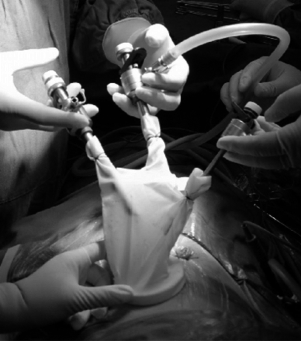
The surgical of domestic single-port retroperitoneal laparoscopic nephron.
Under the laparoscope, the perirenal fascia and fat were vertically dissected, and the kidney was dissociated from the surface of renal parenchyma. Dissociation was finished using the harmonic scalpel as sharply as possible to excise all adhesive tissues. Excessive pulling was not recommended to prevent kidney rupture and bleeding. The renal pedicle could be found between the psoas major muscle and the middle of the renal back, which was often disturbed by the inflammatory swollen lymph node. The renal artery was relatively difficult to be freed. After the adipose connective tissue surrounding the renal pedicle and some vagrant and lymph vessels were dissociated into bundle-like form along their structure, tissues were cut using the harmonic scalpel or after clamping by hem-o-lok clip. When there were no other tissues present, and after clamping of renal artery and vein by hem-o-lok the renal artery and vein could be cut. The ureter was freed downward as much as possible and dissociated at the lowest position after clamping with hem-o-lok clip. If the pipe diameter of the ureter was significantly thick and could not be clamped with hem-o-lok clip, the ureter would be ligatured using the No. 7 thread under laparoscope vision, and then it was dissociated. When there were serious adhesion and errhysis, it was suggested that the back of kidney could be first freed to separate renal artery. After artery was clamped with hem-o-lok clip, other parts of the kidney could be freed carefully. The removed renal specimen was put into a renal bag, and 1 g streptomycin and a laparoscopic indwelling drainage tube were placed on the incomplete cavity.
Open tuberculous nephrectomy
The anesthesia and body position in OTN group were similar with RLTN group. A transverse incision was made below the costal margin of the 12th rib at the left or right upper abdomen. The skin, subcutaneous tissue, obliquus externus abdominis muscle, and obliquus internus abdominis muscle were dissected to find the peritoneum that was pushed open upward. The kidney and renal vessel were freed as the RLTN group.
Statistical methods
In this study, data were analyzed using SPSS version 18.0 (SPSS, Inc., Chicago, IL). Normally distributed data were presented as mean ± standard deviation and abnormally distributed data were presented as median and interquartile range. Variables were compared by independent samples t-test or correspondent nonparametric test. Enumeration data were expressed as n (%) and compared by Pearson's chi-squared test.
Results
Laboratory parameters
The ESR and C-protein level were, respectively, 43 mm/L (range: 6–80) and 15.5 mg/L (range: 4.0–25.0) in all patients. Concerning urine acid-fast bacillus test, there were 99 (38.4%) positive cases and 159 (61.6%) negative cases. Negative results of the urine acid-fast bacillus detection may be due to the fact that test was carried out after the anti-TB therapy.
Detection of TB was done using MycoDotTM three indices test and ICT-TB+ MycoDot+ fast card. Results showed 232 cases (89.9%) with one positive index and 112 cases (43.4%) with three positive indices. Assessment of the preoperative renal function showed a mean level of glomerular filtration rate at 37.45 mL/minute (range: 9.6–65.3) in all patients.
Perioperative period observation
There were 8 (5.1%) patients in the RLTN group that switched to open operation due to intraoperative bleeding. Reason for this was the serious adhesion in the renal region, with six at the right side and three with postcava avulsion. Laparoscopic operation was successfully performed in a total of 150 patients. The ESR of 12 patients (n = 258) still fluctuated at 70–80 mm/L after anti-TB treatment. After nephrectomy, patients were cured and discharged with normal ESR. There were no significant differences between the 2 groups in intraoperative blood loss, operation duration, and intraoperative complications (P > .05). The difference in incision size between the RLTN group and the OTN group was statistically significant (P < .01) (Table 2). Patients in the RLTN group had a shorter length of hospitalization and could eat normally in less time when compared with the OTN group (P < .05). During the operation period, pus overflow due to the rupture of the pus cavity was noted in 6 cases (3.79%) in the RLTN group and 3 cases (3%) in the OTN group. There was 1 patient with abdominal TB infection in the RLTN group after surgery. The retroperitoneal drainage tube was regularly retained during the operation. No tuberculous sinus tract or full-body disseminated TB occurred during the follow-up period.
Operative Parameters in Both Groups
OTN, open tuberculous nephrectomy; RLTN, retroperitoneal laparoscopic tuberculous nephrectomy.
Complications
There was no abdominal organ injury, diaphragm muscle injury, or other complications in both groups. Moreover, 5 patients with subcutaneous emphysema in the RLTN group solved it by self-absorption after operation. There was 1 patient with abdomen TB infection in the RLTN group after operation, which was cured after a month of anti-TB treatment. Drainage tubes were removed 24–72 hours after surgery in both groups, and no postoperative sinus formation was observed. Five cases with fat liquefaction necrosis at the site of incision emerged in the OTN group, which recovered after replacement of dressing.
Follow-up
The anti-TB drugs were continually administered for 6–10 months after surgery. No infection was observed and tuberculous sinus tract occurred in the nephridial pit for 37 months (range: 2–72 months) of follow-up. No full-body disseminated TB occurred.
Pathology
Two-hundred fifty-eight cases of nephrectomy were confirmed, by postoperative pathology, to be renal TB combined with ureter TB.
Discussion
In recent years, due to the increased prevalence of pulmonary TB and the emergence of drug-resistant strains of mycobacterium TB, the incidence of renal TB shows a gradual upward trend.9–11 There are no specific clinical characteristics in patients with renal TB, and that often leads to a delay in diagnosis and treatment. 12 In these cases and until diagnosis is confirmed, TB infection will severely injury renal function and, at the same time, the ureter will also be involved. 13 The conventional long-term conservative treatment with anti-TB drugs is ineffective; thus, the excision of lesions is necessary. 14 Endoscopic excision for renal TB still causes a lot of vice-damage, such as high rate of transfer and opening. With the development of laparoscopic urological surgery technology, RLTN has gradually replaced conventional open surgery.15–17 In this study, RLTN was completed in 158 cases based on the special anatomical characteristics of renal TB. The clinical safety and efficiency of RLTN was similar to that of conventional open surgery procedure. In terms of the incision's size, recovery time of bowel function after surgery, length of hospital stay, and so on, RLTN procedure with small damage and fast recovery was better than OTN. At the same time, a modified single-port laparoscopic operational access was formed with “incision protector and medical gloves,” which was used to complete the single-port RLTN in our study.
Because tuberculous nephrectomy is a contaminated surgery, the access through retroperitoneal space, and not through the abdomen, can significantly reduce abdominal infection. 18 During the retroperitoneal operation, if damage to the peritoneum occurs with rupture of the pus cavity in the tuberculous kidney, the peritoneum must be sutured and repaired (behind the kidney or under the endoscope). This procedure makes sure the peritoneal cavity and the retroperitoneal space remain relatively airtight to avoid abdominal infection. There was 1 case with intraoperative renal cortex rupture and pus overflow in our study, whose peritoneum was damaged seriously. Abdominal TB infection occurred due to the absence of peritoneal suture and repair. C-reactive protein and ESR are important parameters that are expected to reflect the condition of TB infection. Nephrectomy surgery is usually carried out when C-reactive protein and ESR are within the physiological normal range. However, due to renal injury and significantly narrow obstruction of the ureter TB, or even combined with infection, both anti-TB and anti-infection treatment are ineffective. In this condition, when the ESR and C-reactive protein are continually high, there is no need for the nephrectomy surgery until the infection is controlled. In this study, there were 12 patients with uncontrolled infection and serious consumption performed with laparoscopic tuberculous nephrectomy. After surgery, general body state in these patients recovered quickly with a good clinical efficacy. The assessment of the operation in such patients must be careful, because the tissue edema is very serious with a high probability of bleeding and vice-damage. 19
Owing to the enlargement of the pyonephrosis, the retroperitoneal space became much narrowed, leading to a significantly difficult use of surgical instruments and the single-port surgical procedure. In some patients with an end-stage tuberculous kidney, involved kidneys were significantly swollen in size and renal tissues developed completely into encapsulation. Because the tuberculous kidney filled with pus and the cyst walls were very fragile, an air bag with air injected into it was used as the retroperitoneal space dilator. The air bag should not be filled at excessively high pressure, as it can cause tuberculous kidney rupture. Owing to long-term specific infection and secondary mixed infection, there were many swollen lymph nodes at the renal hilus, which may cover the surface of renal pedicle, resulting in a difficulty in identifying the renal vessels and consequently bleeding of the lymph nodes. This situation might lead to a poor view of the surgical field and to a higher difficulty in the separation of renal pedicle vessels. During surgery, dissociation should be done carefully along the anatomic landmark. The upper and inferior pole of kidney should be freed first, which can help in the identification of the renal hilus. After, a local separation of the tissues at the renal hilus should be done carefully. The postcava or the ureter at the inferior pole of the kidney should be identified, and under endoscope, the renal hilus could be freed and identified in an upward direction along the postcava or the ureter. This procedure can be done with a combination of electrocautery and harmonic scalpel, with the electrocautery making anatomy clearer and the harmonic scalpel stopping the bleed with the slow gear. This is a safe and fast way to free the vessel.
When the cortex of the tuberculous kidney is thin, an external excision of the adipose capsule should be adopted to prevent the avulsion of the renal capsule. During laparoscopic surgery, violent squeezing must be avoided to prevent the rupture of the thin wall of the pus second overflow to the fester. As for those who have a particularly serious perirenal organic adhesion, a cutdown should be performed for the renal capsule. The kidney should be freed inside the capsule while the adhesion capsule should be put aside separately. The ureter should be freed as much as possible downward, and first, an occlusion should be made with the harmonic scalpel at the lowest position. All remote vessels should be clamped with hem-o-lok clips. If the pipe diameter of the ureter is too thick to be clamped with hem-o-lok clips, then it should be ligatured with No. 7 thread under laparoscope. Moreover, the resected kidney should be put into the fetching bag. The incision should be protected and the fester of the resected kidney should be drawn out with a syringe to shrink volume, so as to diminish the incision as much as possible. Streptomycin powder (1 g) should be put into the nephridial pit during the operation. A retroperitoneal drainage tube should be conventionally retained and removed 24–72 hours after procedure. Compared with the OTN, RLTN presented many advantages such as clearer vision of the surgical field, faster recovery, less bleeding, and complications. Therefore, RLTN can be used as the first-line surgical procedure for tuberculous nephrectomy. 20
In conclusion, RLTN is the first-line choice for tuberculous nephrectomy with a safe and effective clinical application. There is a relatively long development curve for the surgeon to be able to perform the laparoscopic tuberculous nephrectomy procedure. Specifically, surgeons need superior skills in renal anatomy and laparoscopic use. However, this study has some limitations. Groups were not divided randomly, but according to the performed treatment. Since 2010, with the improvement and progress of laparoscopic technique, laparoscopy approach gradually replaced conventional open surgery. Moreover, this study focused on a summary of operation experience with a low number of cases. Therefore, further studies are required to confirm these results.
Footnotes
Disclosure Statement
The authors declare no conflict of interest.