
Abstract
Background:
There are few studies on postoperative complications after colonoscopic perforation. We aimed to study clinical characteristics and treatment after colonoscopic perforation, and to determine risk factors for postoperative complications by surgical treatment of colonoscopy perforation.
Methods:
Cases with perforation within 7 days after colonoscopy from January 2017 to December 2019 were collected for retrospective analysis. Data regarding demography, clinical information, colonoscopy, perforation, and operation were collected. Single-factor analysis and Spearman correlation analysis were employed to determine the risk factors of postoperative complications.
Results:
A total of 35,243 colonoscopy examinations were performed during the study period, of which 18 cases of colonoscopic perforation were included in the criteria. Most perforations occurred in the rectosigmoid junction (3 cases) and sigmoid colon (11 cases). All perforation patients received operational treatment, and the incidence of postoperative complications was 38.9%, but no deaths. There were 7 patients who developed postoperative complications. Spearman correlation analysis showed that preoperative medication of glucocorticoid and nonrectosigmoid perforation were positively related to postoperative complications (P < .05), while perforation diagnosed immediately and satisfying intestinal cleanliness were negatively related to it (P < .05).
Conclusion:
Perforation is a rare but serious complication of colonoscopy, which mostly occurs in the rectosigmoid junction and sigmoid colon. Laparoscopic primary repair is safe and feasible in resolving colonic perforation due to colonoscopy, and postoperative complications were significantly related to perforation site, preoperative medication of glucocorticoid, perforation diagnosis time, and intestinal cleanliness.
Introduction
The morbidity and mortality of colorectal cancer are increasing year by year. Colonoscopy is widely accepted in screening colorectal diseases such as colorectal cancer, polyps, or colitis. The guidelines for diagnosis and treatment recommend colonoscopy screening for people over the age of 50 who have no alarm symptoms. 1 With the development of the aging society, it can be predicted that the number of colonoscopy will continue to increase, so its safety is very important. Related studies have reported that the incidence of perforation in colonoscopy is 0.03%–2%. Although this complication is relatively rare, it could not be ignored because it may cause serious consequences such as abdominal infection and even septic shock.2,3
At present, there are many reports on the cases of colonoscopic perforation, but there are few studies on the complications after colonoscopic perforation. Therefore, we collected the clinical characteristics and surgical methods of perforation after colonoscopy in the digestive endoscopy center of our hospital from January 2017 to December 2019, and analyzed the possible risk factors of postoperative complications.
Patients and Methods
Patient information
A total of 35,243 patients who underwent colonoscopy in the Endoscopy Center of Zibo Central Hospital from January 2017 to December 2019 were searched (18,017 cases with common colonoscopy and 17,226 cases with anesthetic colonoscopy). Colonoscopy in the conscious state is defined as common colonoscopy. Colonoscopy with propofol anesthesia as sedation mode is defined as anesthetic colonoscopy. The inclusion criteria were as follows: (1) those with perforation within 7 days after diagnostic or therapeutic colonoscopy; (2) the symptoms of abdominal pain or peritonitis after colonoscopy; (3) free gas was found in the abdominal cavity by standing abdominal plain film or abdominal computed tomography; (4) Surgical treatment for patients with perforation.
Demographic data (age, sex), clinical data (complications, medication history of glucocorticoid, length of hospital stay), colonoscopy-related information (examination type, anesthesia, intestinal cleanliness [according to Boston scale, nonexcellent and good patients all think that intestinal preparation is poor]) of perforation cases, perforation-related information (perforation site, perforation diagnosis time), and operation-related information (laparotomy or laparoscopy, operation methods, postoperative complications), etc., and in-depth description and summary of the clinical characteristics of perforation cases were collected. The surgical cases were divided into two groups according to laparotomy and laparoscopy, and the effects of surgical treatment were compared and analyzed. According to the occurrence of postoperative complications, the patients were divided into two groups, and the possible risk factors of postoperative complications were compared and analyzed.
Surgical technique
The patients in the laparoscopy group were given general anesthesia by endotracheal intubation in the supine split-leg position. Pneumoperitoneum was established in a small umbilical incision, the pressure was maintained at 12 mmHg, and Trocar was placed as a laparoscopic observation hole. If the patient's abdominal cavity is explored first, the location of the perforation can be determined if the colon surface is bleeding or covered with pus, for those with difficulty in locating perforation, colonoscopy could be used. The main operating hole was set on the opposite side of the intestinal perforation, and the second operating hole was located at 45°–135° of the connection between the two, which was convenient for intraoperative suture and knot. A 10 mm Trocar was chosen both for the main operating hole and the observation hole, and as for the secondary operation hole a 5 mm Trocar was used. The position of the operating bed was adjusted according to the position of the intestinal perforation so that the perforation was exposed to the surgical field of vision. After disinfecting the perforation with iodophor gauze, a 3-0 absorbable thread was selected to repair the perforation with double suture. The first layer was intermittent full-layer suture of the intestinal wall, and the second layer was intermittent seromuscular suture. The abdominal cavity was rinsed with saline 5000–10,000 mL, and a modified double-lumen irrigation–suction tube was placed for those with severe abdominal contamination, followed by continuous negative pressure irrigation and drainage after operation (Fig. 1). After the operation, the patients were given broad-spectrum antibiotic anti-infective treatment, fasting, and parenteral nutrition support at the same time, and the abdominal drainage tube was removed after the recovery of gastrointestinal function. In patients undergoing laparoscopic surgery, there was no obvious incision in the abdomen, and only one drainage tube was placed (Fig. 2). Laparotomy was performed with traditional exploratory laparotomy, perforation suture with double-layer suture, and abdominal irrigation with normal saline, and the intraoperative operation was the same as that in the laparoscopy group. During the study period, all operations were performed by the same team, and all surgeons had extensive experience in open and laparoscopic colorectal surgery.

Modified double-lumen irrigation–suction tube consists of an irrigation catheter
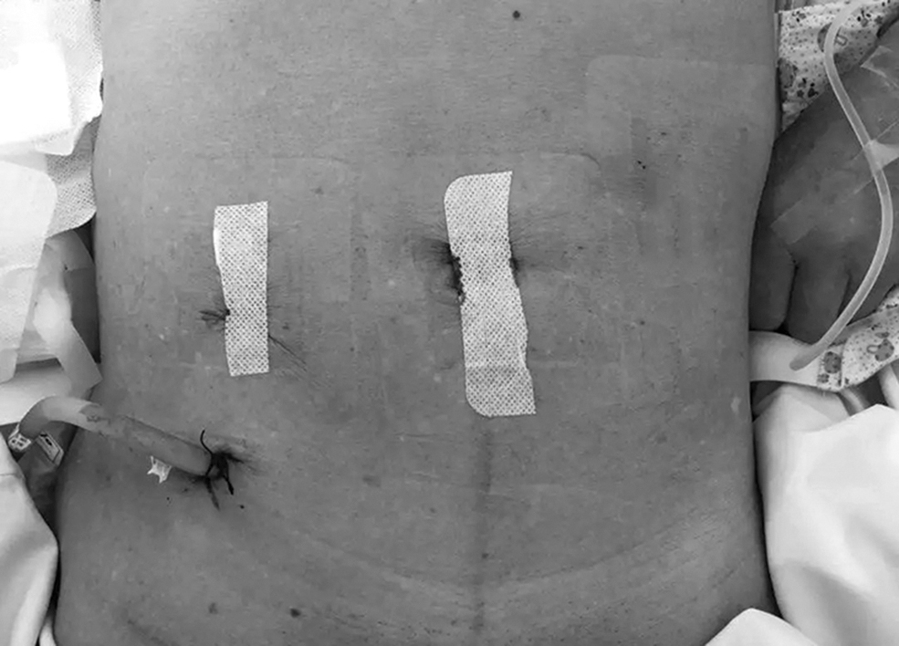
No obvious incision in the abdomen, and only one modified double-lumen irrigation–suction tube was placed.
Statistical analysis
Using SPSS 25.0 statistical software, the counting data were expressed by the number of cases and percentage, the differences between groups were compared by
Results
Cases of colonoscopic perforation
Perforation occurred in 18 cases (0.05%) within 7 days after diagnostic and therapeutic colonoscopy. Details of perforation cases are shown in Table 1. Among the perforation cases, 9 cases(50%) were male, from 25 to 76 years of age, with an average age of 58 years. There were 5 cases of common colonoscopy and 13 cases of anesthetic colonoscopy, and the difference in the incidence of perforation was statistically significant (0.028% versus 0.075%, P = .047). Perforation mostly occurred in the rectosigmoid junction (3 cases) and sigmoid colon (11 cases), accounting for 77.8%, and perforation of terminal ileum (2 cases), cecum (1 case), and ascending colon (1 case). The perforation was found immediately in 9 cases (50.0%), and was at latest discovered 48 hours after the end of the operation. All the patients who received surgical treatment were successfully recovered and discharged from the hospital.
Baseline Characteristics of Eighteen Patients with Colonoscopic Perforation
EMR, endoscopic mucosal resection; ESD, endoscopic submucosal dissection; F, female; Lap, laparoscopic; M, male; OS, open surgery.
Surgical correlation analysis
Among the cases of perforation, all cases were treated by operation, including laparoscopic operation in 13 cases and laparotomy in 5 cases. Repair of perforation was performed in 14 cases, intestinal resection and anastomosis were performed in 2 cases, and a proximal colostomy was performed in 2 cases. Eighteen patients who underwent operation were divided into two groups according to laparoscopy and laparotomy, and the incision size, operation time, intraoperative blood loss, postoperative exhaust time, and hospital stay were compared between the two groups, as shown in Table 2. There were significant differences in incision length, intraoperative blood loss, postoperative exhaust time, and postoperative hospital stay between the laparoscopy group and the laparotomy group. There was no significant difference in operation time between the two groups. There was a statistically significant difference in the incidence of complications between the laparoscopy group and the laparotomy group (23.1% versus 80%, P = .047).
Comparison of Operative Outcomes for Laparoscopic Surgery Group Versus Open Surgery Group in Patients with Colonoscopic Perforation
Values are presented as number (%) or mean (range).
LG, laparoscopic surgery group; OG, open surgery group.
Risk factors of postoperative complications
The incidence of postoperative complications was 38.9%. There were 4 cases of incision infection, 2 cases of abdominal infection, and 1 case of postoperative inflammatory intestinal obstruction. The hospitalization time of perforated patients with postoperative complications was significantly longer than those without complications, with a statistically significant difference ([30.0 ± 11.4] days versus [12.9 ± 3.0] days, P = .007). Eighteen patients who received surgical treatment were divided into two groups according to the occurrence of complications. There were 4 males in the noncomplication group and 5 males in the complication group. There was no significant difference in sex and age between the two groups (68 ± 7 versus 45 ± 18). The differences of possible risk factors between the two groups, such as medication history of glucocorticoid, anesthesia, intestinal preparation, perforation site (rectosigmoid junction and sigmoid colon or not), diagnosis time of perforation (early or nonearly), surgical treatment (resection or nonresection), and other possible risk factors, as shown in Table 3.Univariate analysis showed that the use of glucocorticoid before surgery and perforation in nonrectosigmoid perforation may increase the risk of postoperative complications (P < .05), while the immediate diagnosis of perforation and higher intestinal cleanliness were associated with lower risk (P < .05). Spearman correlation analysis showed that preoperative medication history of glucocorticoid and nonrectosigmoid perforation were positively correlated with postoperative complications (P < .05), while perforation diagnosed immediately and satisfying intestinal cleanliness were negatively correlated with it (P < .05).
Analysis of Risk Factors of Postoperative Complications in Patients with Colonoscopic Perforation (%)
Unilateral P value.
Bilateral P value.
RS, rectosigmoid.
Discussion
According to literature reports, the incidence of perforation in diagnostic colonoscopy is 0.03%–0.8%, while that in therapeutic colonoscopy is 0.15%–2%.3–5 The perforation rate of endoscopic central colonoscopy in our hospital was 0.05%, which was similar to that reported abroad. Previous population-based studies have shown that anesthetic colonoscopy slightly increases the risk of perforation, and the perforation rate of nontherapeutic anesthetic colonoscopy is similar to that of common colonoscopy.6,7 In our study, the perforation rate of anesthetic colonoscopy was significantly higher than that of common colonoscopy. Seven cases of anesthetic colonoscopy perforation in our hospital had complex complications (2 cases of intestinal obstruction, 2 cases of Crohn's disease, 1 case of ulcerative colitis, 1 case of ovarian cancer, and 1 case of connective tissue disease). The difficulty of endoscopic access was significantly increased. Therefore, if there are definite comorbidities, the safety of common colonoscopy may be higher.
About the effect of anesthesia on the risk of colonoscopic perforation, it is necessary to further increase the number of observed cases; on the other hand, it has been previously reported that the risk factors for colonoscopic perforation include female, advanced age, low body mass index, severe comorbidities (such as colon obstruction, colon cancer, inflammatory bowel disease, etc.), history of abdominal and pelvic surgery, and therapeutic measures (such as dilatation of narrow intestinal segment, polypectomy, etc.).8,9
Perforation is most common in the sigmoid colon and the rectosigmoid junction; because of the tortuosity, stenosis, and dissociation of the intestinal canal, the free intestine is easy to be elongated or the endoscopic body is easy to form a loop, and the pressure of the endoscopic body on the intestinal wall increases. If the sigmoid colon is associated with ulcers, diverticula, tumors, or severe inflammation, the intestinal wall is fragile and prone to perforation. 10 Other perforations are mostly related to complications or treatment procedures 11 : in this study, 3 cases of perforation occurred in the ileocecal part, and it was confirmed that severe local adhesion caused by complications was the main cause of perforation.
In many cases, perforation is ignored during colonoscopy, and patients do not have clinical manifestations such as elevated body temperature, abdominal pain, peritoneal irritation sign, and leukocytosis until a few hours after colonoscopy. 2 The management of colonoscopic perforation includes simple conservative treatment, endoscopic closure of perforation, and surgical treatment. As the smaller perforation is quickly covered by colonic fat droop or adjacent mesentery, conservative treatment can be tried in patients with small perforation, clean intestines, limited abdominal pain, and mild peritonitis, but the symptoms and abdominal signs should be closely monitored; if there is high fever, shock, or aggravation of abdominal pain, surgery should be performed in time. 12 In recent years, with the development of endoscopic technology, it has become a treatment option to close perforation immediately after finding perforation under endoscope. It is reported that the success rate of endoscopic closure of perforation is 70%–90%. Compared with surgery, the hospital stay is shorter and the cost is lower.5,13,14 However, it is mostly suitable for perforation <1 cm (especially perforation caused by treatment) and intestinal cleaning, and has high technical requirements for endoscopic physicians. 15 In addition, there is still a high probability of abdominal and pelvic abscess after endoscopic closure perforation, which requires close observation. 16
The most commonly used treatment for colonoscopic perforation is surgery, and the choice of surgical procedure depends on the patient's condition, perforation site and size, intestinal cleanliness, complicated diseases of the damaged intestine, and the time from perforation to surgery. 4 Laparoscopic repair or resection of damaged intestine is gradually underway.17,18 In this study, the majority of patients with perforation were treated by laparoscopy (72.2%). If the perforation is small or located on the mesangial side, it is suggested that: (1) to determine the most seriously contaminated area to reduce the scope of exploration; (2) there is usually local bleeding after intestinal perforation, individual formation of small hematoma, can play a prompt role. If it is found in the late stage, with severe abdominal contamination, significant intestinal edema and poor systemic condition, loop colostomy at the perforated site is feasible. 19 Our results showed that laparoscopic primary repair of colonoscopic perforation was safe and effective, with short incision length, less blood loss, short postoperative hospital stay, rapid recovery of intestinal function, and fewer complications, avoiding the significant increase in operation time. Due to the continuous irrigation and drainage through the double-lumen irrigation–suction tube after operation, there were no complications such as abdominal infection in the laparoscopy group.
In this study, the postoperative complications of surgical treatment of perforation include incision infection, abdominal infection, and postoperative inflammatory intestinal obstruction, of which incision infection is the most common, which was basically consistent with previous reports.3,20 In this study, it was found that the site of perforation, history of hormone use, diagnosis time of perforation, and intestinal cleanliness were significantly correlated with the risk of postoperative complications. Nonrectosigmoid perforation (ileocecal part, ascending colon) is often caused by intestinal adhesion caused by complications, and the perforation injury is large, which will definitely increase the risk of postoperative complications.3,21
In this study, there were 5 cases of perforation with medication history of glucocorticoid (4 cases of inflammatory bowel disease and 1 case of connective tissue disease): glucocorticoid significantly increased the risk of complications after perforation in patients with inflammatory bowel disease. 22 The diagnosis time of perforation and intestinal cleanliness are directly related to the degree of abdominal contamination after perforation; if the diagnosis time of perforation is delayed, or more intestinal feces enter the abdominal cavity, it will directly increase the risk of infection complications, and even cause intraabdominal tissue and intestinal tube edema and brittleness due to acute inflammation, making it difficult to complete primary repair or anastomosis. Previous studies have also pointed out that postoperative complications of surgical treatment within 24 hours after perforation were significantly reduced.20,23 In addition, factors that may affect postoperative complications include smoking, failure of conservative treatment, and American Society for Anesthesiologists grade ≥3.3,21,24
There are some shortcomings in this study. First, this study is a single-center retrospective study, because colonoscopic perforation cases are relatively rare and fewer cases are included, so there is a selection bias. Prospective multicenter registration studies are also needed to obtain information on colonoscopy complications in a broad sense. Second, the exact endoscopic treatment information of all cases was not obtained, and the demographic and clinical information of nonperforation cases were not obtained. It is difficult to use multiple regression analysis to find the possible independent risk factors of perforation. Finally, in the process of exploring the risk factors of postoperative complications, due to the limited population and positive cases, multifactor regression could not be carried out, but through the method of correlation analysis, there are statistical deficiencies.
In conclusion, perforation is a rare but serious complication of colonoscopy, which mostly occurs in rectosigmoid junction and sigmoid colon. Surgical intervention is often required in the management of perforation. Laparoscopic primary repair is safe and feasible in resolving colonic perforation due to colonoscopy, and postoperative complications were significantly related to perforation site, preoperative medication of glucocorticoid, perforation diagnosis time, and intestinal cleanliness.
Footnotes
Disclosure Statement
No competing financial interests exist.
Funding Information
No funding was received for this article.