
Abstract
Background:
The umbilicus is commonly used as an access site to perform minimally invasive colorectal surgery. Umbilical stomas are becoming an attractive option as an alternative site for temporary stomas since they reduce the need for additional abdominal incisions.
Methods:
We retrospectively evaluated patients who underwent umbilical stoma creation after colorectal resection for diagnosis of rectal cancer, diverticulitis, or ulcerative colitis between January 2020 and July 2021. Surgical technique, clinical and perioperative outcomes, complications, and cosmetic end results were described.
Results:
A total of 11 patients underwent umbilical stoma creation. There were no major surgical complications. Peristomal skin irritation was secondary to difficulties with the management of the pouching system. One patient presented with ostomy prolapse. Three patients experienced incisional hernia after stoma reversal. There were no wound infections.
Conclusion:
Umbilical stomas appear to be a safe alternative to conventional stomas and provide superior cosmetic outcomes.
Introduction
Commonly in minimally invasive and robotic surgical techniques, the umbilicus is used as an access port to perform abdominal procedures. In colorectal resections, temporary diverting stomas (e.g., ileostomy and colostomy) are frequently used for high-risk anastomosis with the goal of minimizing the morbidity and septic complications associated with anastomotic leaks. 1
A traditional stoma site transverses the rectus abdominis muscle in the right or left paramedian region of the abdominal wall and is associated with additional scar formation, as well as increased wound-related complications. 2 This decreases the overall cosmetic outcome gained by using a minimally invasive surgical approach.
Umbilical stomas are an attractive option as an alternative site for temporary stomas. They are proven to be both safe and feasible in practice, do not require a special procedure for its construction, and take advantage of the already created umbilical access incision, therefore, reducing the need for additional abdominal incisions.3,4
In this case series, we present 11 patients who underwent creation of an umbilical stoma after colorectal resection with the aim of describing the procedure and documenting the clinical outcomes, complications, and cosmetic end results.
Materials and Methods
A retrospective review of a cohort of patients who underwent colorectal resection with creation of umbilical stomas at a single institution between January 2020 and July 2021 was conducted after Institutional Review Board approval. Patients who required colorectal resection for benign or malignant disease and underwent creation of a temporary diverting ostomy through the umbilical incision were included.
Electronic medical record data were used to obtain patient demographics including age, gender, body mass index (BMI), American Society of Anesthesiologists (ASA) score, medical comorbidities (including hypertension, chronic obstructive pulmonary disease, diabetes mellitus, smoking, renal insufficiency, or immunosuppressant medication use), and indication for surgery. Patient characteristics included diagnosis, procedure, type of stoma, length of hospital stay, days with stoma, and stoma reversal. Stoma-related complications were evaluated.
Surgical technique
The patient was placed on the operating room table in supine position (lithotomy for rectal resections) under general anesthesia. A transumbilical incision was then carried down through the skin and subcutaneous tissues to the fascia. The peritoneal cavity was entered under direct vision and a single site laparoscopic trocar was introduced and the abdomen insufflated to 15 mmHg of carbon dioxide. After completion of the colectomy or ileoanal pouch creation attention was then directed to creation of a transumbilical diverting loop colostomy or ileostomy.
For creation of a diverting loop ileostomy/colostomy, first the umbilical port site fascia was reapproximated using 2.0 polydioxanone (PDS) sutures to ensure a fascial defect that was 5 cm in length for creation of the diverting ostomy. Then a loop of ileum 15 cm from the ileocecal valve, or 15 cm from the afferent limb of the J-pouch, or the mid transverse colon were brought up through the umbilicus to the level of the skin without tension. Using 3-0 Silk sutures the loop of bowel was sutured to the abdominal fascia using seromuscular thickness stitches. The bowel was then opened transversely, and the stoma was matured in the standard Brooke manner using the 3-0 Vicryl sutures. In many cases to avoid skin retraction the ostomy was matured using the skin at the area of the umbilical ring (not the cutting edge of the skin).
One patient had an end ileostomy created at the level of the umbilicus. After completion of a laparoscopic total abdominal colectomy, the terminal ileum was then brought out through the umbilical port site, proper orientation was confirmed. The fascial defect was approximated with 2-0 PDS sutures ensuring a 3 cm fascial defect for creation of the stoma. The terminal ileum was then secured to the fascia with 3-0 Silk sutures to prevent stoma prolapse, the stoma was then matured in the standard Brooke manner with interrupted 3-0 Vicryl sutures (Figs. 1 and 2).

Umbilical stomas. Color images are available online.

Umbilical stoma. Color images are available online.
Stoma care
All patients underwent preoperative ostomy teaching, and 1 had a previous ostomy in a different location. General ostomy management education, including preoperative marking in the event the umbilicus is not feasible at the time of operation, care, and prevention of complications such as dehydration, skin irritation, and proper pouching was provided routinely. All patients had follow-up appointments with ostomy nurses who performed stoma assessments and checked that patients learned how to appropriately manage the pouching system. Patient teaching interventions included pouch preparation, application and emptying, nighttime pouching system use, fluid/dietary principles, and potential ostomy complications.
Stoma closure
Stoma closures were performed through a circumferential incision around the stoma. The bowel was separated from the surrounding soft tissues using sharp dissection. After mobilization of the bowel a functional end-to-end stapled anastomosis was created. The abdominal wall fascial defect was then inspected. Defects that could come together without tension were closed primarily with continuous running 2-0 PDS suture. For fascial defects that were under tension on attempted closure, a mesh repair was utilized for closure.
For mesh repair, the rectus sheath was opened bilaterally, and flaps were raised in the retrorectus space, the posterior rectus sheath was then closed in a running manner with 2-0 PDS suture. A biologic mesh (SurgiMend®) was trimmed to size and placed in the retrorectus space; anterior rectus sheath was then reapproximated with another 2-0 running PDS suture. The dermal layer was then fixed to the fascia with 3-0 Vicryl sutures. Skin was loosely approximated with interrupted 3-0 Monocryl sutures to allow for wound drainage.
Results
A total of 11 patients underwent umbilical stoma creation from January 2020 to July 2021. Median age was 58 years (26–71) and median BMI 25.3 (21.3–33.4). There were 5 (45.5%) female and 6 (54.5%) male patients. Indications for surgery included rectal cancer (63.6%), diverticulitis (18.2%), and ulcerative colitis (18.2%). Patient characteristics are summarized in Table 1. Detailed and individual patient characteristics, including diagnosis, operative intervention performed, and stoma type, are displayed in Table 2.
Patient Characteristics
Comorbidities: hypertension, chronic obstructive pulmonary disease, diabetes mellitus, smoker, renal insufficiency, on immunosuppressors.
ASA, American Society of Anesthesiologists; BMI, body mass index.
Baseline, Operative, and Postoperative Characteristics
End converted to loop stoma after 91 days.
IPAA, ileal pouch-anal anastomosis; LAR, low anterior resection; LOS, length of hospital stay; TAC, total abdominal colectomy.
The median postoperative length of stay was 5 days (3–14). There was no mortality. One patient presented on postoperative day 54 with stoma prolapse, this required revision and relocation of the ostomy to the right lower quadrant.
The median time to stoma reversal was 118 days (54–288). Ten (91%) of the stomas were reversed; one is awaiting reversal due to the patient requiring dilation of the coloanal anastomosis after low anterior resection. A patient with ulcerative colitis underwent a three-stage surgical management; he had a total abdominal colectomy with end ileostomy, which was converted into a diverting loop ileostomy 91 days later after completion proctectomy and ileal pouch-anal anastomosis (IPAA) (Table 2) this patient's stoma was then reversed.
Another patient who underwent completion proctectomy with IPAA required creation of an umbilical stoma due to a shortened mesentery that would place the stoma under significant tension in the right lower quadrant. The umbilical stoma was chosen in this situation due to a thinner abdominal wall at the umbilicus. This patient did develop stoma retraction and difficulty with the pouching system that led to significant skin irritation (Fig. 3).
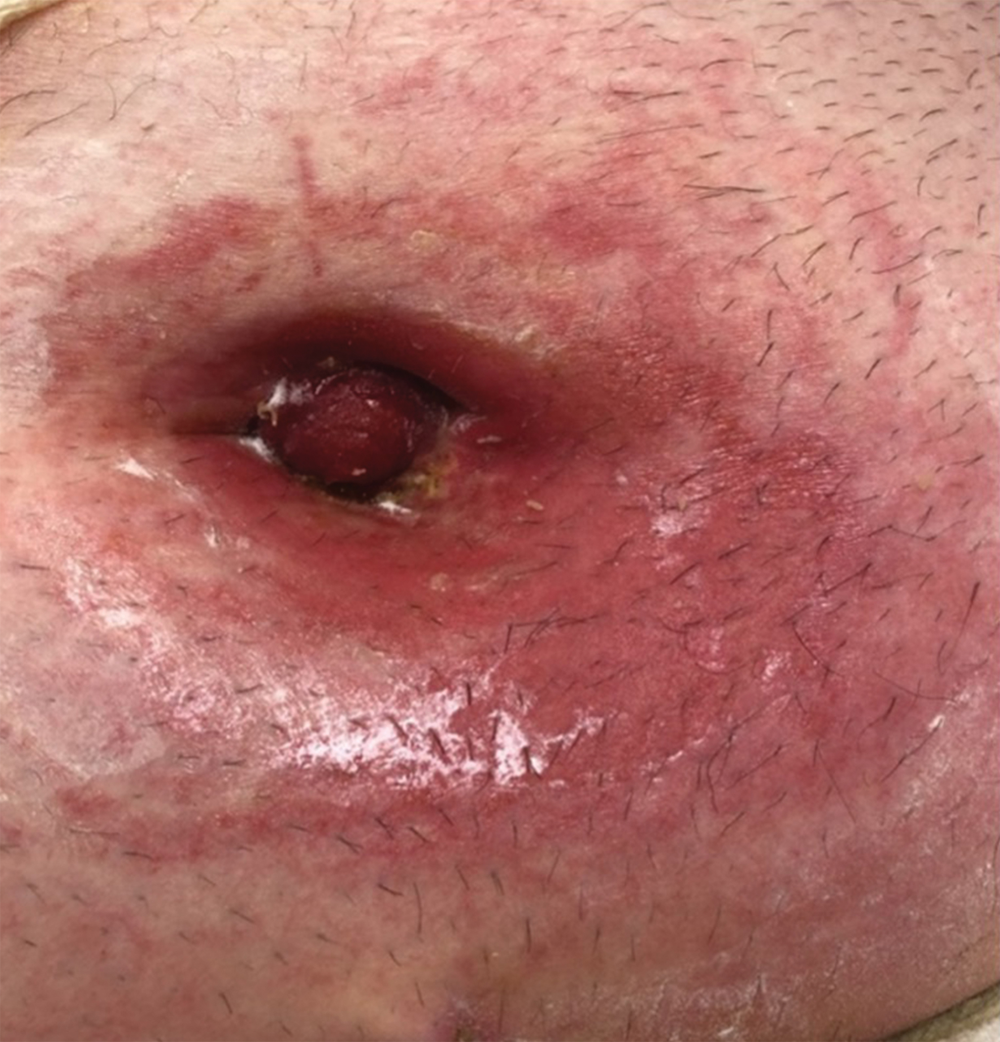
Umbilical stoma retraction and skin irritation. Color images are available online.
Most of the patients who developed peristomal skin irritation was secondary to difficulties with the management of the pouching system (Fig. 4). No surgical site infections occurred after umbilical stoma reversal. After closure of the umbilical ostomy, 3 (27.3%) of the patients developed an incisional hernia, with a median time of 142 days (113–142) after stoma reversal.
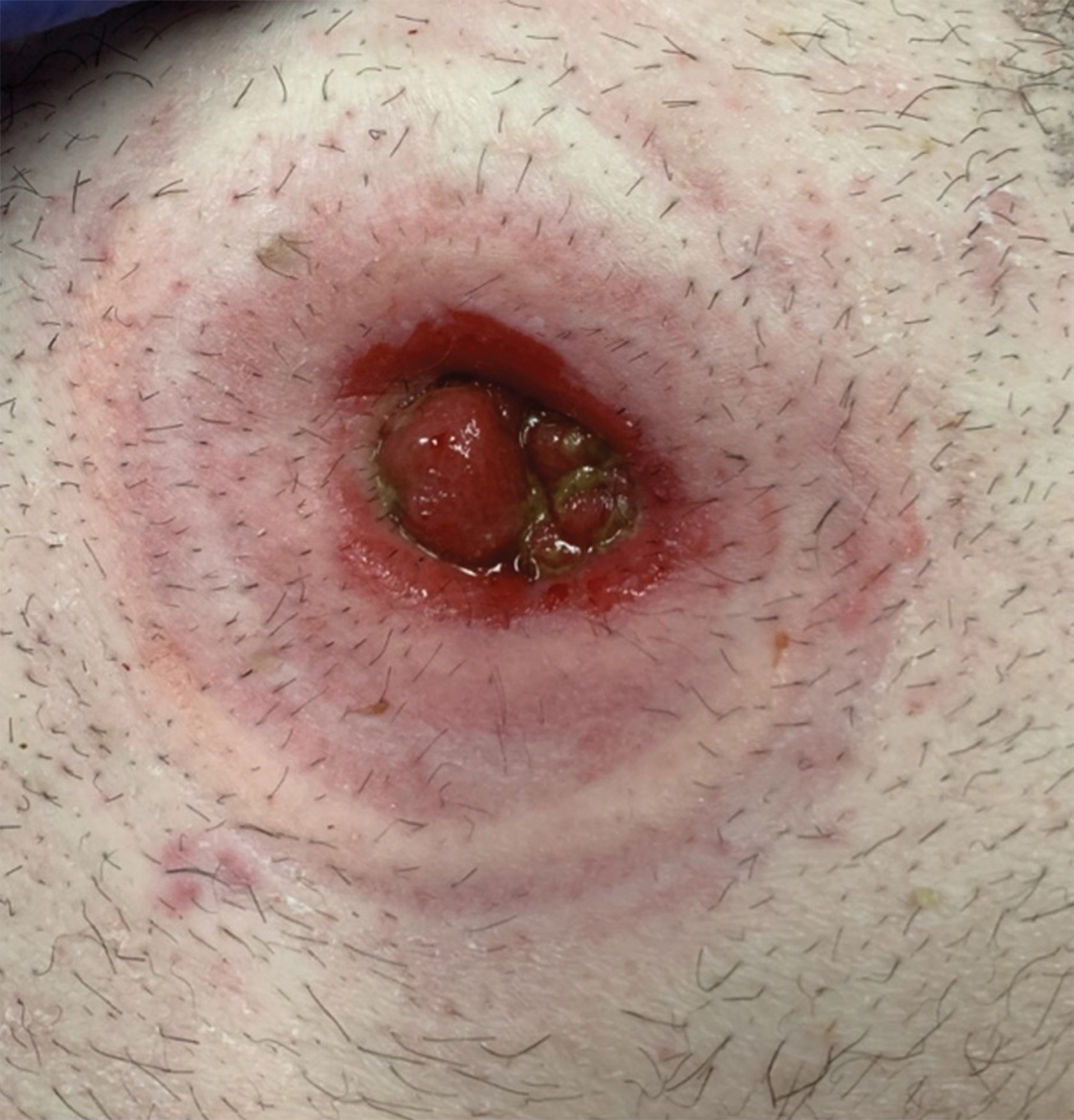
Peristomal skin irritation. Color images are available online.
Figure 5 depicts an example of the final cosmetic appearance of the umbilical site after stoma closure.

Cosmetic result of the umbilicus after stoma reversal. Color images are available online.
Discussion
The use of umbilical stomas was first described by Cameron and Lau in the 1980s, as a site for a temporary colostomy in infants with Hirschsprung's disease or imperforate anus, with the purpose of improving the long-term cosmetic results. 5 Since its initial description, there have been several reports regarding its use in the adult population; however, this practice is not routinely implemented.4,6,7
In laparoscopic and robotic surgery, the umbilicus is used as an access port for instruments and for specimen extraction. By constructing a temporary diverting stoma in this location, additional incisions and scars can be avoided, thus one of the biggest advantages of umbilical stomas is the improved cosmetic result since the incision is hidden in the umbilicus.
Traditional ostomies are brought out through the rectus abdominis muscle, in this technique the bowel traversed two layers of fascia and the rectus muscle. The pressure difference between the bowel and abdominal wall can predispose to stoma complications such as obstruction. Furthermore, when reversing the ostomy, adhesions of the intestinal tract to the muscle are common, which can lead to a more difficult dissection and increased risk of bowel injury; this predisposes patients to wound infections and delayed healing. 8
Umbilical stomas are brought up through a midline incision, avoiding the rectus muscle. This makes stoma reversal easier, safer, and faster since adhesions to the rectus muscle will not be encountered. Likewise, since the abdominal wall is thinner in the midline at the umbilicus (only fascia and subcutaneous fat), access to the abdominal cavity is easier, requiring less bowel mobilization for stoma reversal. In this case series, we did not encounter any bowel injuries during stoma reversal nor closure site wound infections.
Utilization of the umbilicus for stoma creation does, however, present with common disadvantages. Patients frequently encountered difficulties with the pouching system, resulting in increased incidence of skin irritation, compared with patients with conventional stoma sites. In our case series, a common trouble with the pouching system due of the abdominal fold creating a poor seal and often resulting in leaks that generated skin irritation and dermatitis, which in some cases was severe.
The incidence of incisional hernia in conventional ostomy sites is reported to be up to 30%. 9 Three of our initial umbilical stoma patients developed incisional hernia after umbilical stoma reversal; to prevent this complication, in the last three cases a prophylactic biologic mesh was used during stoma reversal. Recent studies have shown that the use of prophylactic mesh during stoma reversal decreases the rate of incisional hernias; therefore, biologic mesh reinforcement should be considered in these patients. 10
Umbilical stomas provide an excellent cosmetic option for temporary stoma creation in patients undergoing colorectal resection who will require a temporary diverting ileostomy or colostomy. When compared with traditional stoma location through the rectus sheath, there is no increased rates of parastomal or incisional hernia after stoma reversal. In addition, there were no surgical site infections; patients, however, encountered difficulty with stoma management, which resulted in skin irritation and dermatitis.
Although the umbilicus serves as an excellent option for temporary diverting stomas, it should not be chosen for permanent stoma creation. An ideal operative candidate for umbilical stoma selection would be a young patient undergoing restorative proctocolectomy, since they are expected to undergo two to three procedures within 5 months and will require a temporary stoma. These patients, usually <40 years old, end up having a more cosmetic and barely visible umbilical scar after the completion of all the procedures (Fig. 6).
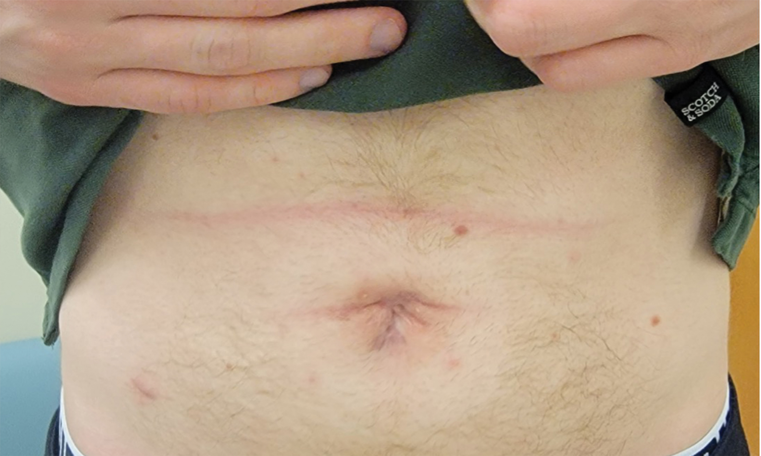
Twenty six-year-old male, status postrestorative proctocolectomy with J-pouch and umbilical stoma, now 2 months after stoma reversal. Color images are available online.
Conclusion
Diverting umbilical stomas appear to be a safe alternative with superior cosmetic outcomes to conventional stomas, provided the area around the umbilicus falls within the criteria for stoma marking such as avoiding skin folds, irregular contours, and patient is able visualize the stoma. 11 Larger prospective studies are needed to evaluate their long-term outcomes and superiority when compared with conventional stomas.
Footnotes
Disclosure Statement
No competing financial interests exist.
Funding Information
No funding was provided.